Copyright
©The Author(s) 2025.
World J Radiol. Sep 28, 2025; 17(9): 110906
Published online Sep 28, 2025. doi: 10.4329/wjr.v17.i9.110906
Published online Sep 28, 2025. doi: 10.4329/wjr.v17.i9.110906
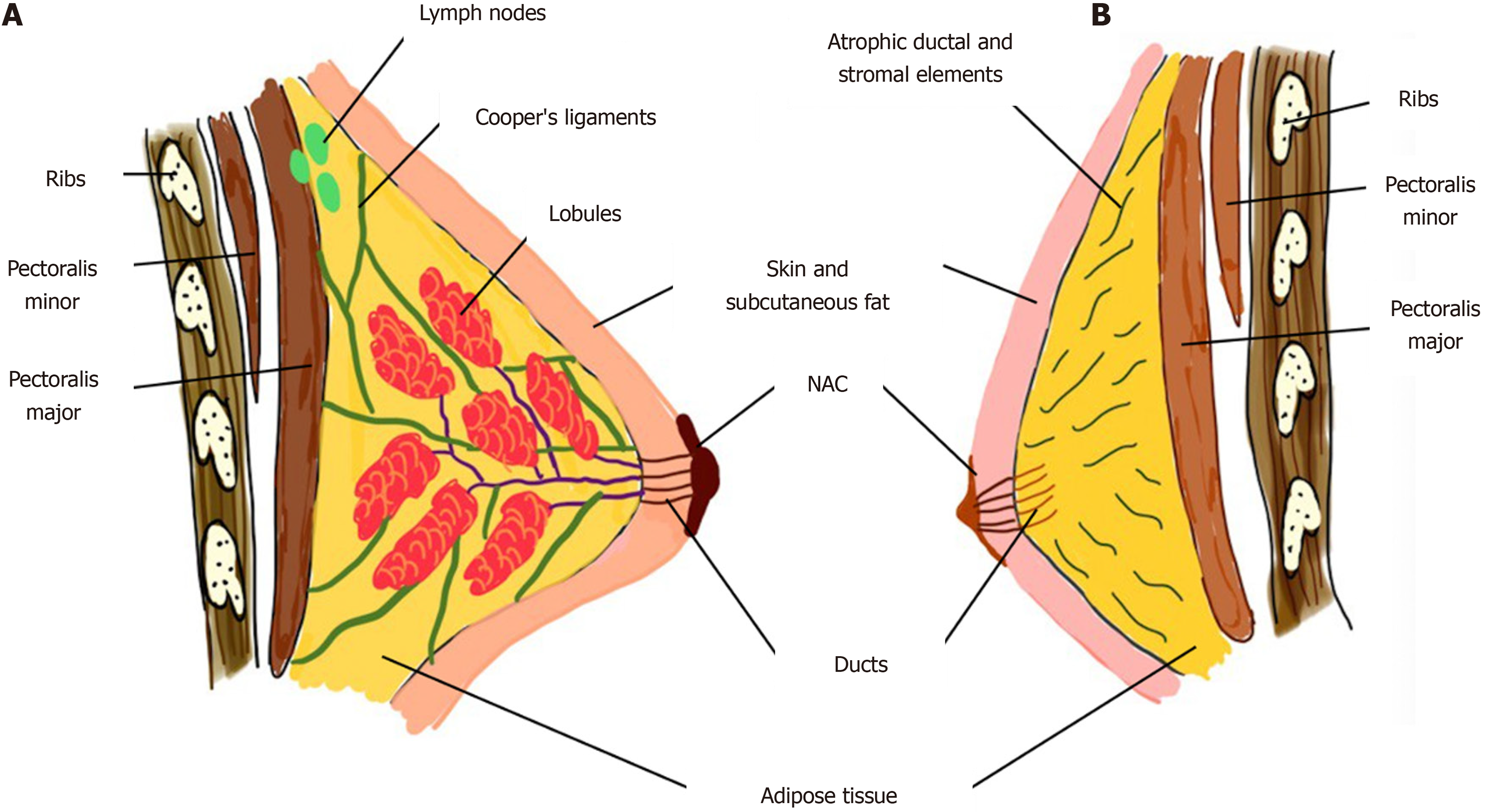
Figure 1 Diagrammatic representation of male vs female breast anatomy.
NAC: Nipple areola complex.
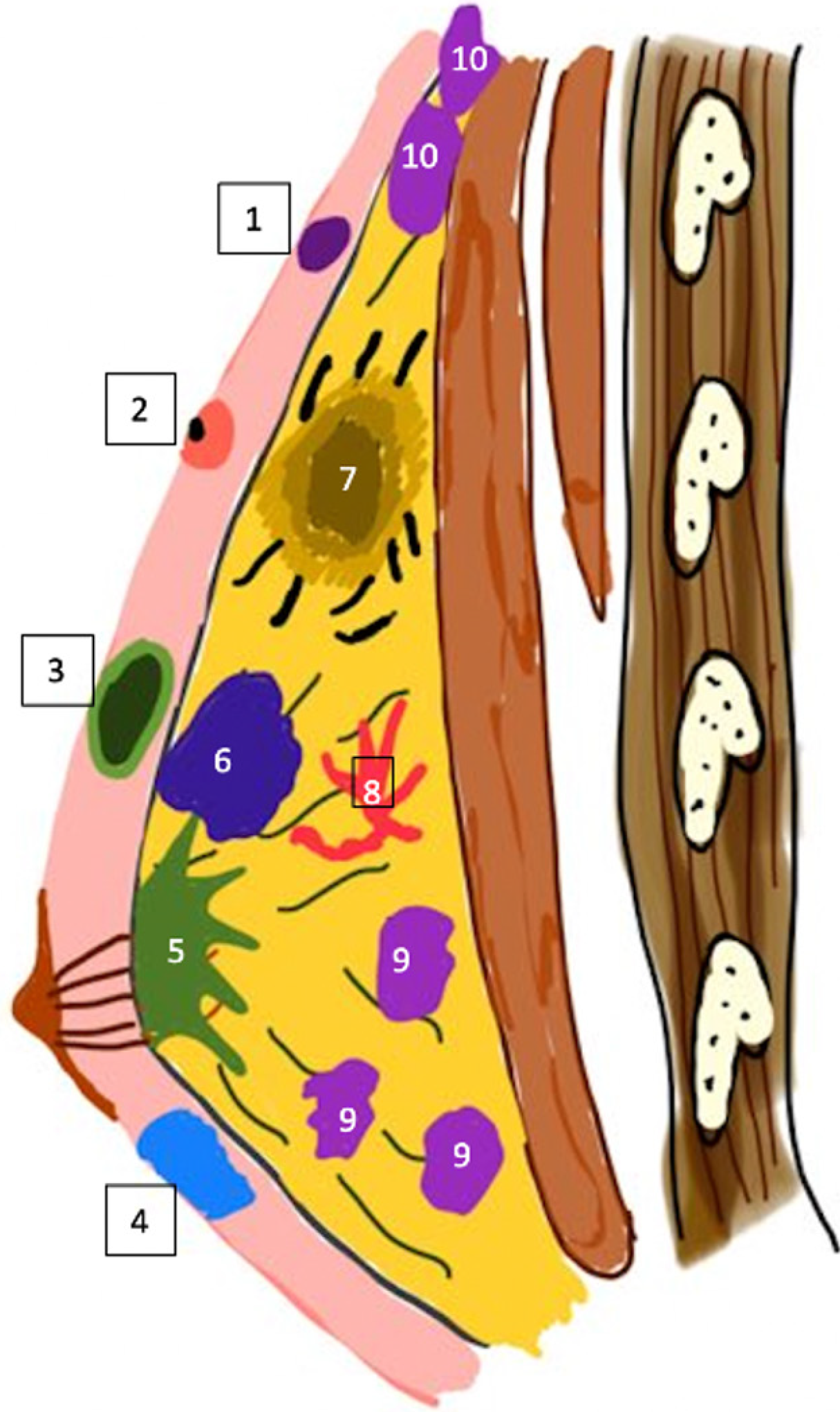
Figure 2 Diagrammatic representation of various male breast lesions according to their location.
1: Epidermal inclusion cyst; 2: Sebaceous cyst with punctum; 3: Lipoma with capsule; 4: Hematoma; 5: Gynecomastia; 6: Carcinoma breast; 7: Mastitis with abscess and surrounding trabecular thickening; 8: Vascular malformation; 9: Lymphoma; 10: Axillary lymph nodes.
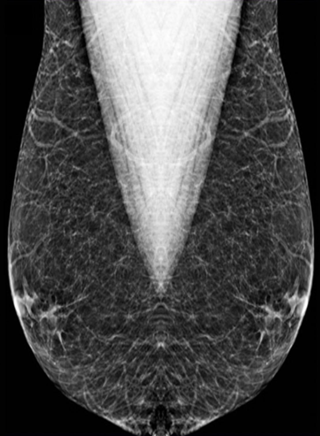
Figure 3
Pseudogynecomastia: Mediolateral oblique mammography view of a 17-year-old obese male showing a diffuse bilateral increase in fat density.
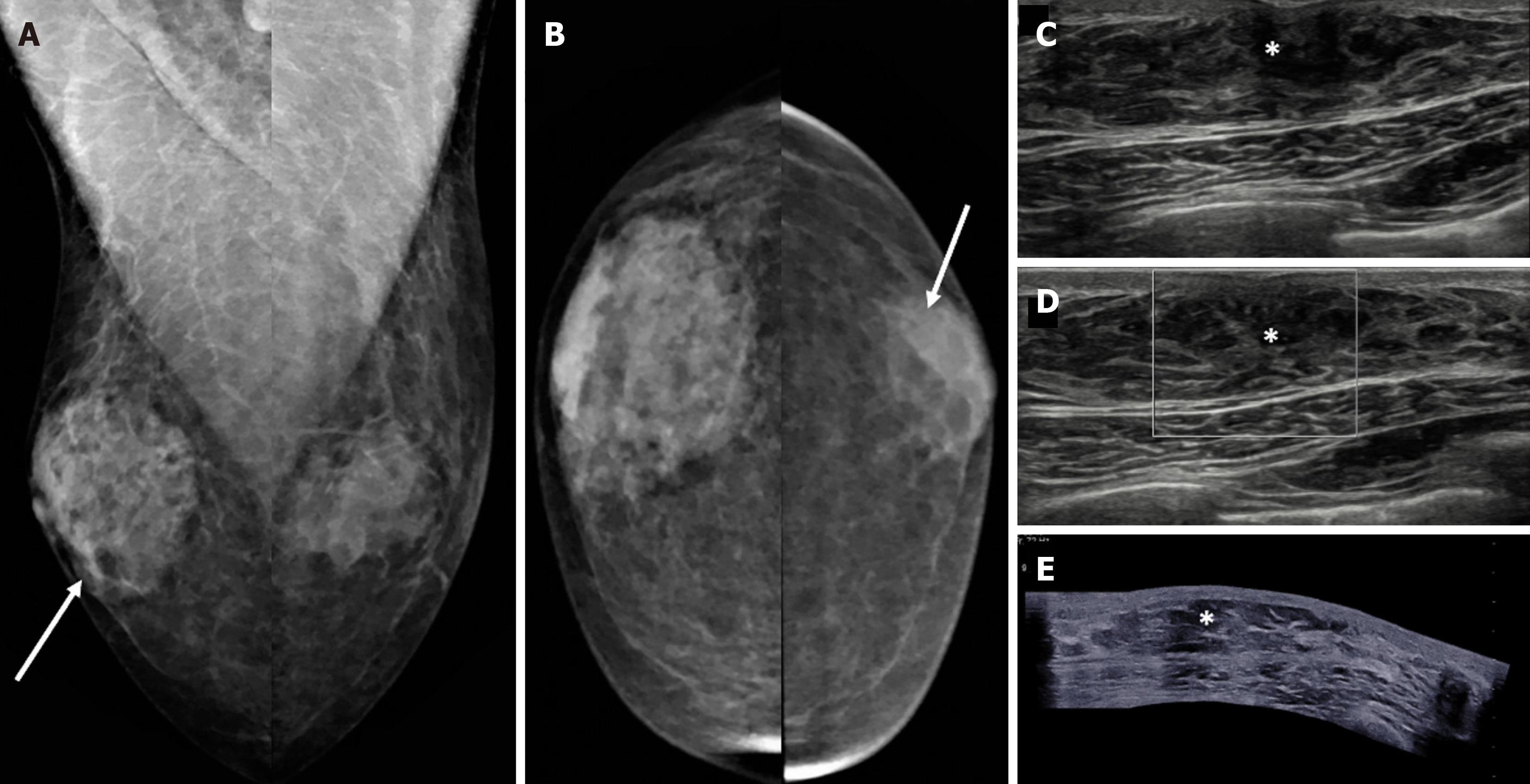
Figure 4 Nodular gynecomastia in a 62-year-old male with tender subareolar masses in bilateral breasts.
A: Mediolateral oblique; B: Craniocaudal mammography views show oval equal density masses in the retroareolar locations of both breasts (white arrows); C-E: Ultrasound images [C, D (color Doppler) and E (panoramic views)] demonstrate an oval parallel indistinct hypoechoic area (denoted by asterisks) in the retroareolar location with no evident vascularity, which is consistent with gynecomastia.
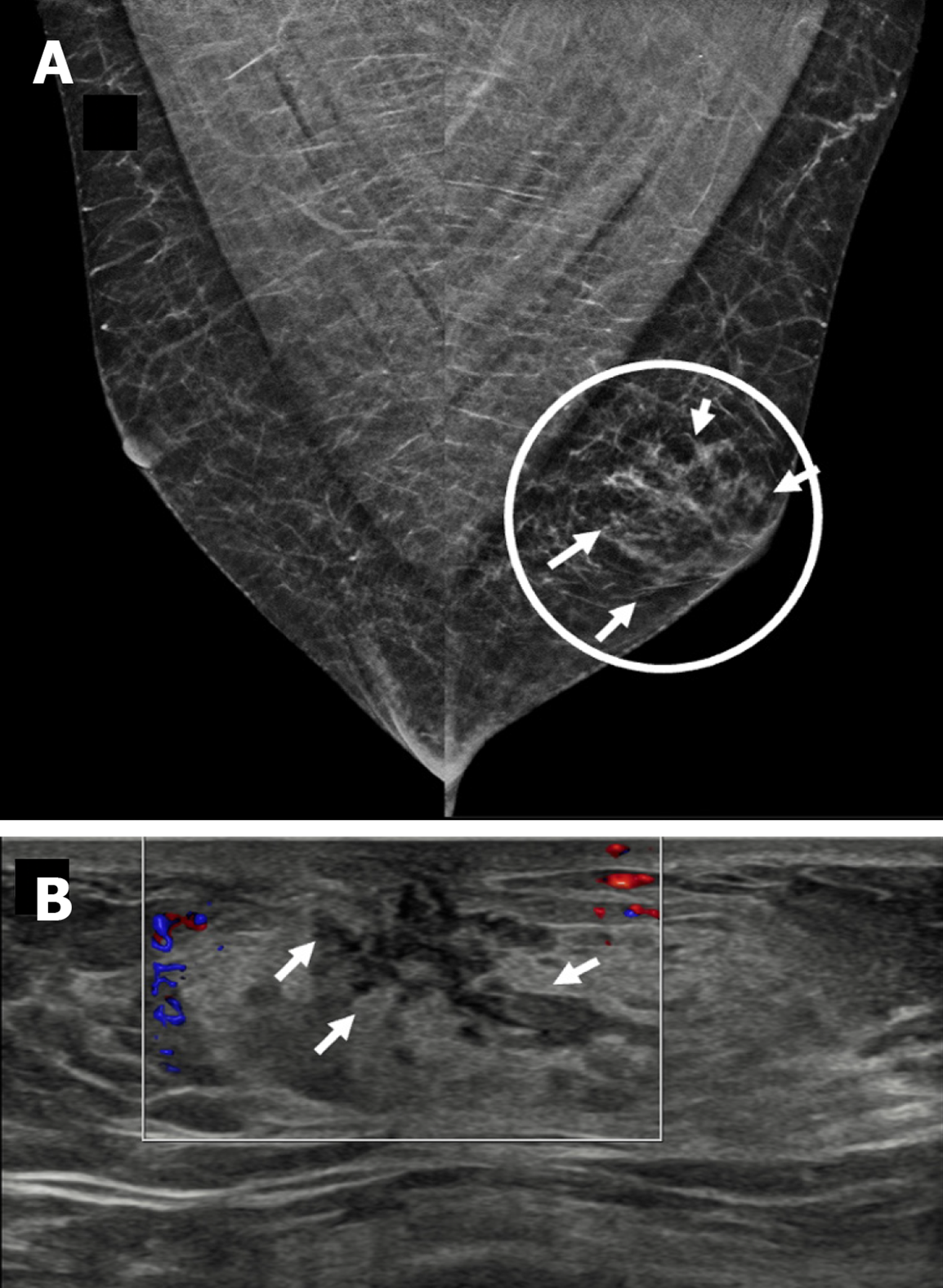
Figure 5 Dendritic gynecomastia in a 69-year-old male with a tender soft mass in the left breast.
A: Mediolateral oblique mammography view showing a triangular flame-shaped area of increased density in the retroareolar location of the left breast (encircled); B: Ultrasound image showing an irregular hypoechoic retroareolar lesion with dendritic projections inside the underlying glandular fat (small white arrows).
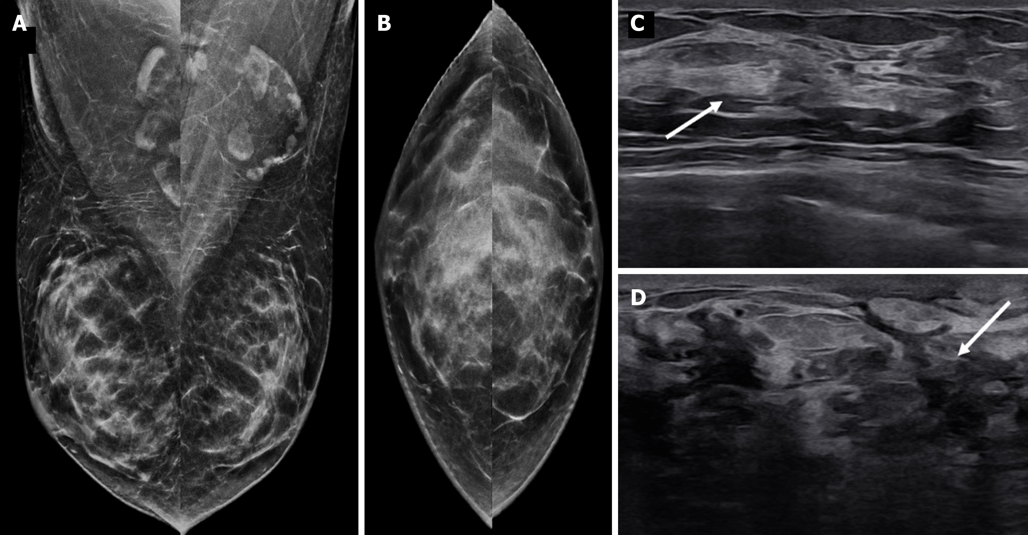
Figure 6 Diffuse gynecomastia in a 42-year-old male on estrogen therapy.
A: Mediolateral oblique; B: Craniocaudal mammography images showing a diffuse increase in breast density, with the male breast resembling the female breast; C and D: Ultrasound images showing diffuse deposition of glandular tissue (white arrows).
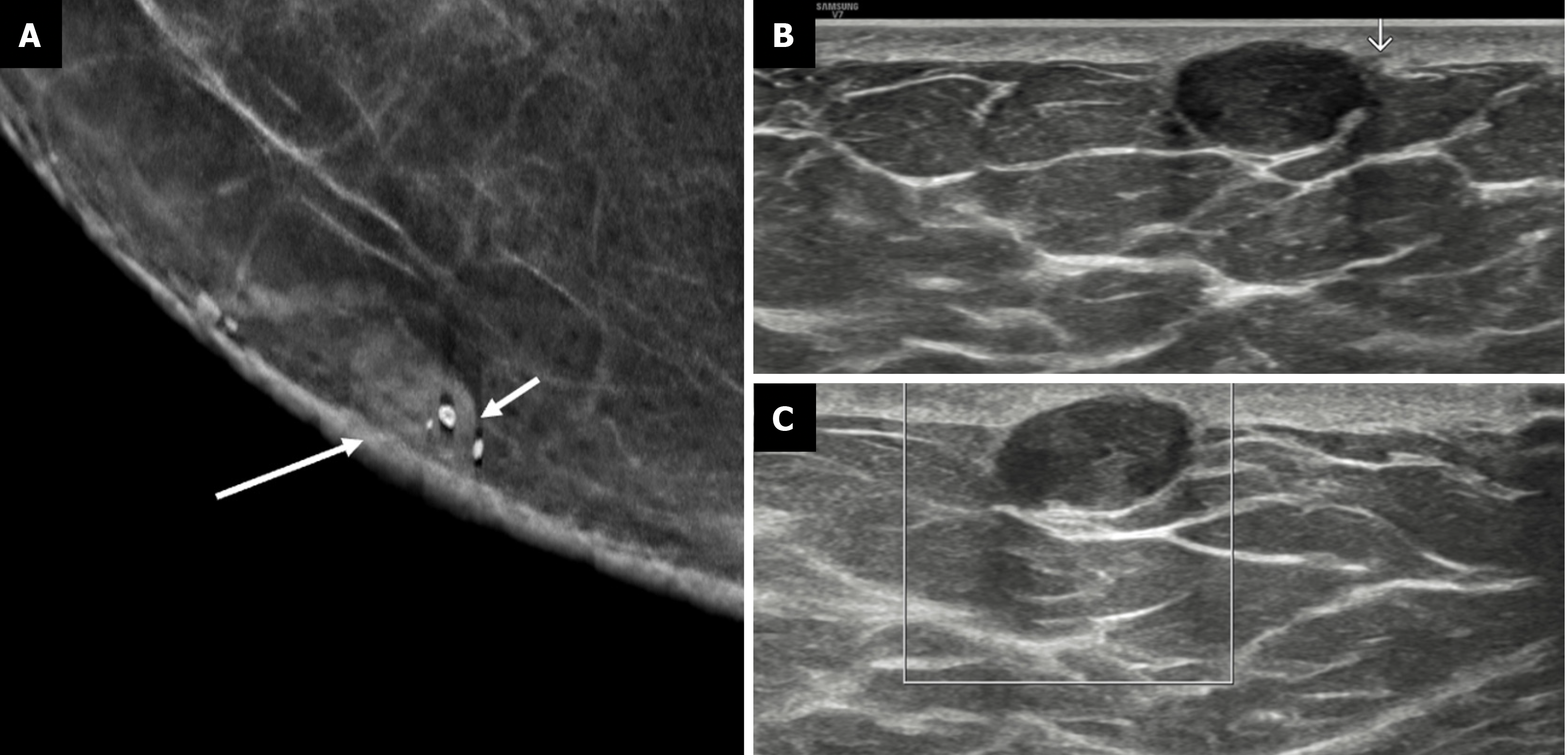
Figure 7 Epidermal inclusion cyst breast in a 23-year-old male with a palpable nodule and a history of blunt trauma to the chest.
A: Mag
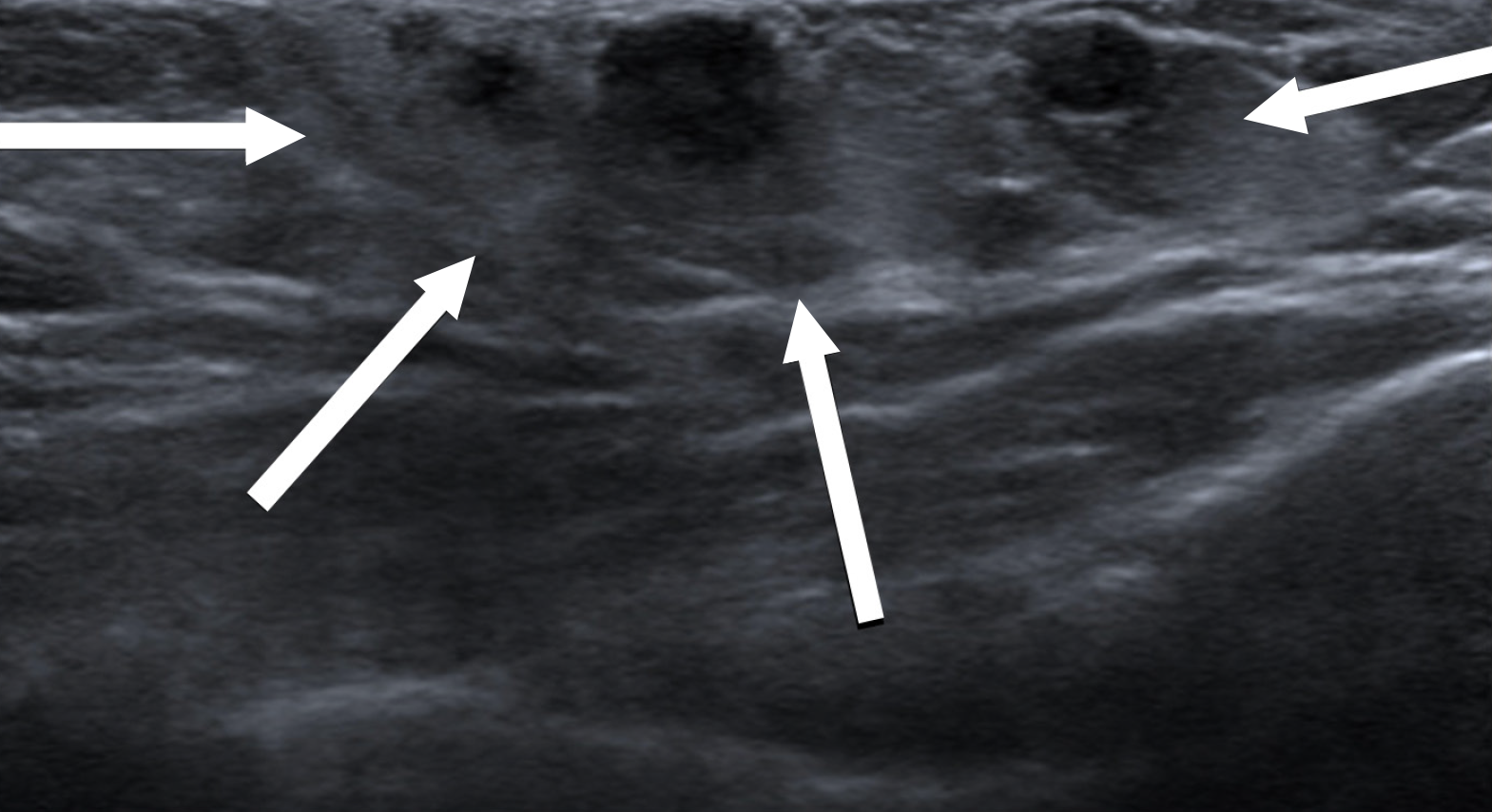
Figure 8
Fat necrosis in a 23-year-old male who suffered trauma to the right breast by football shows an irregular hyperechoic area with indistinct margins (white arrows) containing few anechoic areas.
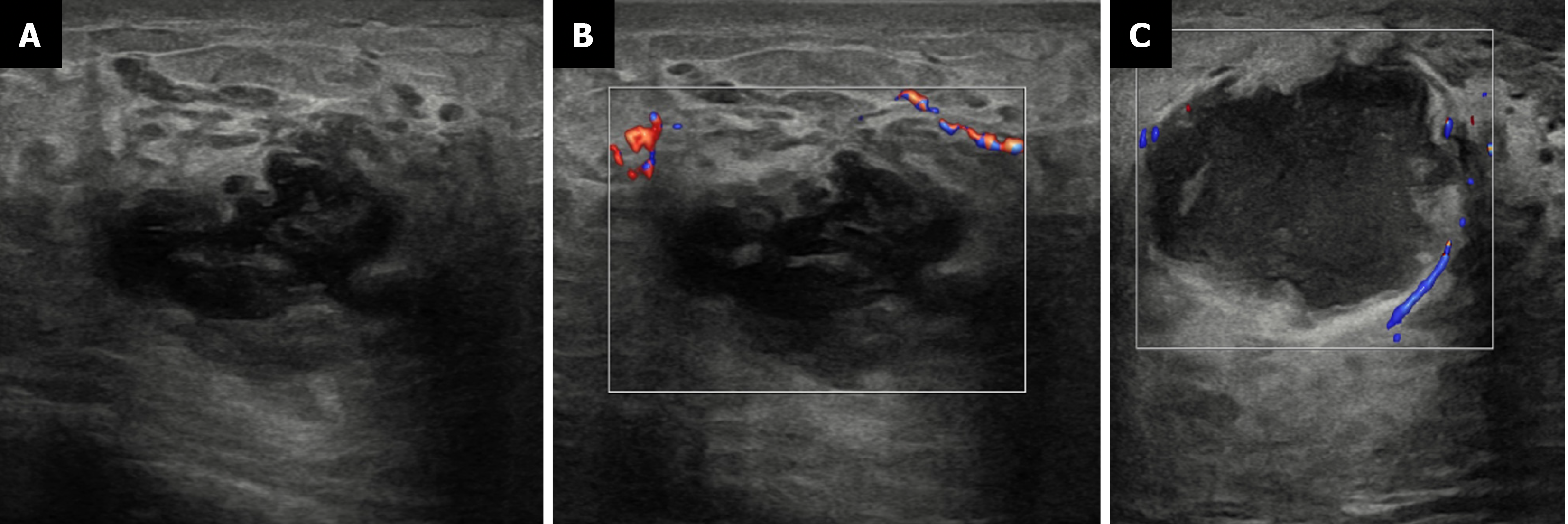
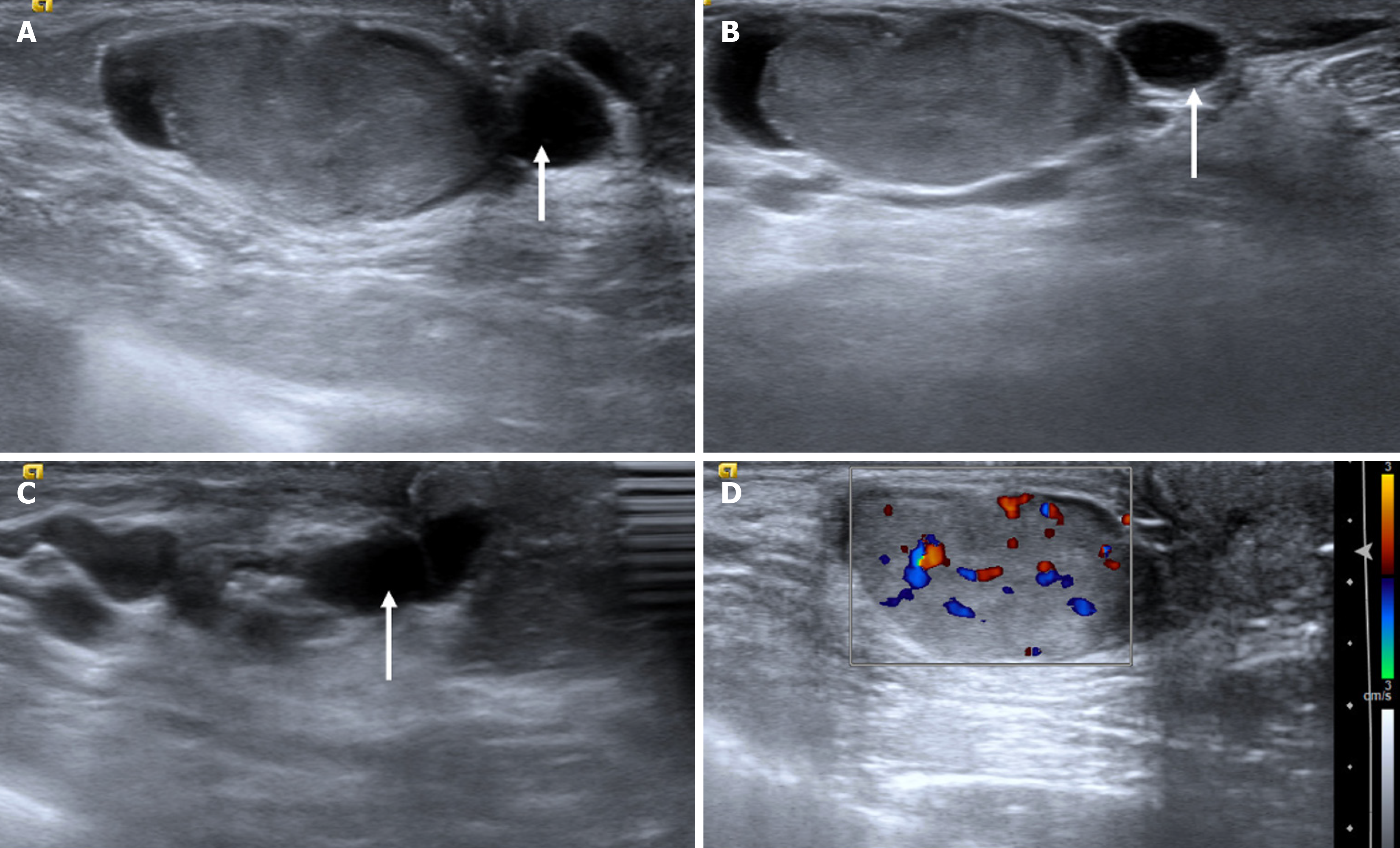
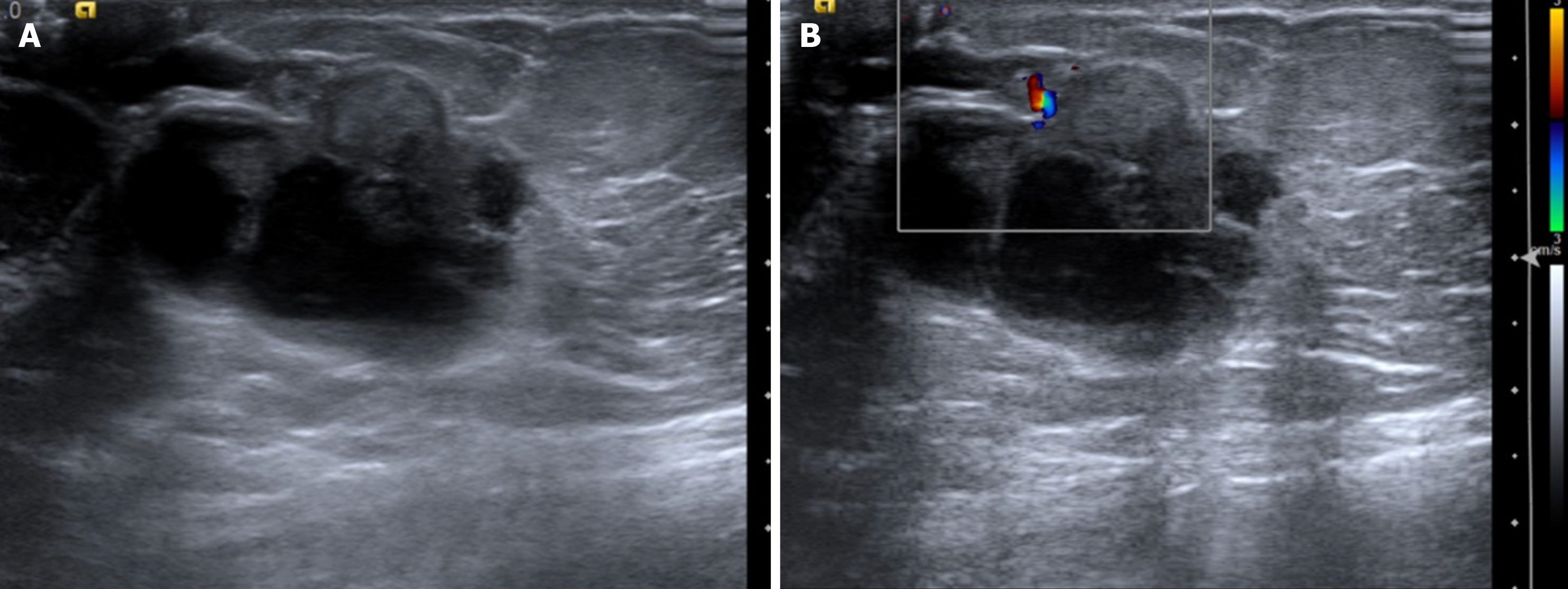
Figure 9 Mastitis with abscess in a 61-year-old diabetic male with a tender enlarged breast.
A: Ultrasound; B and C: Color Doppler images showing an irregular cystic collection with internal debris and peripheral vascularity. The surrounding fat appeared echogenic, suggesting inflammatory changes.
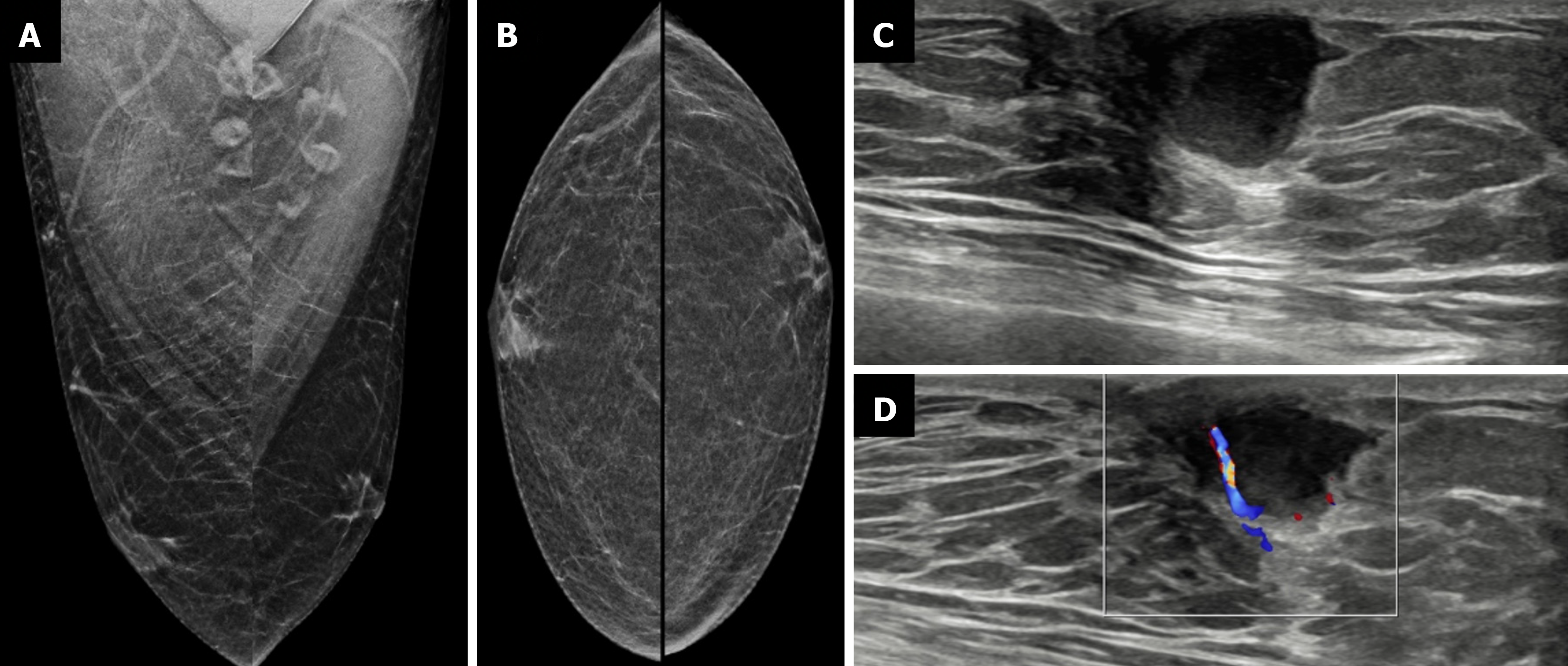
Figure 10 Infective collection from a 23-year-old male with a history of high-grade fever after trauma to the left breast.
A: Mediolateral oblique; B: Craniocaudal mammography views showing no obvious abnormality; C: Ultrasound; D: Color Doppler images revealing a well-defined anechoic collection with peripheral internal vascularity.
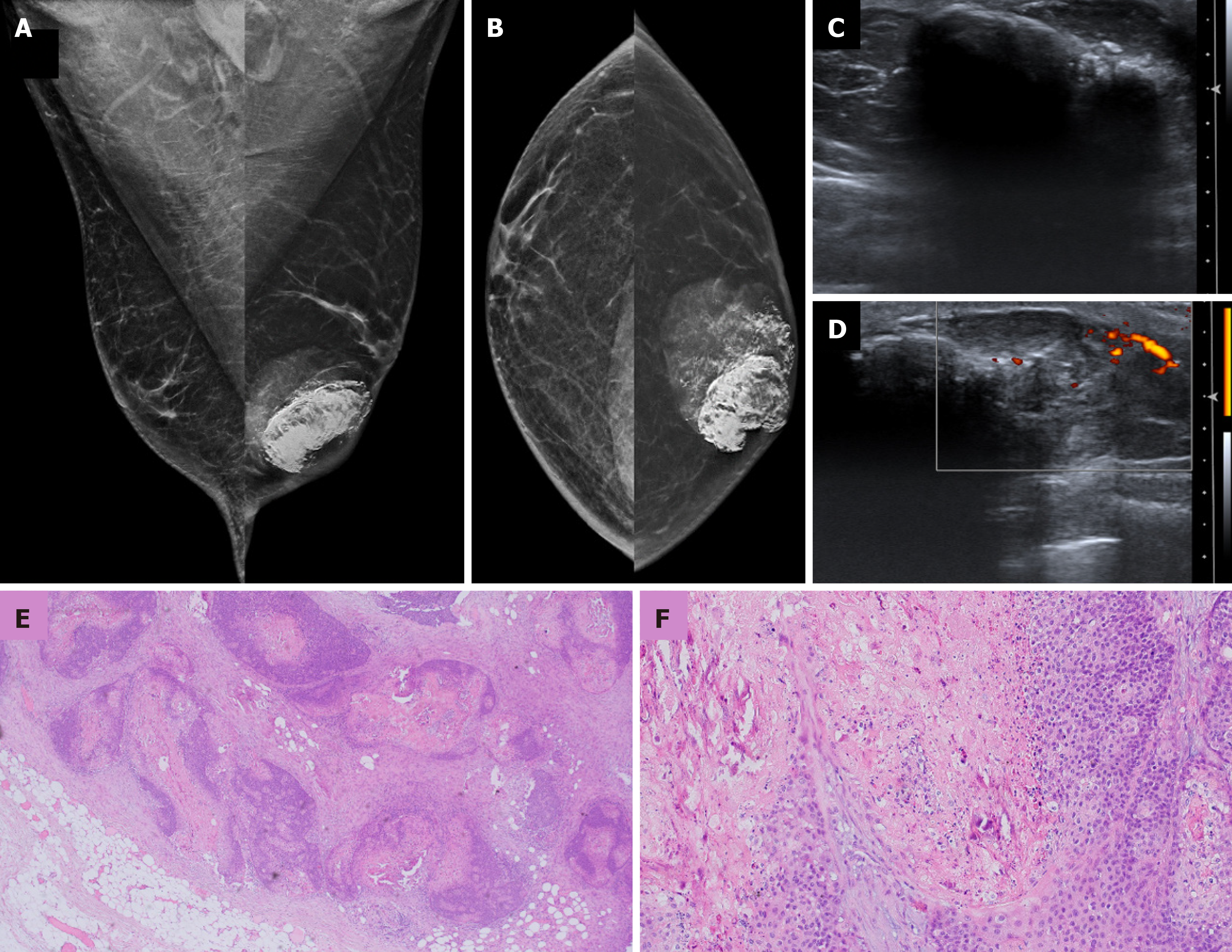
Figure 11 Pilomatricoma in a 33-year-old male with a palpable left breast mass.
A: Mediolateral oblique and B: Craniocaudal mammography images showing a large irregular high-density mass with circumscribed margins and coarse calcifications in the retroareolar location; C: Ultrasound; D: Color Doppler images showing an irregular lesion with extensive posterior acoustic shadowing and some internal vascularity; E and F: Histopathology images (hematoxylin and eosin, × 40) showing a circumscribed lobulated mass in subcutaneous tissue and islands of basaloid cells exhibiting abrupt keratinization without an intervening granular layer along with shadow cells and central calcification.
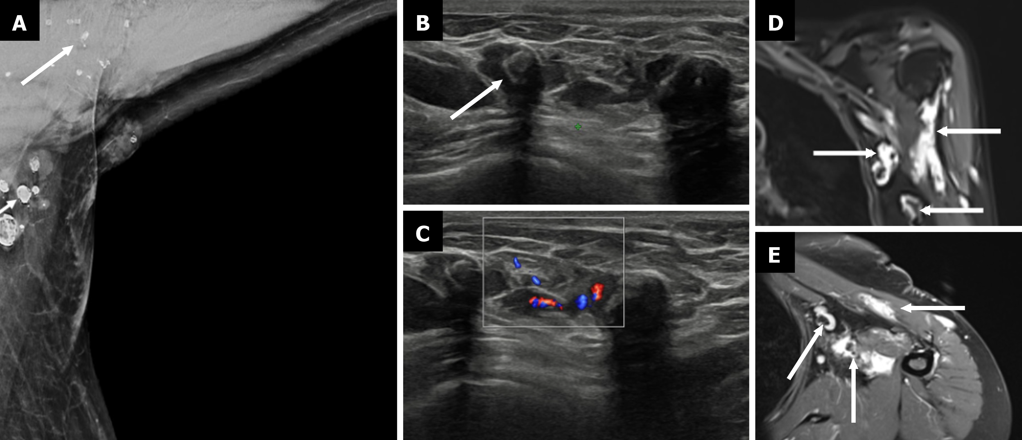
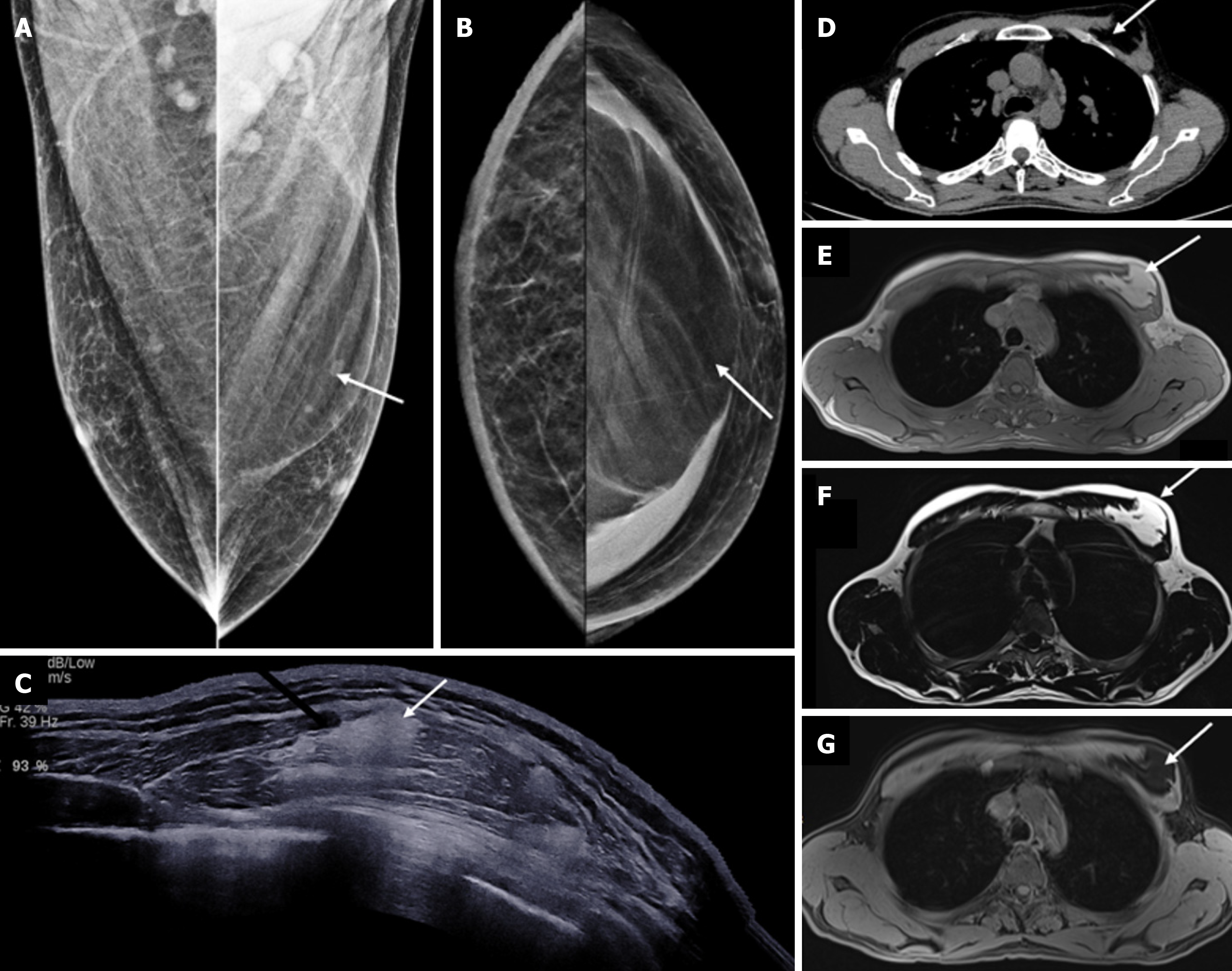
Figure 12 Venolymphatic malformation.
A: Mediolateral oblique mammogram with an axillary view shows multiple well-defined, lobulated densities in the axilla (arrow), some containing coarse calcifications; B: Grayscale ultrasound reveals multiple anechoic and hypoechoic compressible tubular channels in the subcutaneous plane with echogenic phleboliths (arrow), which is consistent with dilated venous and lymphatic components; C: Color Doppler ultrasound demonstrates slow flow within some of the vascular channels; D: Coronal T2-weighted fat-saturated magnetic resonance image (MRI) shows hyperintense lobulated masses (arrows) in the axilla; E: Postcontrast axial T1-weighted fat-suppressed MRI shows enhancement of the lesion (thin arrows), with nonenhancing phleboliths, which is consistent with a benign low-flow vascular malformation.
Figure 13 Lipoma in a 34-year-old male with a palpable mass in the axilla.
A: Ultrasound; B: Elastography images showing a circumscribed oval homo
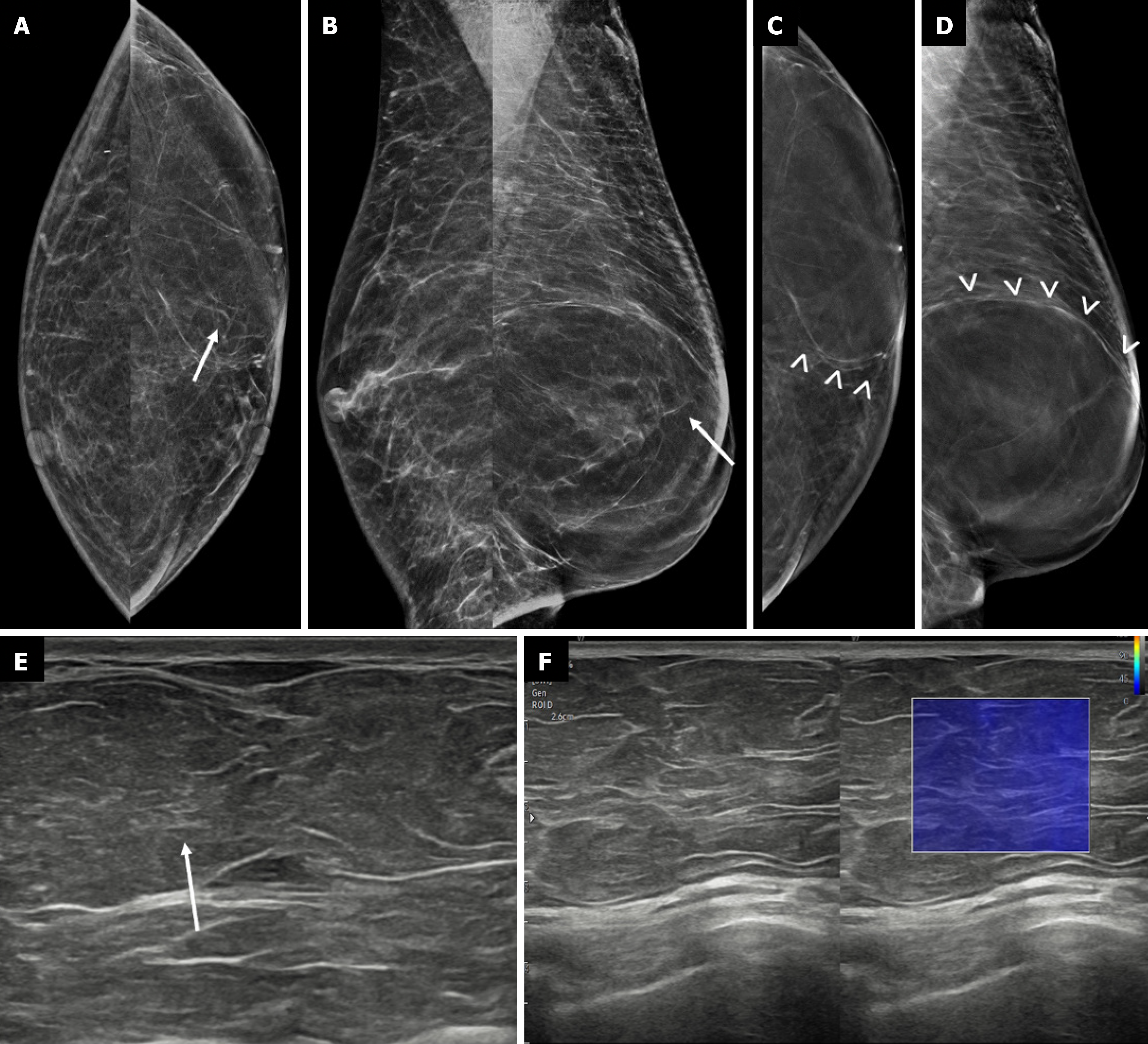
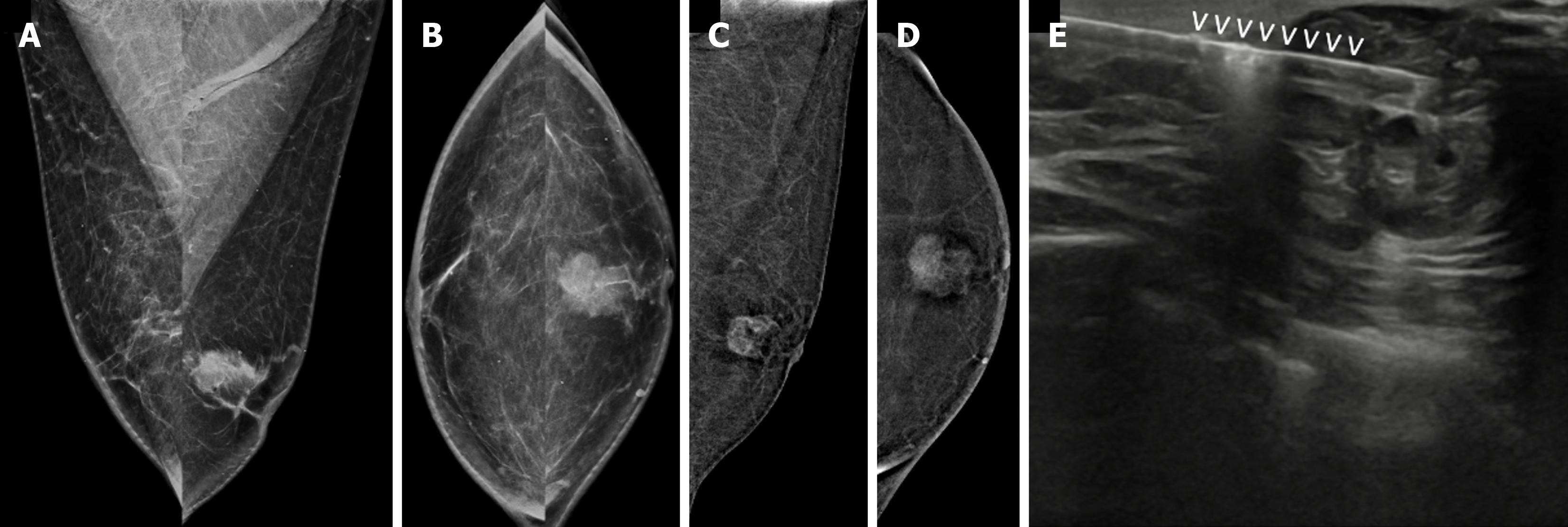
Figure 14 Giant lipoma in a 52-year-old male with a palpable nontender mobile mass in the left breast.
A: Mediolateral oblique; B: Craniocaudal mammography views showing a large, circumscribed round fat density mass in the left breast; C and D: Digital breast tomosynthesis slices demonstrating the lipoma capsule (denoted by vvv); E: Ultrasound; F: Elastography images showing a fat-containing isoechoic mass that appears soft on elastography.
Figure 15 Intrapectoral lipoma in a 45-year-old male with a palpable mass in the left breast.
A: Mediolateral oblique; B: Craniocaudal mammo
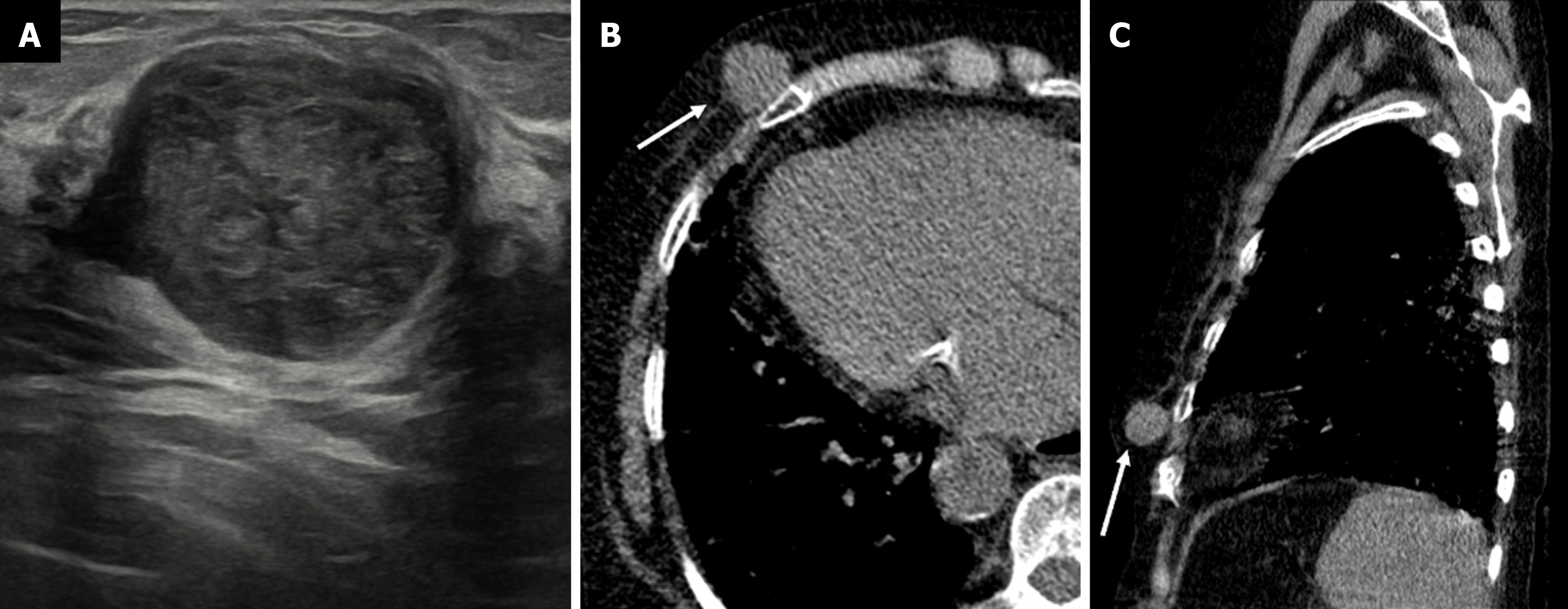
Figure 16 Myofibroblastoma in a 56-year-old male with a palpable mass in the right breast.
A: Ultrasound image showing a round circumscribed heterogeneously hypoechoic mass; B: Axial; C: Sagittal noncontrast computed tomography sections demonstrating a well-defined hypoattenuating soft tissue density mass in the right breast (white arrows) with no calcifications or chest wall invasion.
Figure 17 Intraductal papilloma in a 46-year-old male with a palpable abnormality.
A-C: Ultrasound; D: Color Doppler images showing a circumscri
Figure 18 Ductal carcinoma in situ in a 69-year-old male with a palpable lump in the left breast.
A: Mediolateral oblique (MLO) view; B: Cra
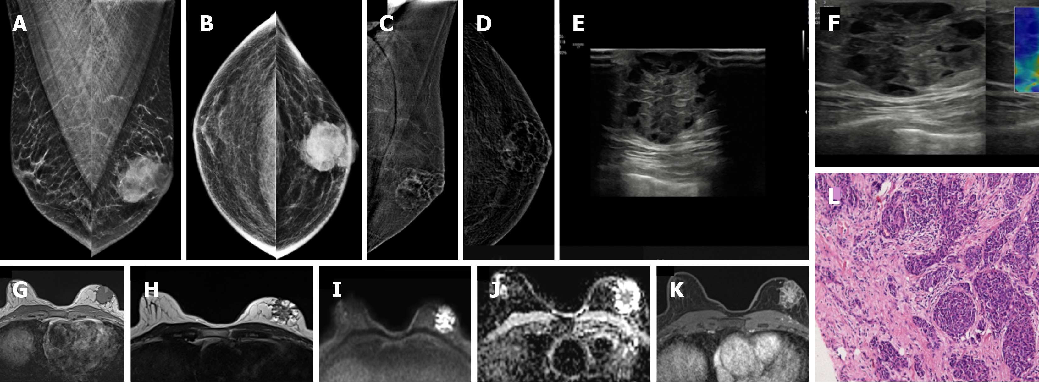
Figure 19 Invasive breast cancer in a 74-year-old male with a hard lump in left breast.
A: Mediolateral oblique (MLO) view; B: Craniocaudal (CC) view showing an irregular, high-density mass; C and D: Contrast-enhanced mammography; C: MLO and D: CC views of the recombined images showing heterogeneous enhancement; E: Ultrasound; F: Elastography images showing an irregular heteroechoic mass with microlobulated margins that appear hard on elastography; G-J: Magnetic resonance images showing an irregular mass; G: T1-weighted image showing a hypointense signal; H: T2-weighted image showing a heterogeneously hyperintense signal; I and J: DWI-ADC images showing central diffusion restriction; K: Postcontrast image showing heterogeneous septal enhancement; L: Histopathological examination image (hematoxylin–eosin, × 40) showing invasive breast cancer (no special type) composed of tumor cells arranged in clusters and tubules.
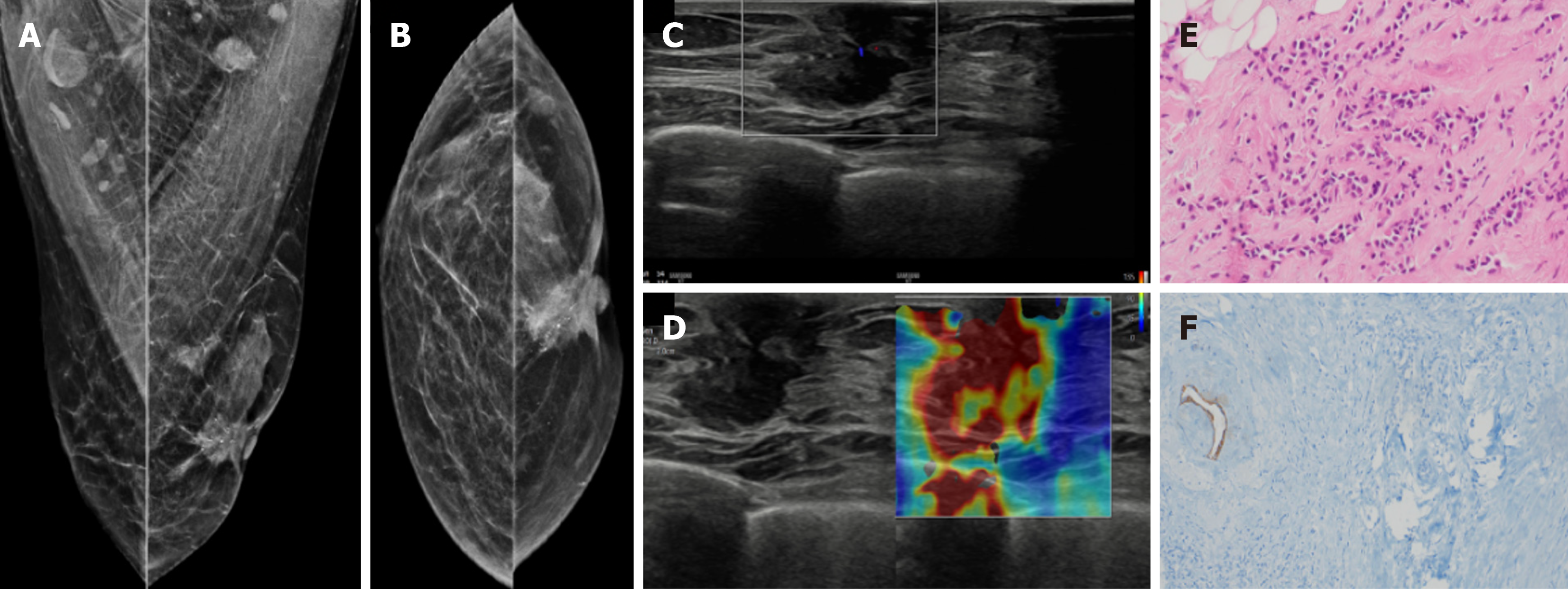
Figure 20 Invasive lobular carcinoma in a 65-year-old male with a hard retroareolar lump.
A: Mediolateral oblique; B: Craniocaudal mammography views showing an irregular high-density mass with spiculated margins, few punctate calcific foci and surrounding architectural distortion. Nipple retraction and overlying skin thickening are also observed; C: Ultrasound; D: Elastography images show an irregular mass with angular margins, which appears hard on elastography; E: Histopathology (hematoxylin and eosin, × 40); F: E-cadherin images show medium to small dyscohesive cells that lack E-cadherin expression.
Figure 21 Papillary carcinoma in a 54-year-old male with a hard mass in left breast.
A: Ultrasound; B: Color Doppler images showing an irregular solid-cystic mass with an eccentric solid component, with minimal peripheral vascularity.
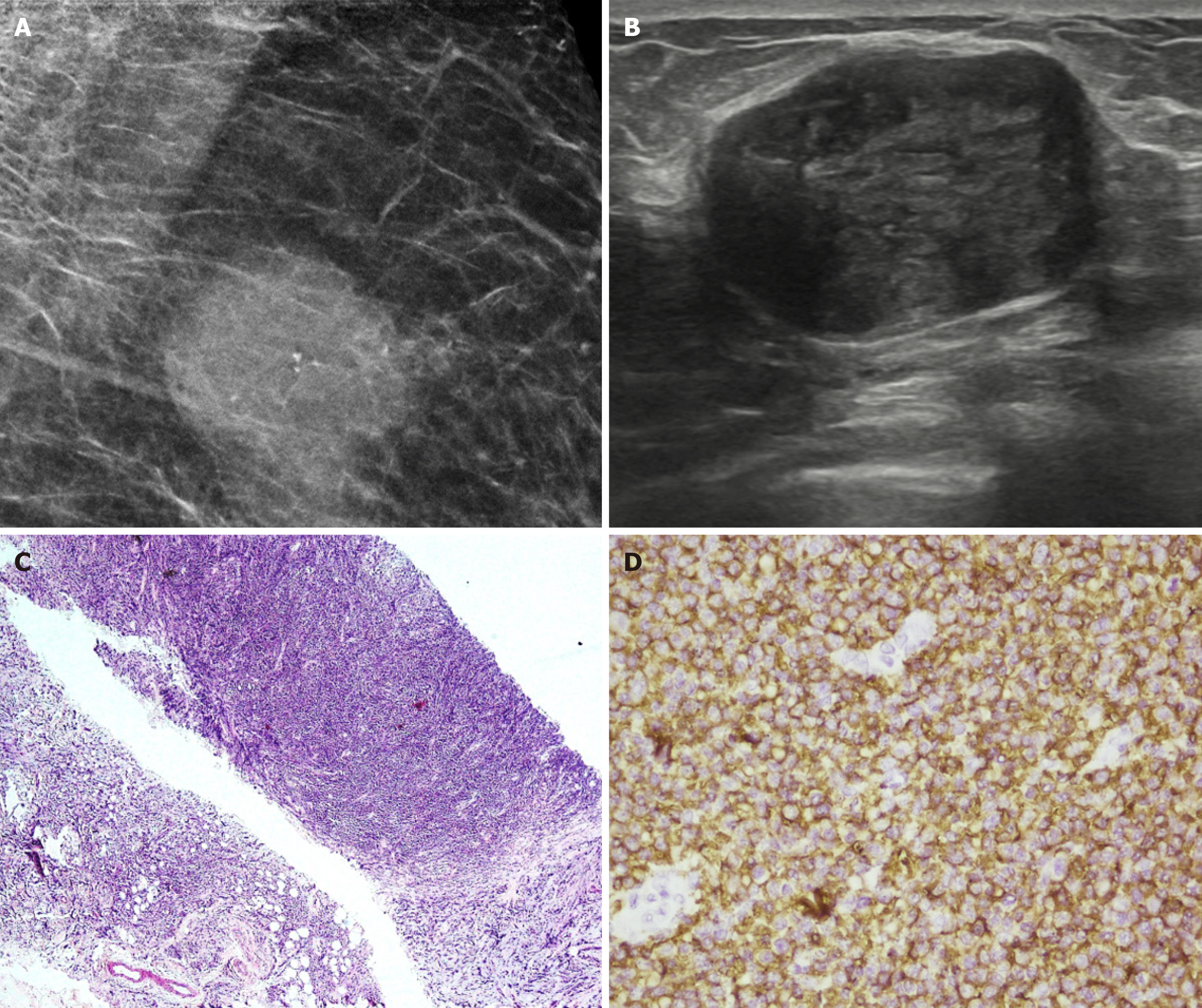
Figure 22 Lymphoma in a 43-year-old male after 3 cycles of chemotherapy.
A: Magnified mediolateral oblique view of the right breast showing a round high-density mass with partially indistinct margins and few punctate calcifications. B: Ultrasound image showing an oval hypoechoic parallel mass with circumscribed margins. C: Histopathology (hematoxylin and eosin, × 40). D: CD20 immunohistochemical staining revealed diffuse large B-cell lymphoma with sheets of atypical lymphoid cells that were positive for CD20.
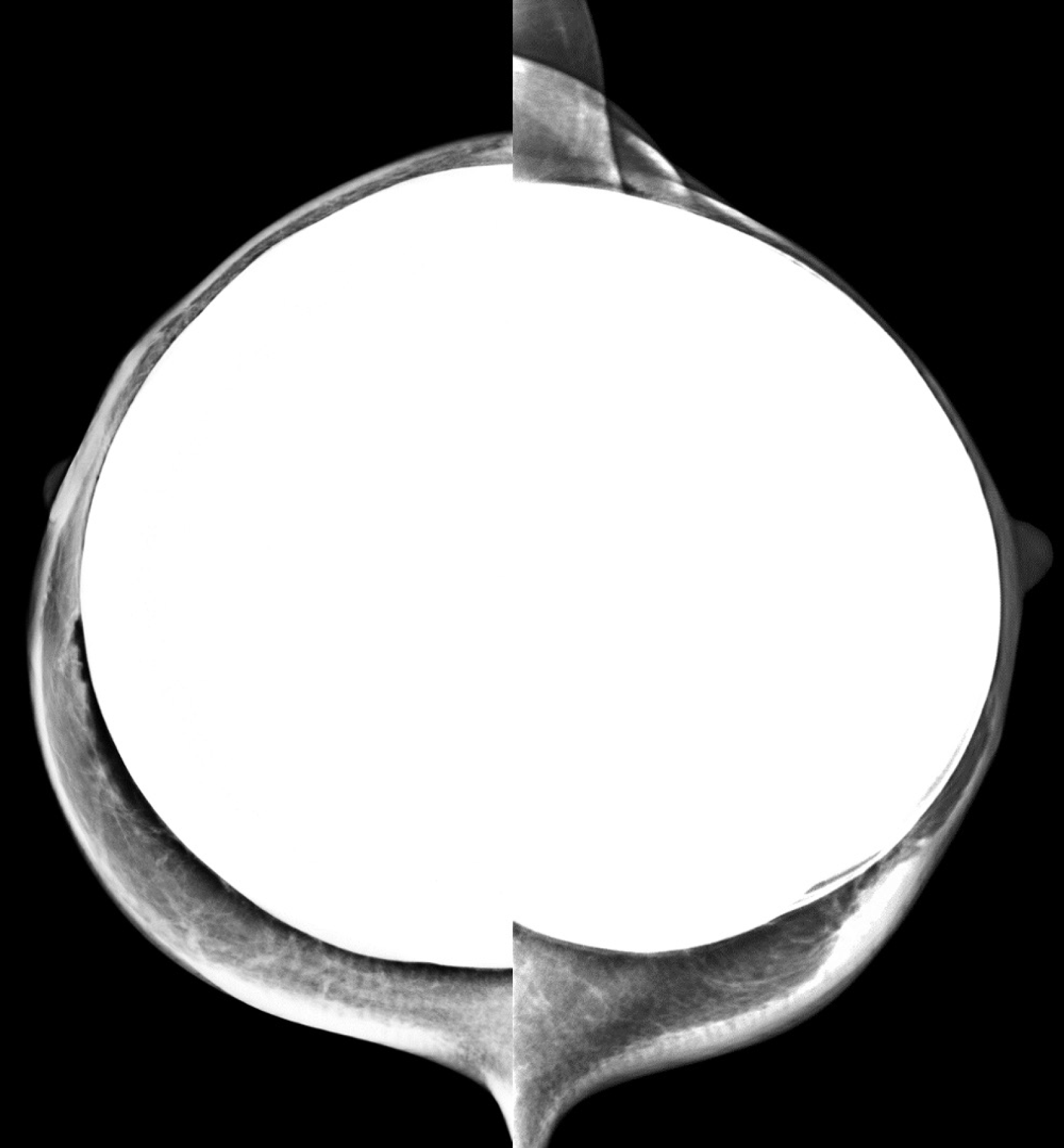
Figure 23
Craniocaudal mammogram of a transfeminine (male to female) individual showing bilateral silicone implants with smooth margins and no signs of rupture.
- Citation: Singla V, Bhatia H, Garg D, Bal A, Sekar A. A compendium of male breast imaging: The road less traveled. World J Radiol 2025; 17(9): 110906
- URL: https://www.wjgnet.com/1949-8470/full/v17/i9/110906.htm
- DOI: https://dx.doi.org/10.4329/wjr.v17.i9.110906


