Copyright
©The Author(s) 2025.
World J Radiol. Oct 28, 2025; 17(10): 112271
Published online Oct 28, 2025. doi: 10.4329/wjr.v17.i10.112271
Published online Oct 28, 2025. doi: 10.4329/wjr.v17.i10.112271
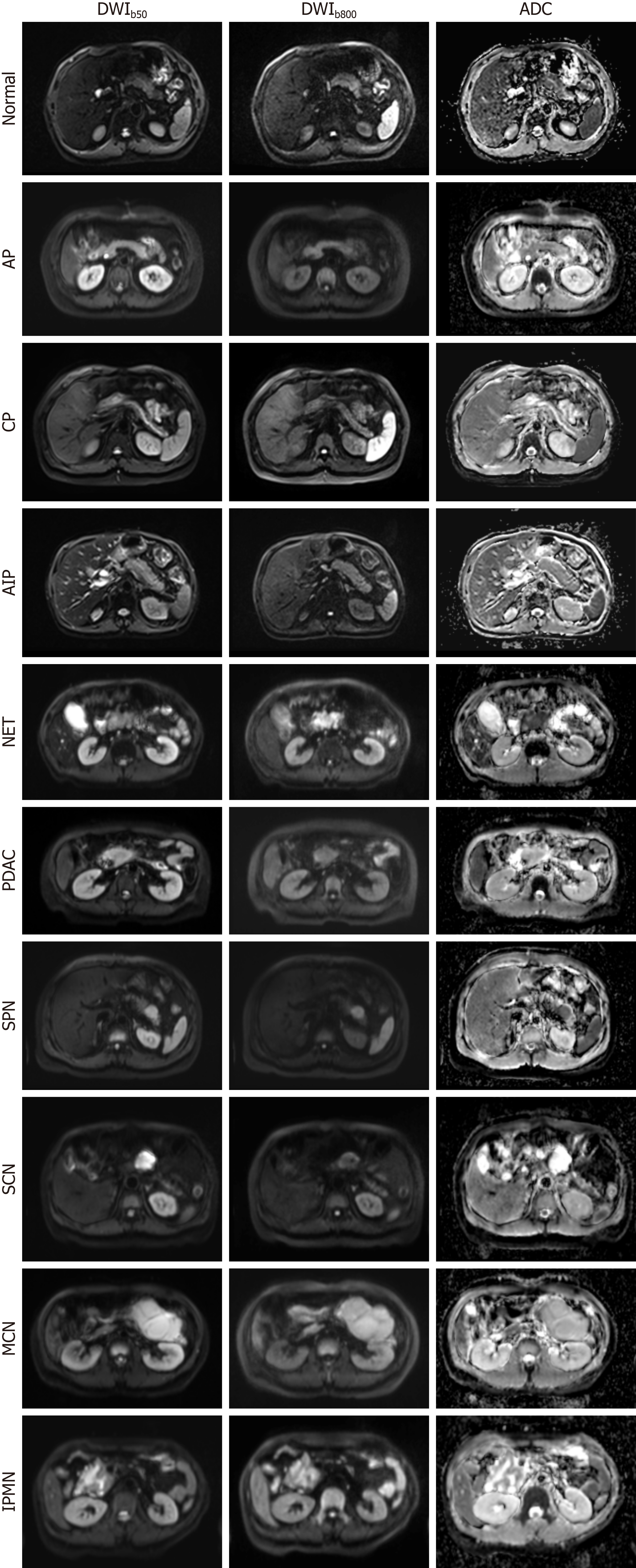
Figure 1 Diffusion-weighted imaging and corresponding apparent diffusion coefficient maps of the normal pancreas and nine repre
- Citation: Gao QY, Wang LJ, Ma C. Diffusion-weighted magnetic resonance imaging of the pancreas: A narrative review. World J Radiol 2025; 17(10): 112271
- URL: https://www.wjgnet.com/1949-8470/full/v17/i10/112271.htm
- DOI: https://dx.doi.org/10.4329/wjr.v17.i10.112271


