Copyright
©The Author(s) 2024.
World J Radiol. Apr 28, 2024; 16(4): 72-81
Published online Apr 28, 2024. doi: 10.4329/wjr.v16.i4.72
Published online Apr 28, 2024. doi: 10.4329/wjr.v16.i4.72
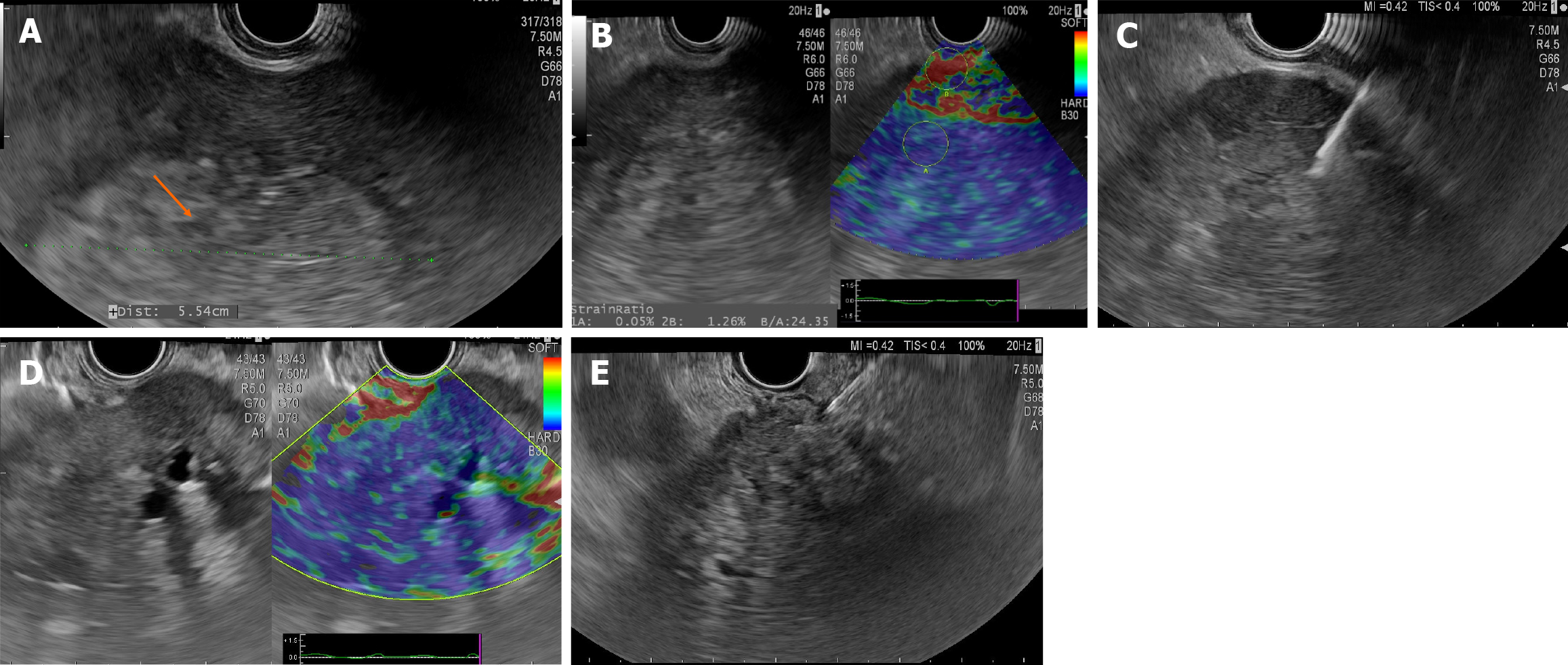
Figure 1 Endoscopic ultrasound.
A and B: Endoscopic ultrasound view of a right lobe hepatocarcinoma. Large hyperechoic tumor mass with halo segments V-VIII (A), Ultrasound elastography showing high strain ration predicting malignant character of the lesion (B); C: Endoscopic ultrasound guided fine needle biopsy from a left lobe hepatocarcinoma in a patient with liver cirrhosis; D and E: Endoscopic ultrasound view of a left lobe cholangiocarcinoma. Large inhomogeneous tumor mass with intratumoral left bile duct dilatations (D); Endoscopic ultrasound guided fine needle aspiration from the tumor mass (E).
- Citation: Tantău A, Sutac C, Pop A, Tantău M. Endoscopic ultrasound-guided tissue acquisition for the diagnosis of focal liver lesion. World J Radiol 2024; 16(4): 72-81
- URL: https://www.wjgnet.com/1949-8470/full/v16/i4/72.htm
- DOI: https://dx.doi.org/10.4329/wjr.v16.i4.72


