©The Author(s) 2024.
World J Radiol. Oct 28, 2024; 16(10): 616-620
Published online Oct 28, 2024. doi: 10.4329/wjr.v16.i10.616
Published online Oct 28, 2024. doi: 10.4329/wjr.v16.i10.616
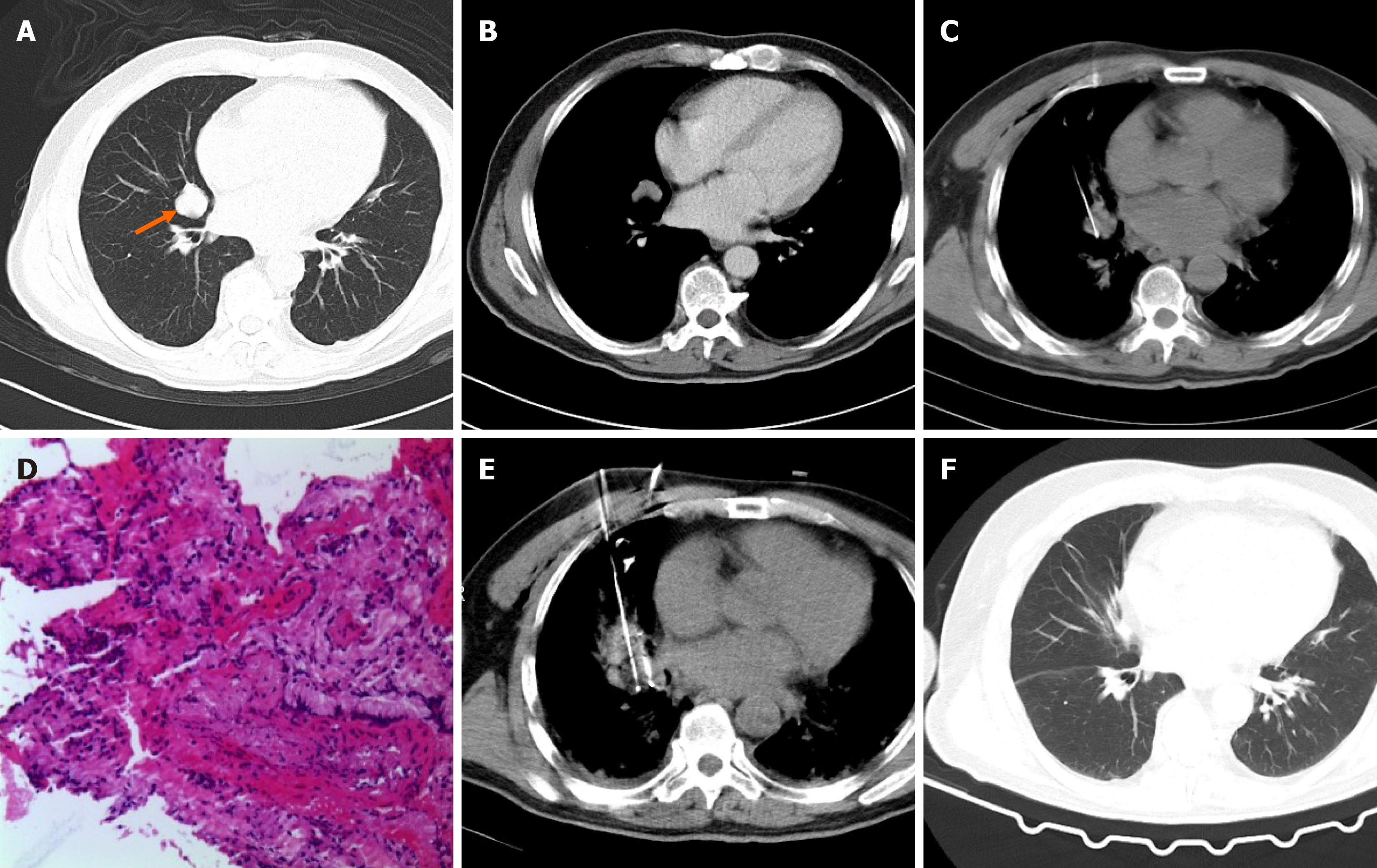
Figure 1 Intrapulmonary bronchial cysts treated with cryoablation and follow-up computed tomography images.
A: lung window computed tomography (CT) image, medial segment of the middle lobe of the right lung with a cyst of about 2.7 cm × 2.4 cm in size (arrow); B: Mediastinal window-enhanced CT image; C: Biopsy image; D: Pathological examination (HE stain); E: During cryoablation; F: Complete response was achieved at 3 months after cryoablation.
- Citation: Li ZH, Ma YY, Niu LZ, Xu KC. Cryoablation for intrapulmonary bronchial cyst: A case report. World J Radiol 2024; 16(10): 616-620
- URL: https://www.wjgnet.com/1949-8470/full/v16/i10/616.htm
- DOI: https://dx.doi.org/10.4329/wjr.v16.i10.616


