
















©The Author(s) 2024.
World J Radiol. Oct 28, 2024; 16(10): 537-544
Published online Oct 28, 2024. doi: 10.4329/wjr.v16.i10.537
Published online Oct 28, 2024. doi: 10.4329/wjr.v16.i10.537
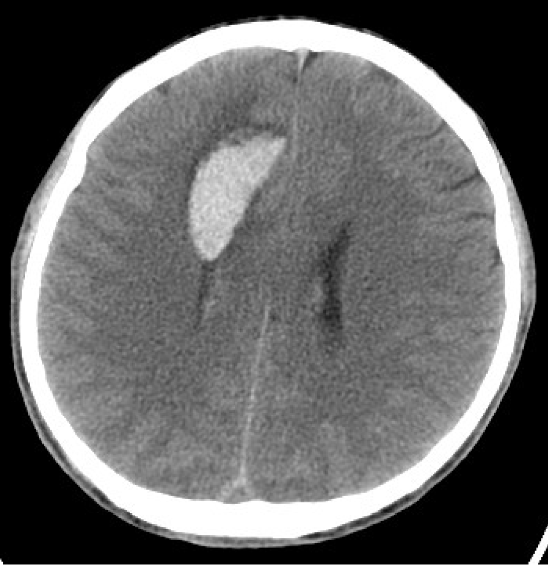
Figure 1
Preoperative computed tomography showing a lamellar hyperdense area in the right frontal lobe; lamellar hyperdense shadows can be seen in the ventricles, while the right lateral ventricle was narrowed by compression, with a slightly leftward deviation of the midline structure.
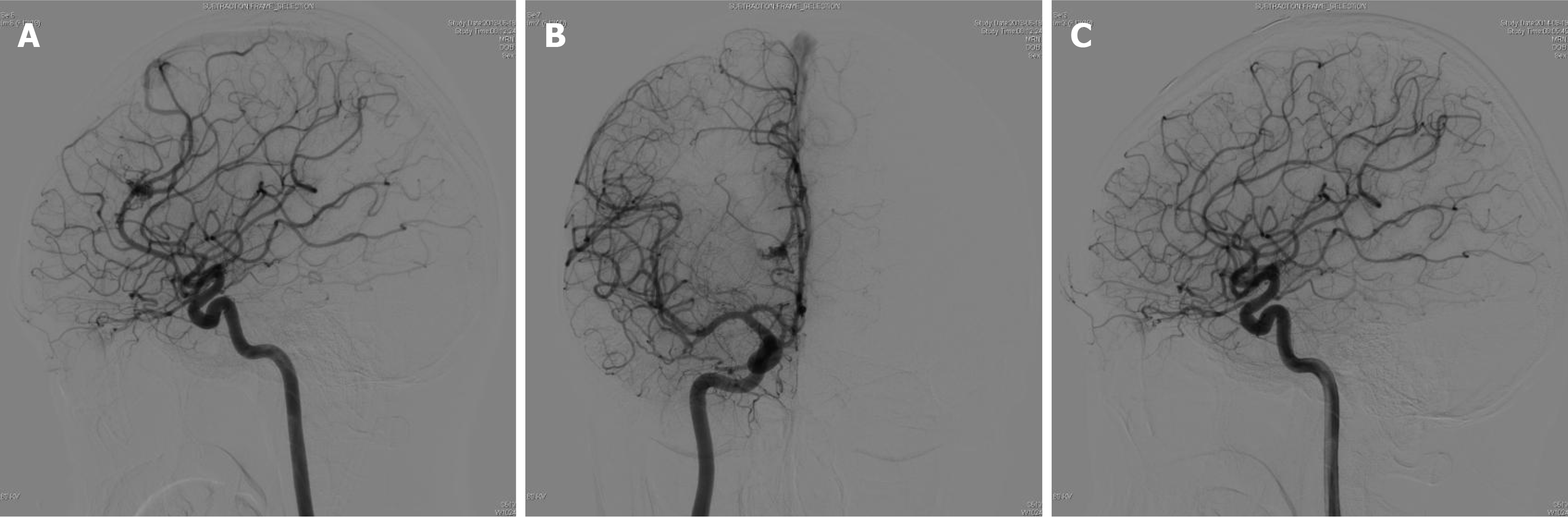
Figure 2 Right internal carotid artery angiography.
A: Preoperative internal carotid artery angiography lateral cut and; B: An orthogonal slice showing 1 arteriovenous malformation (AVM) in the right frontal lobe, with the supplying artery originating from the anterior cerebral artery; C: Digital subtraction angiography performed one-year postoperatively showing complete AVM resection.
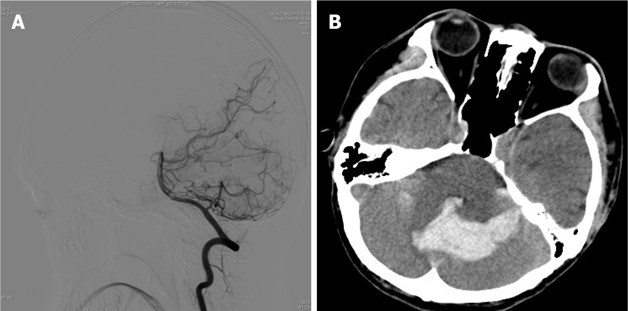
Figure 3 Right vertebral artery angiography and Preoperative computed tomography.
A: Right vertebral artery angiography performed before the first arteriovenous malformation resection showing no cerebellar abnormalities; B: Preoperative computed tomography showing multiple patchy hyperdense shadows in the 3rd and 4th ventricles, indicating bilateral cerebellar hemorrhage.
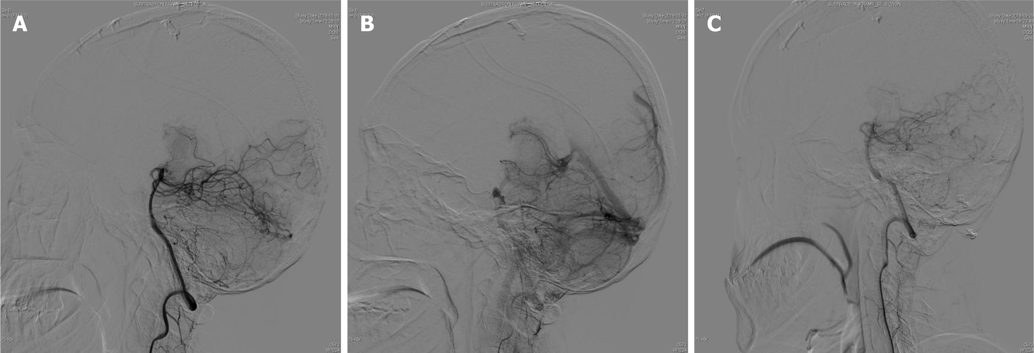
Figure 4 Left vertebral artery angiography.
A: Image in the preoperative lateral view showing a cerebellar arteriovenous malformation fed by the left superior cerebellar artery; B: The venous phase of the angiogram showing the draining veins into the transverse sinus; C: Follow-up imaging at 7 months showing no evidence of a residual malformation.
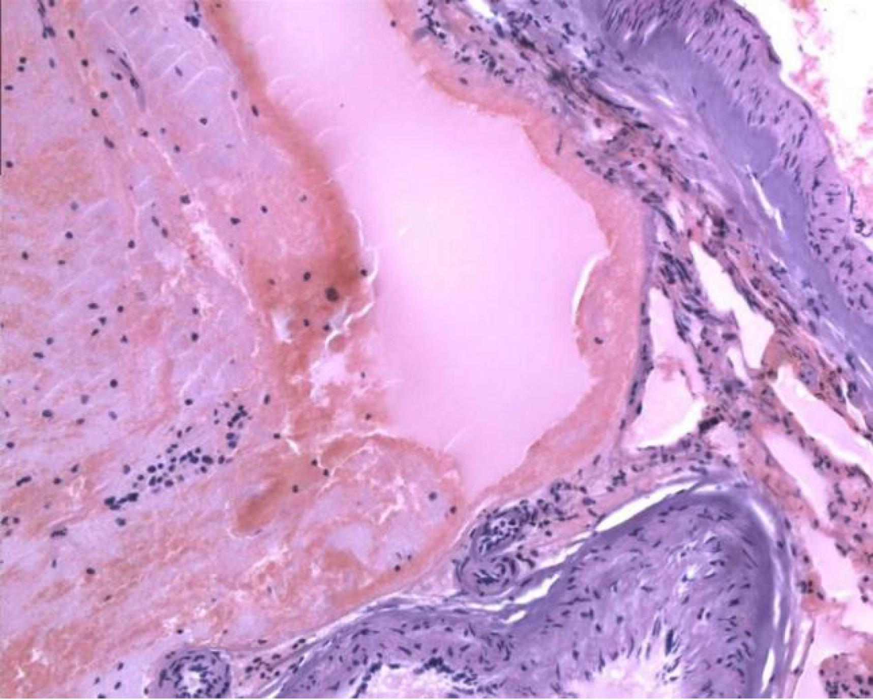
Figure 5 Postoperative pathological examination.
Large amounts of vascular tissue with crooked and tortuous lumens can be observed in the resected tissue, consistent with the presentation of arteriovenous malformation.
- Citation: Cao WY, Li JP, Guo P, Song LX. Ectopic recurrence following treatment of arteriovenous malformations in an adult: A case report and review of literature. World J Radiol 2024; 16(10): 537-544
- URL: https://www.wjgnet.com/1949-8470/full/v16/i10/537.htm
- DOI: https://dx.doi.org/10.4329/wjr.v16.i10.537