Copyright
©The Author(s) 2023.
World J Radiol. Jul 28, 2023; 15(7): 226-233
Published online Jul 28, 2023. doi: 10.4329/wjr.v15.i7.226
Published online Jul 28, 2023. doi: 10.4329/wjr.v15.i7.226
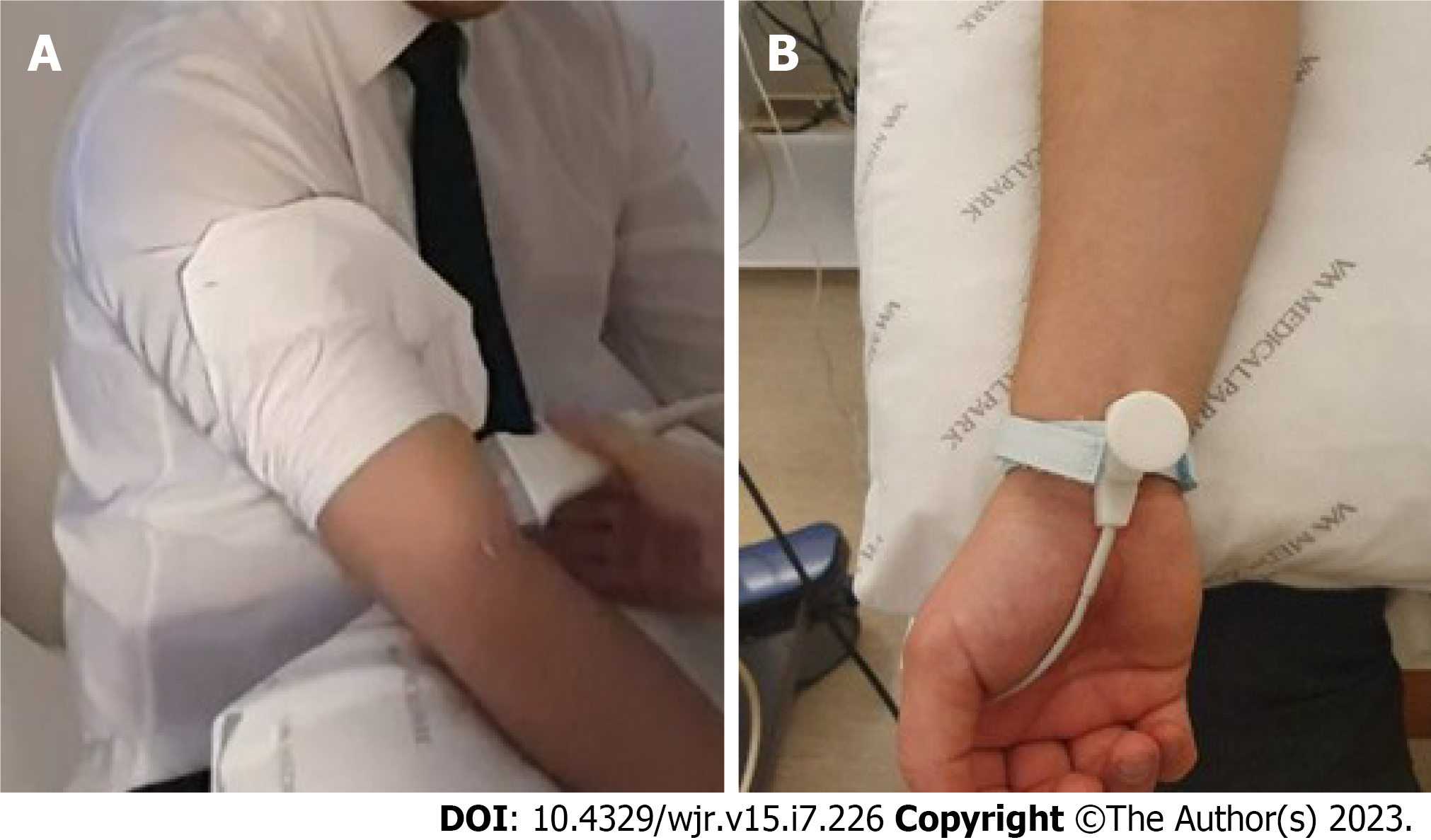
Figure 1 Positioning of the Doppler ultrasound probe and bipolar stimulus electrode.
A: Positioning of the Doppler ultrasound probe, 2 cm above the antecubital fossa using a 9 Hz linear probe; B: Positioning of the bipolar stimulus electrode.
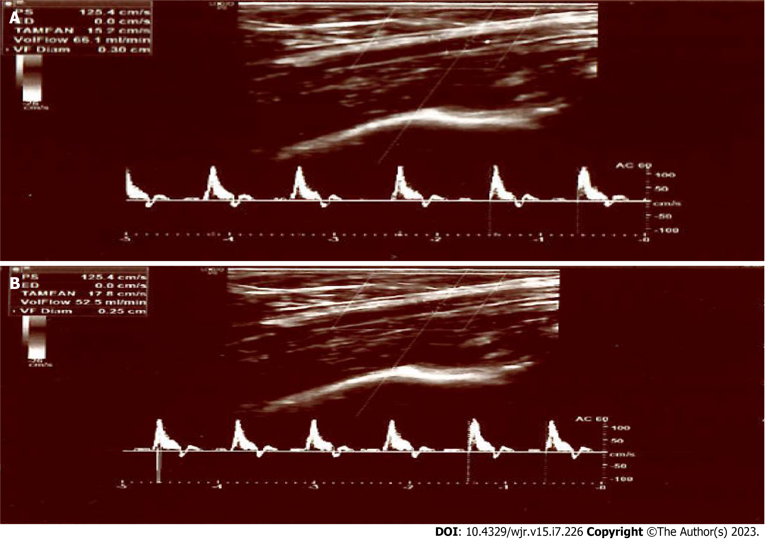
Figure 2 Before and after electrical stimulation, the brachial artery flow/diameter parameters at control participant.
A: Before electrical stimulation, the brachial artery volume flow rates/diameter values of a 45-year-old healthy control participant were measured to be 66.1 mL/min, 3 mm; B: The post-stimulation values of the same patient were 52.5 mL/min, 2.5 mm.
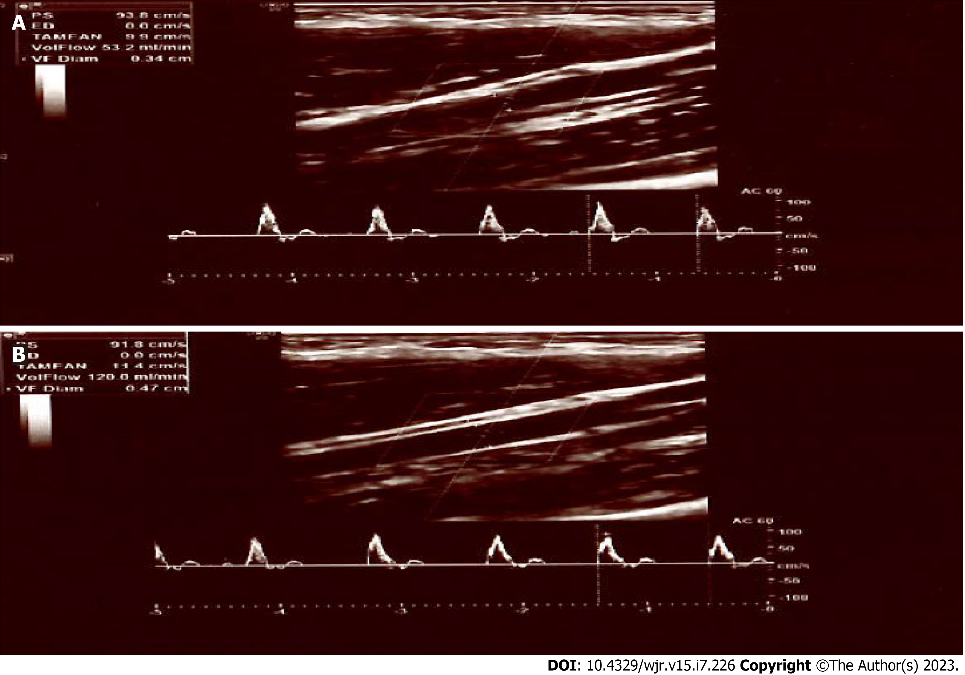
Figure 3 Before and after electrical stimulation, the brachial artery flow/diameter parameters in a patient diagnosed with irritable bowel syndrome.
A: Before electrical stimulation, the brachial artery flow/diameter parameters of a 38-year-old female patient diagnosed with irritable bowel syndrome were assessed to be 53.2 mL/min, 3.2 mm; B: The post-stimulation values of the same patient were 120 mL/min, 4.7 mm.
- Citation: Kazci O, Ege F, Aydemir H, Kazci S, Aydin S. Can the change of vasomotor activity in irritable bowel syndrome patients be detected via color Doppler ultrasound? World J Radiol 2023; 15(7): 226-233
- URL: https://www.wjgnet.com/1949-8470/full/v15/i7/226.htm
- DOI: https://dx.doi.org/10.4329/wjr.v15.i7.226


