















©The Author(s) 2022.
World J Radiol. Jan 28, 2022; 14(1): 19-29
Published online Jan 28, 2022. doi: 10.4329/wjr.v14.i1.19
Published online Jan 28, 2022. doi: 10.4329/wjr.v14.i1.19
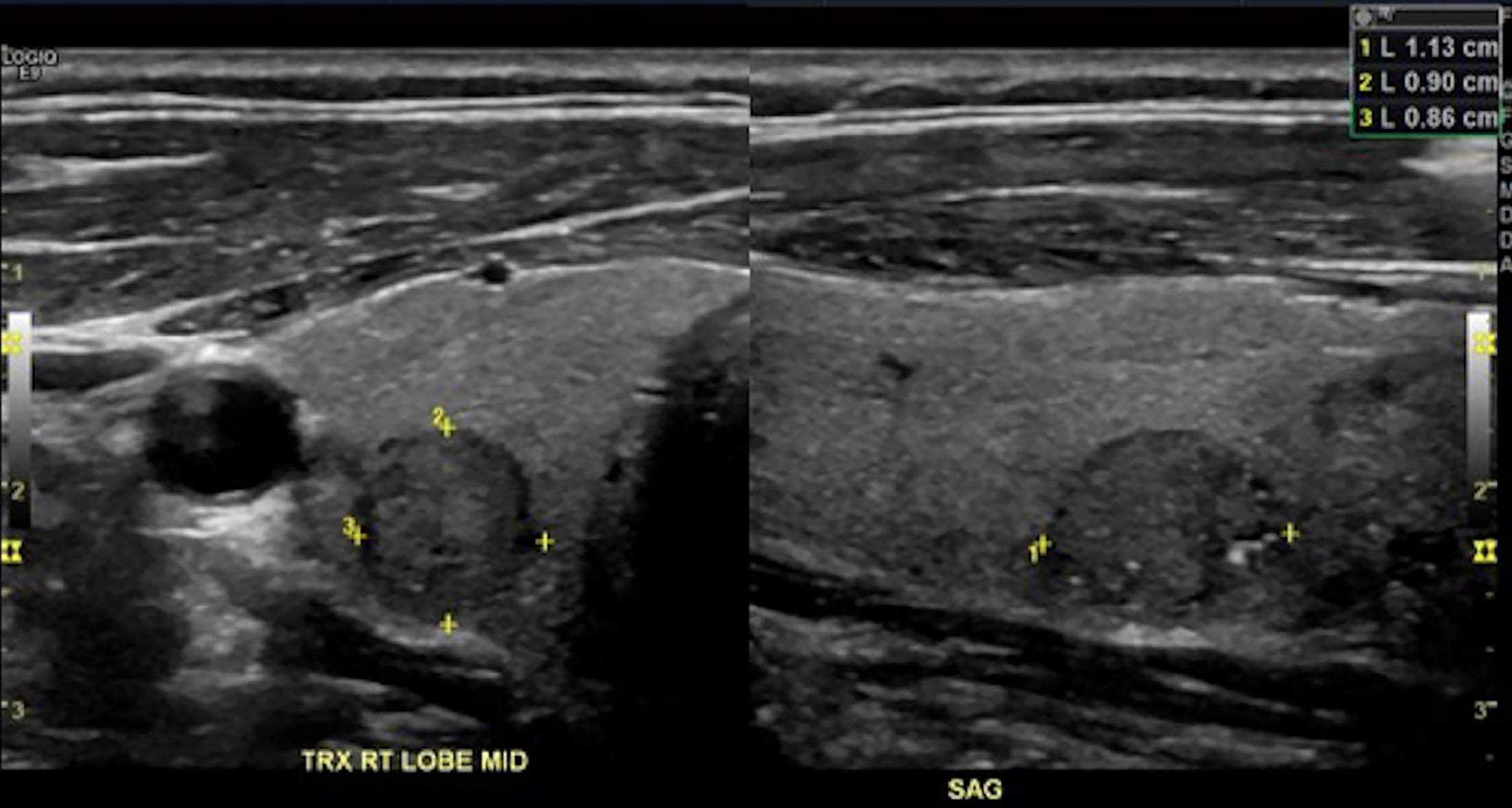
Figure 1 A 51-year-old female with a 1.
1 cm × 0.9 cm × 0.9 cm right mid pole thyroid nodule. This nodule was classified correctly with perfect concordance by all 3 readers as solid (+ 2 points), hypoechoic (+ 2 points), taller-than-wide (+ 3 points), smooth margins (+ 0 points), and with punctate echogenic foci (+ 3 points). This had a total points of 10 and a Thyroid Imaging Reporting and Data System level of TR5.
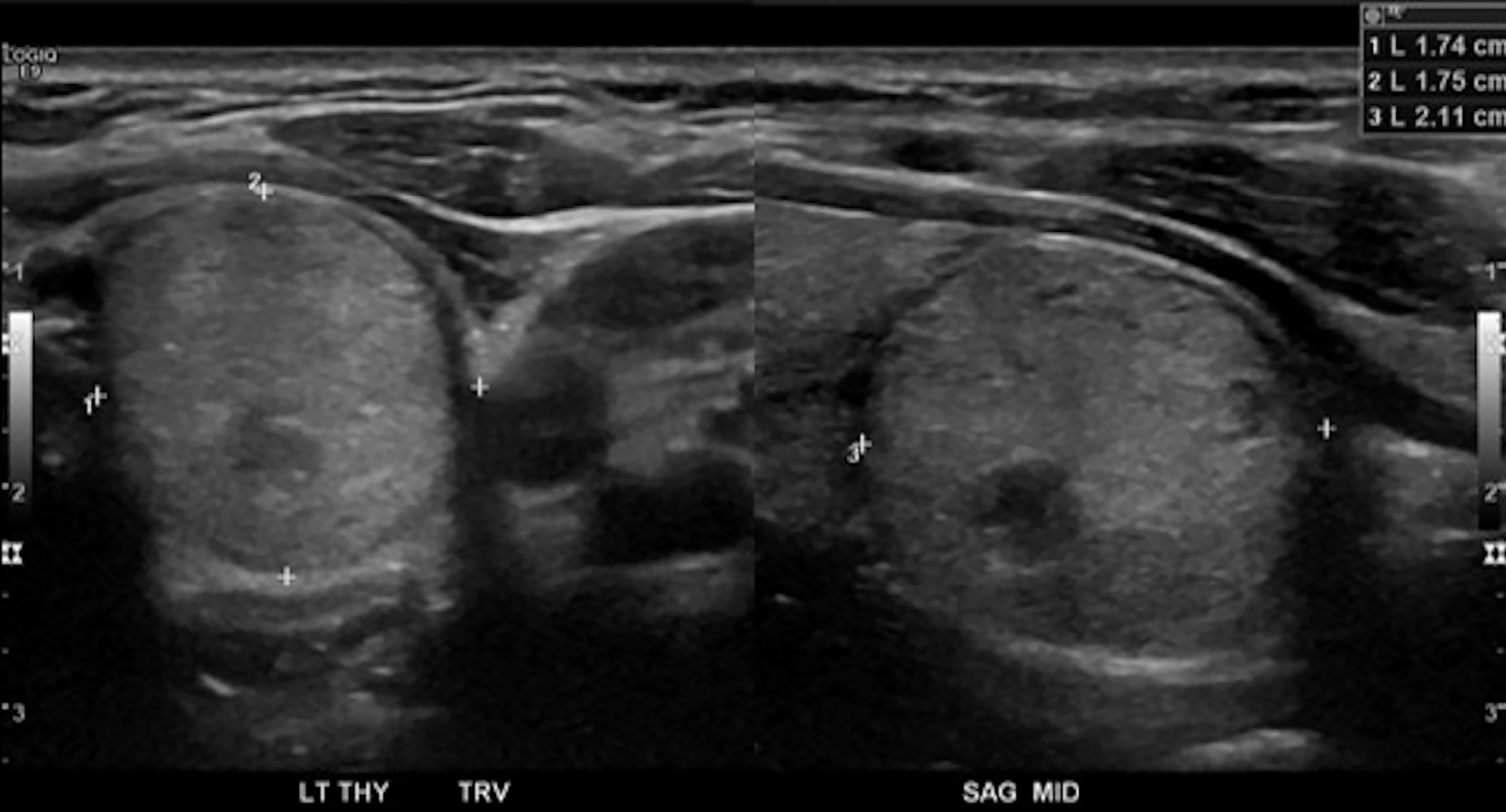
Figure 2 A 45-year-old female with a 1.
7 cm × 1.8 cm × 2.1 cm left mid pole thyroid nodule. This nodule was classified by first two readers as Thyroid Imaging Reporting and Data System (TI-RADS) level TR4 and by the third reader as TI-RADS level TR5. The first two readers classified the nodule as solid (+ 2 points), isoechoic (+ 2 points), taller-than-wide (+ 3 points), smooth margins (+ 0 points) and with no echogenic foci (+ 0 points) for a total points of 6 and a TI-RAD level of TR4. For the third reader, a single discrepancy in the scoring of echogenicity as hypoechoic (+ 2 points) rather than isoechoic (+ 1 point) as in the other 2 readers, resulted in a total points of 7 and a TI-RADS level of TR5. As can be seen in the images, the nodule has mixed echogenicity although most of the nodule is isoechoic making this the preferred option.
- Citation: Du Y, Bara M, Katlariwala P, Croutze R, Resch K, Porter J, Sam M, Wilson MP, Low G. Effect of training on resident inter-reader agreement with American College of Radiology Thyroid Imaging Reporting and Data System. World J Radiol 2022; 14(1): 19-29
- URL: https://www.wjgnet.com/1949-8470/full/v14/i1/19.htm
- DOI: https://dx.doi.org/10.4329/wjr.v14.i1.19