©The Author(s) 2022.
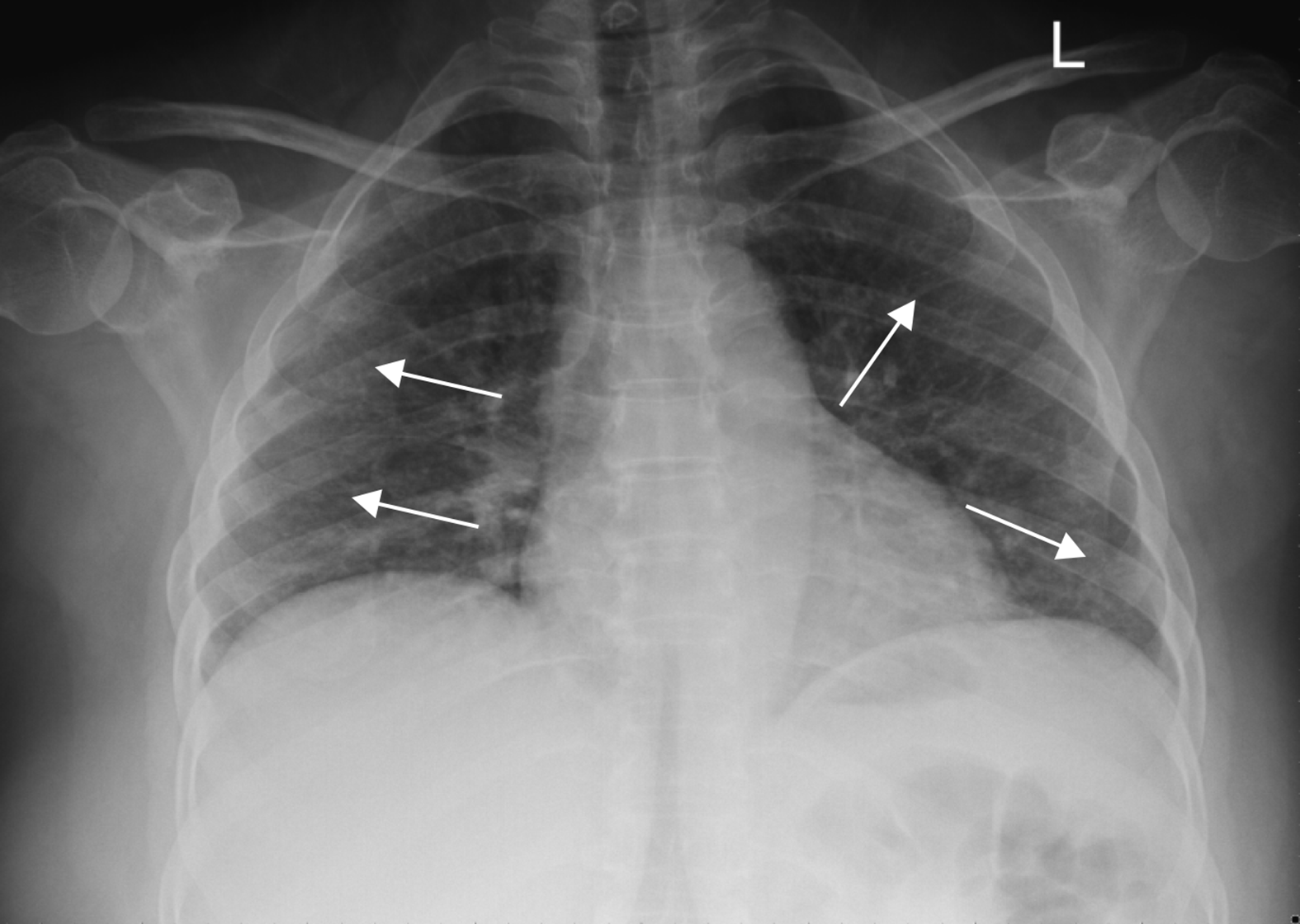
Figure 1 Postero-anterior chest X-RAY in one asymptomatic patient with coronavirus disease 2019 pneumonia from our institution.
It shows Interstitial infiltrates and ill-defined, patchy, peripheral opacities in bilateral lung fields.
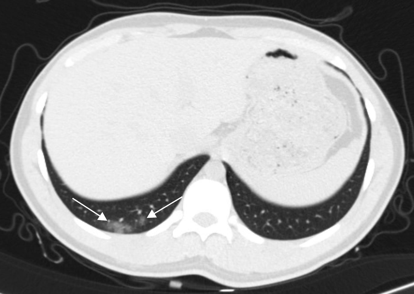
Figure 2 Axial-basal chest cut in urinary tract computed tomography in a patient presenting with renal colic at our institution who was diagnosed with asymptomatic coronavirus disease 2019 due to the presence of peripheral small focal areas of ground glass veiling.
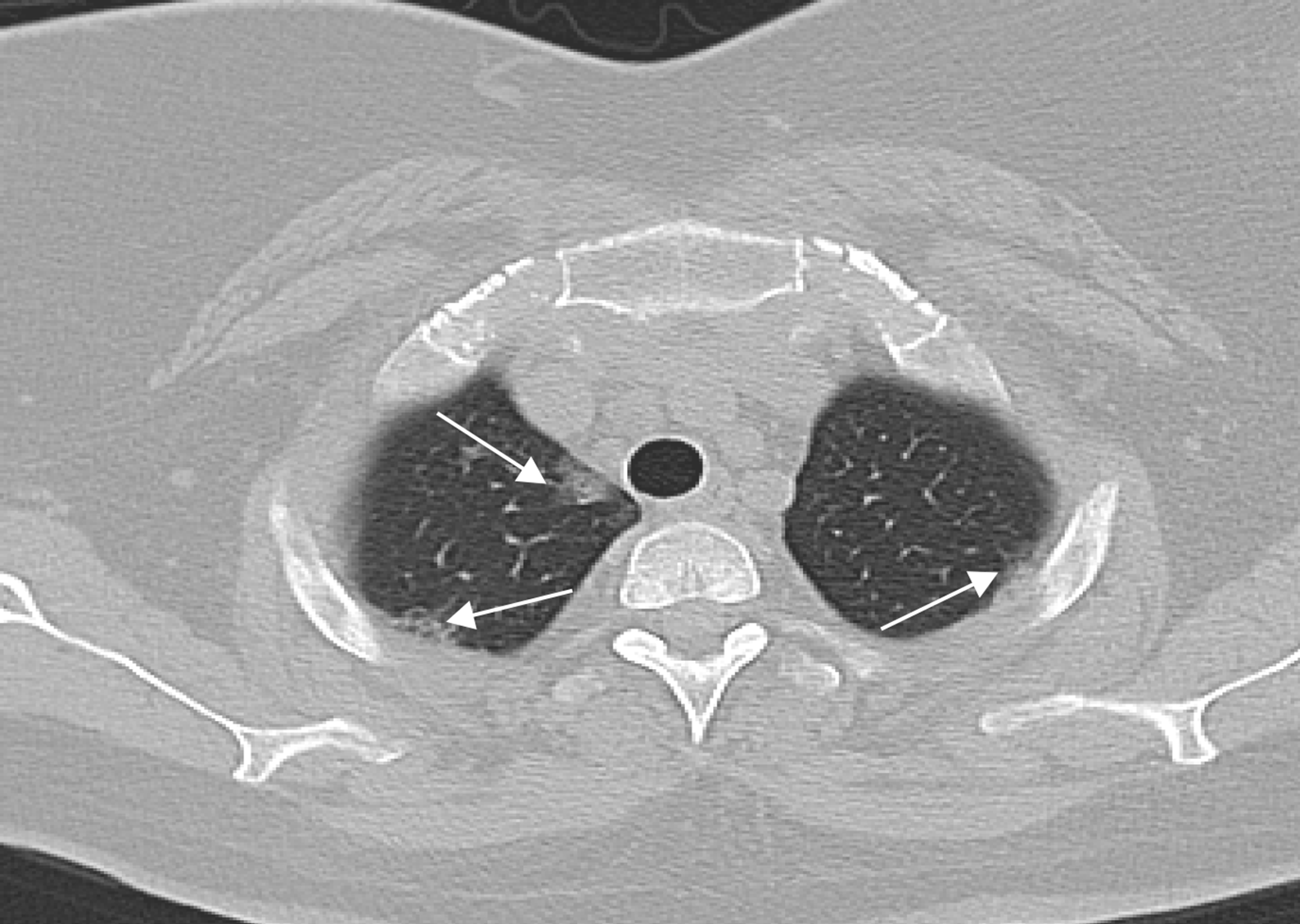
Figure 3 Axial-apical chest cut in brain computed tomography in a patient presenting with head trauma at our institution who was diagnosed with asymptomatic coronavirus disease 2019 due to the bilateral presence of multiple peripheral small foci of ground glass veiling with mild interstitial thickening.
Figure 4 Cardiac magnetic resonance images of a patient with coronavirus disease 2019 who presented to our institute for a viability study showing multifocal peripheral areas of abnormal signal in both lungs that appear as high signal intensity areas localized in the coronal plane (A), high T2 signals (B), and faint heterogenous enhancement in post-contrast sequences (C).
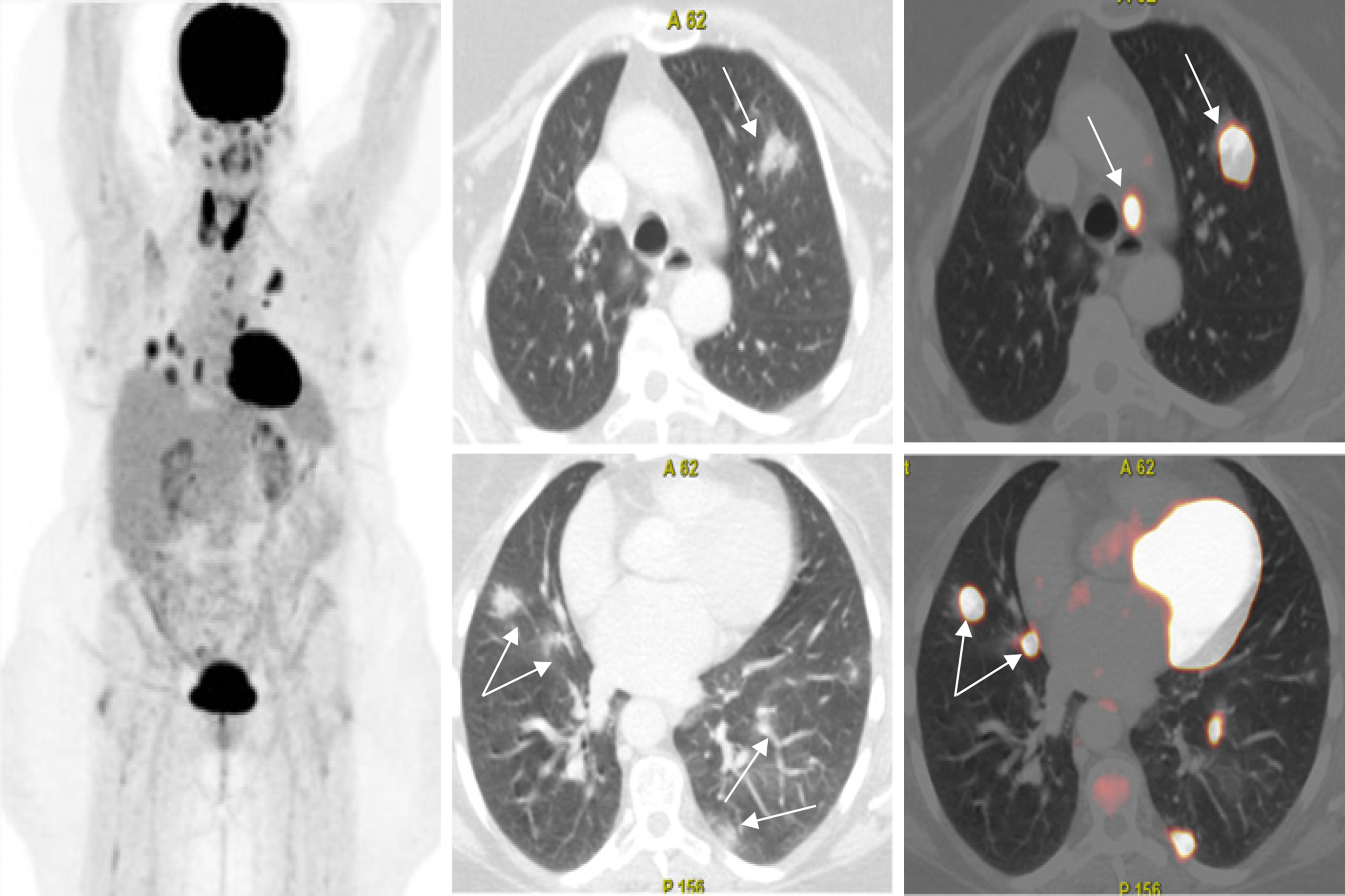
Figure 5 Axial fused thoracic 18Fluorodeoxyglucose-positron emission tomography-computed tomography showing multiple variable-sized metabolically active and mainly subpleural subsegmental consolidative lesions with an SUVmax of up to 10.
9 as well as metabolically active lymph node seen in the aorto-pulmonary window in a patient with thyroid cancer and asymptomatic coronavirus 2019.
- Citation: Romeih M, Mahrous MR, El Kassas M. Incidental radiological findings suggestive of COVID-19 in asymptomatic patients. World J Radiol 2022; 14(1): 1-12
- URL: https://www.wjgnet.com/1949-8470/full/v14/i1/1.htm
- DOI: https://dx.doi.org/10.4329/wjr.v14.i1.1


