Copyright
©The Author(s) 2025.
World J Gastrointest Surg. Sep 27, 2025; 17(9): 106258
Published online Sep 27, 2025. doi: 10.4240/wjgs.v17.i9.106258
Published online Sep 27, 2025. doi: 10.4240/wjgs.v17.i9.106258
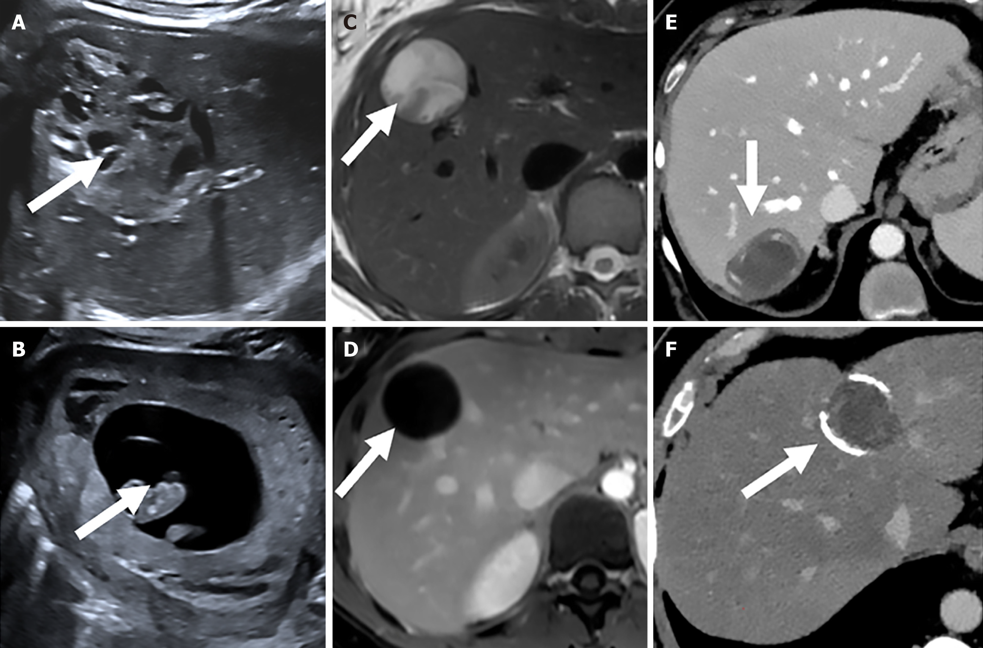
Figure 1 Imaging of hepatic cystic echinococcosis.
A and B: Ultrasound; C and D: Magnetic resonance imaging; E and F: Computed tomography.
Figure 2 Schematic drawings representation of cystic echinococcosis and pericystectomy as well as the postoperative outcome.
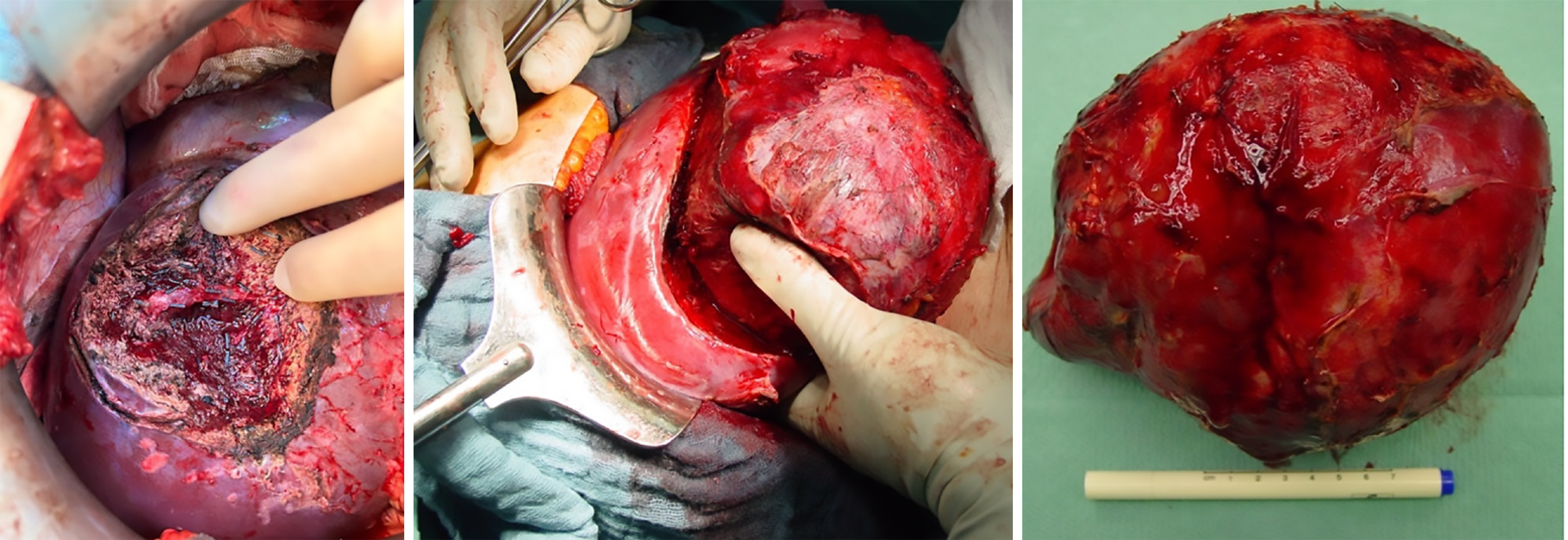
Figure 3 Intraoperative documentation of the cystic echinococcus and pericystectomy.
- Citation: Wagner T, Struck S, Persigehl T, Nierhoff D, Schmidt T, Hummels M, Bruns CJ, Stippel DL, Thomas MN. Long-term outcomes after open total pericystectomy for cystic echinococcosis. World J Gastrointest Surg 2025; 17(9): 106258
- URL: https://www.wjgnet.com/1948-9366/full/v17/i9/106258.htm
- DOI: https://dx.doi.org/10.4240/wjgs.v17.i9.106258


