Copyright
©The Author(s) 2025.
World J Gastrointest Surg. Jul 27, 2025; 17(7): 107046
Published online Jul 27, 2025. doi: 10.4240/wjgs.v17.i7.107046
Published online Jul 27, 2025. doi: 10.4240/wjgs.v17.i7.107046
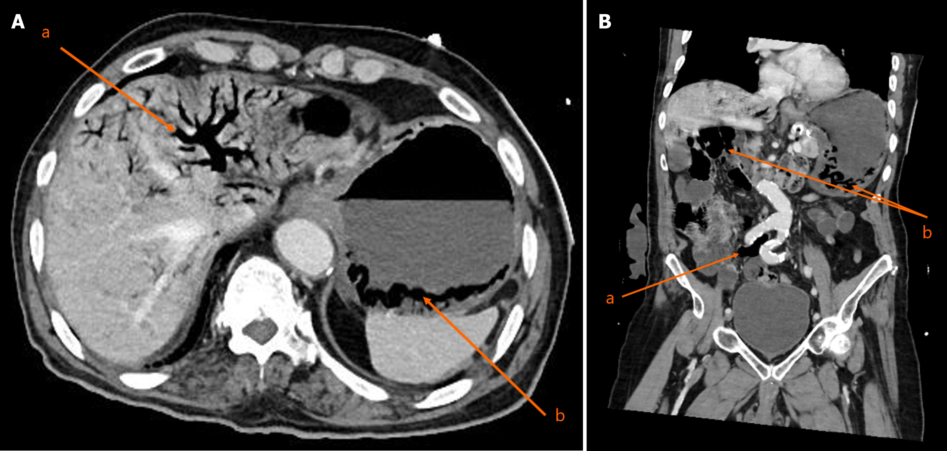
Figure 1 Contrast enhanced computed tomography.
A: Contrast enhanced axial computed tomography (CT) revealed extensive pneumatosis portalis and air within the short gastric veins, indicative of ischemia (a) and0 intramural gastric gas within the gastric wall, a hallmark finding of emphysematous gastritis (b); B: Contrast enhanced coronal CT revealed additional findings of gas in the mesenteric veins and features suggestive of small bowel ischemia (a) and extensive pneumatosis portalis and air within the gastric wall consistent with emphysematous gastritis (b).
- Citation: Alshahwan N, Alqarzaie AA, Aldeligan SH, Alqusiyer AA, Alnumay A, Mashbari H, Alkanhal A. Successful conservative management of emphysematous gastritis in an elderly patient with multiple comorbidities: A case report. World J Gastrointest Surg 2025; 17(7): 107046
- URL: https://www.wjgnet.com/1948-9366/full/v17/i7/107046.htm
- DOI: https://dx.doi.org/10.4240/wjgs.v17.i7.107046


