Copyright
©The Author(s) 2025.
World J Gastrointest Surg. Oct 27, 2025; 17(10): 110540
Published online Oct 27, 2025. doi: 10.4240/wjgs.v17.i10.110540
Published online Oct 27, 2025. doi: 10.4240/wjgs.v17.i10.110540
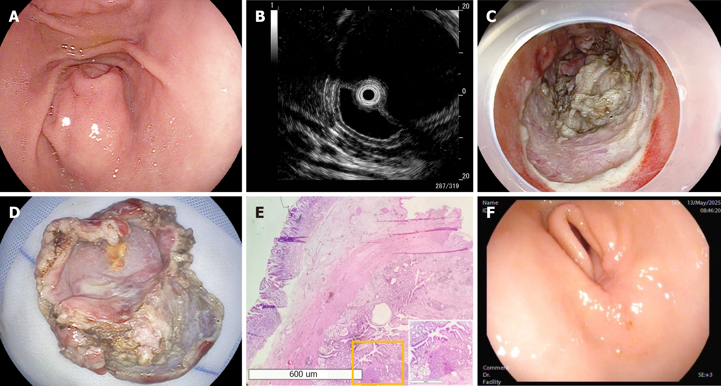
Figure 1 Imaging examination results and follow-up endoscopic findings.
A: Endoscopic findings: On September 18, 2024, endoscopy showed a centrally depressed surface without ulceration, covered by normal mucosa; B: Endoscopic ultrasound findings: Endoscopic ultrasound view showing the cystic lesion in the antrum. The lesion arises from the muscularis mucosa, presenting as a well-defined, homogeneous hypoechoic cystic structure; C: Postoperative wound presentation: Due to firm adhesion between the cyst base and the muscularis propria, a small portion of the cyst wall was intentionally left unresected; D: Gross examination revealed a cystic specimen measuring 4.0 cm × 5.0 cm; E: Pathological findings: Histopathology confirmed a gastric duplication cyst: Gastric mucosa overlying smooth muscle layer (hematoxylin and eosin, × 20) with ectopic pancreatic tissue in the cyst wall (boxed area, × 100); F: Follow-up endoscopic findings: Six-month postoperative follow-up gastroscopy revealed complete healing of the surgical site, with the residual cyst wall demonstrating benign histological features consistent with gastric antral mucosa.
- Citation: Zhu N, Chen MY, Li KQ, Zhang YM, Li FL, Li P, Wu J, Zou BC. Subtotal resection of gastric duplication cysts using endoscopic submucosal dissection: A case report. World J Gastrointest Surg 2025; 17(10): 110540
- URL: https://www.wjgnet.com/1948-9366/full/v17/i10/110540.htm
- DOI: https://dx.doi.org/10.4240/wjgs.v17.i10.110540


