Copyright
©The Author(s) 2024.
World J Gastrointest Surg. Nov 27, 2024; 16(11): 3538-3545
Published online Nov 27, 2024. doi: 10.4240/wjgs.v16.i11.3538
Published online Nov 27, 2024. doi: 10.4240/wjgs.v16.i11.3538
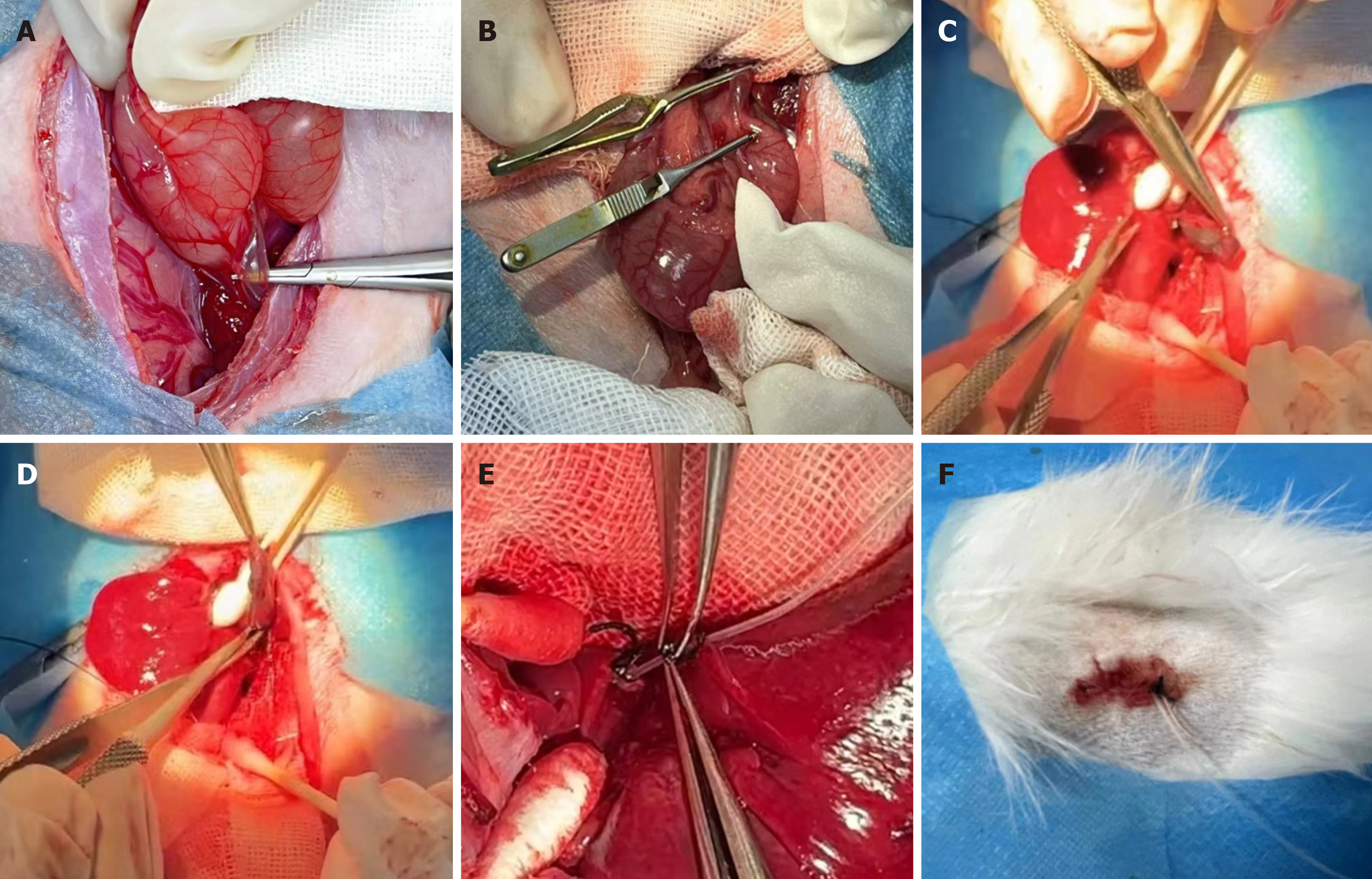
Figure 1 Establishment of a rabbit model of benign biliary stricture.
A: Identification of the common bile duct; B: Injury to the common bile duct; C: Ligation of the cystic duct; D: Cholecystectomy; E: Placement of a drainage tube; F: Percutaneous back fixation.
Figure 2 Serological results of the two groups detected using a biochemical analyzer.
A: Total bilirubin content; B: Conjugated bilirubin content; C: Alkaline phosphatase content; D: Alanine aminotransferase content; E: γ-glutamyl transferase content. aP < 0.0001; bP < 0.005. TBIL: Total bilirubin; DBIL: Direct bilirubin; ALT: Alanine aminotransferase; ALP: Alkaline phosphatase; GGT: γ-glutamyl transferase.
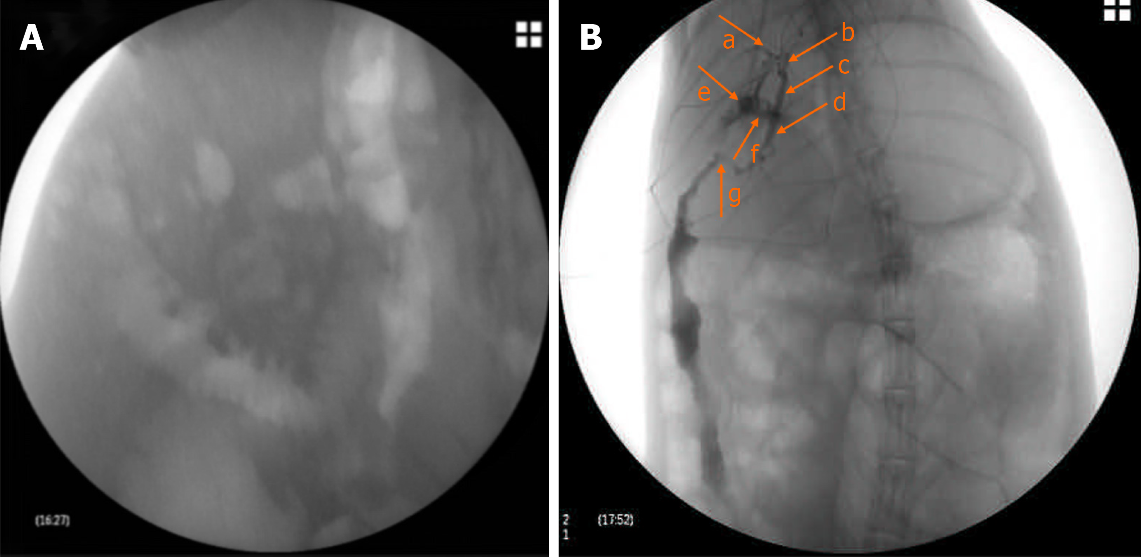
Figure 3 Angiographic evaluation of the biliary drainage tube.
A: No contrast medium was infused; B: Contrast medium was injected through the drainage tube for visualization. a: Right hepatic duct; b: Left hepatic duct; c: Ductuli hepaticus communis; d: Common bile duct; e: Gallbladder stump; f: Cystic duct; g: Lower common bile duct stenosis.
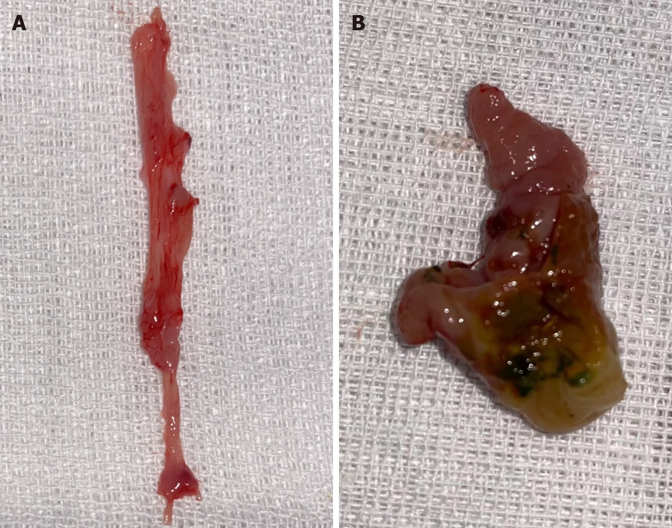
Figure 4 Morphological changes of the bile duct.
A: Healthy control group; B: Model group.
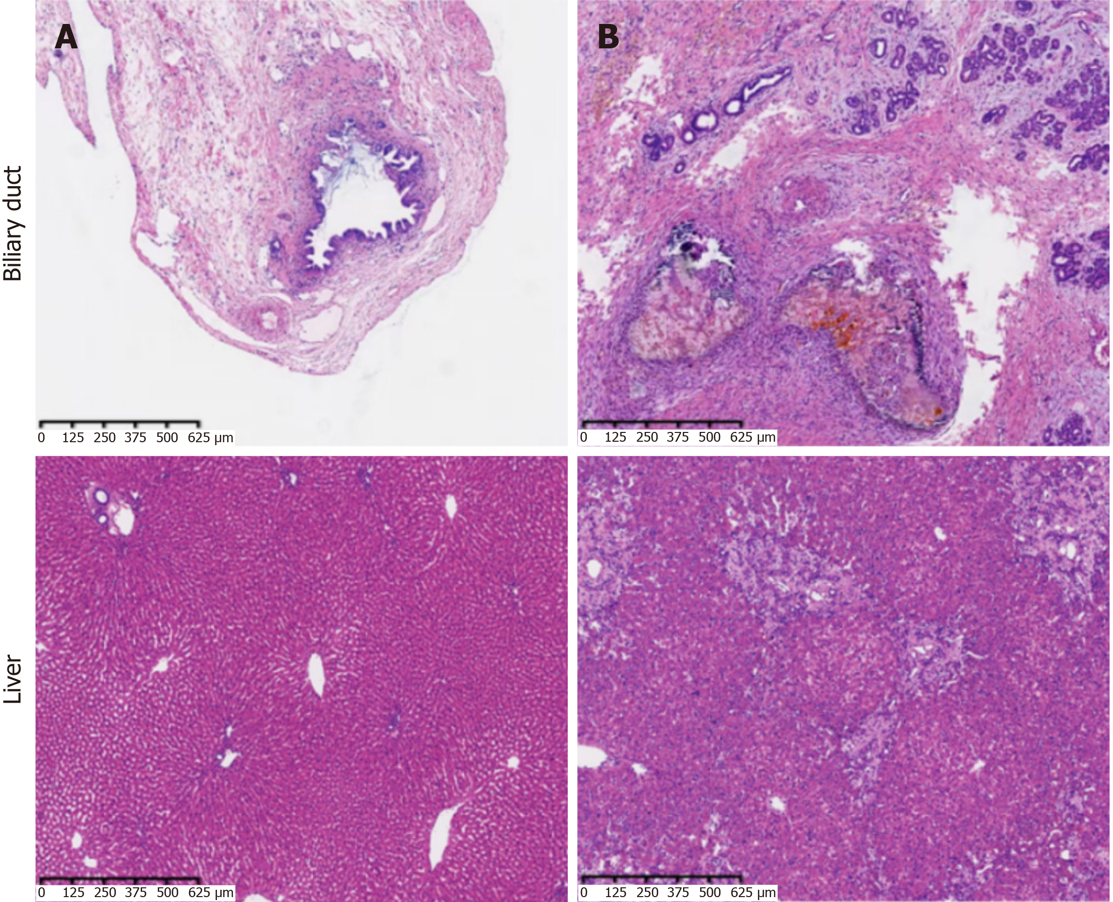
Figure 5 Hematoxylin and eosin staining of bile duct and liver tissues in the two groups (magnification, 4 ×).
A: Healthy control group; B: Model group.
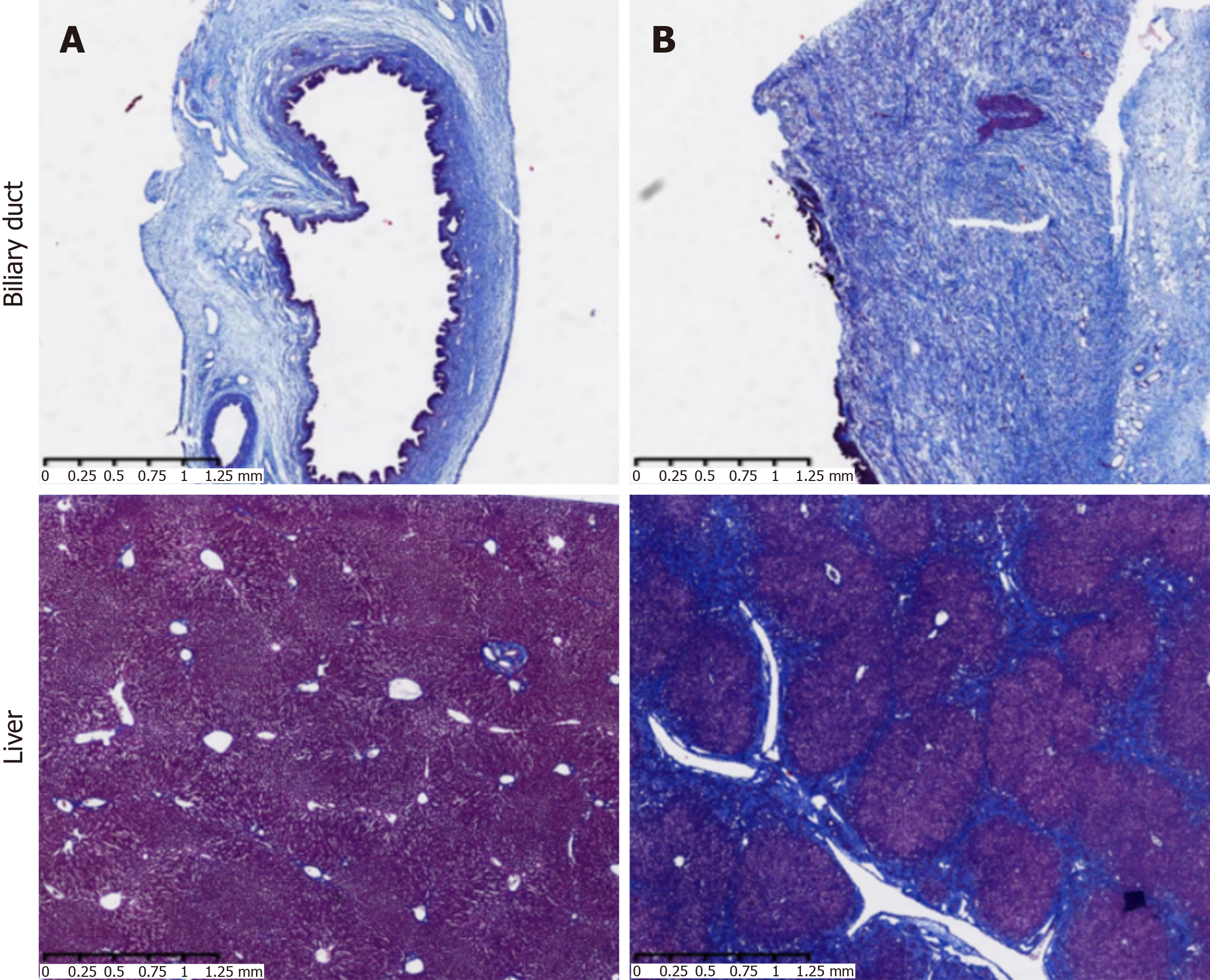
Figure 6 Masson staining of bile duct and liver tissues in the two groups (magnification, 2 ×).
A: Healthy control group; B: Model group.
- Citation: Sun QY, Cheng YM, Sun YH, Huang J. New rabbit model for benign biliary stricture formation with repeatable administration. World J Gastrointest Surg 2024; 16(11): 3538-3545
- URL: https://www.wjgnet.com/1948-9366/full/v16/i11/3538.htm
- DOI: https://dx.doi.org/10.4240/wjgs.v16.i11.3538


