©The Author(s) 2022.
World J Gastrointest Surg. Aug 27, 2022; 14(8): 849-854
Published online Aug 27, 2022. doi: 10.4240/wjgs.v14.i8.849
Published online Aug 27, 2022. doi: 10.4240/wjgs.v14.i8.849
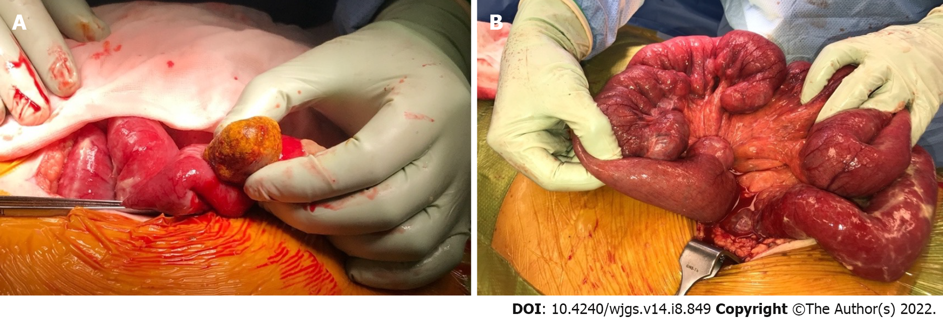
Figure 1 Intraoperative photographs during the patient’s initial laparotomy.
A: Offending enterolith removed via longitudinal enterotomy; B: Extensive jejunal diverticulosis.
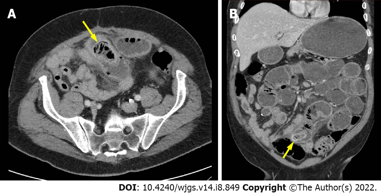
Figure 2 Obstructing enterolith on computed tomography of abdomen and pelvis.
A: Axial image of offending enterolith (yellow arrow); B: Coronal image of offending enterolith (yellow arrow).
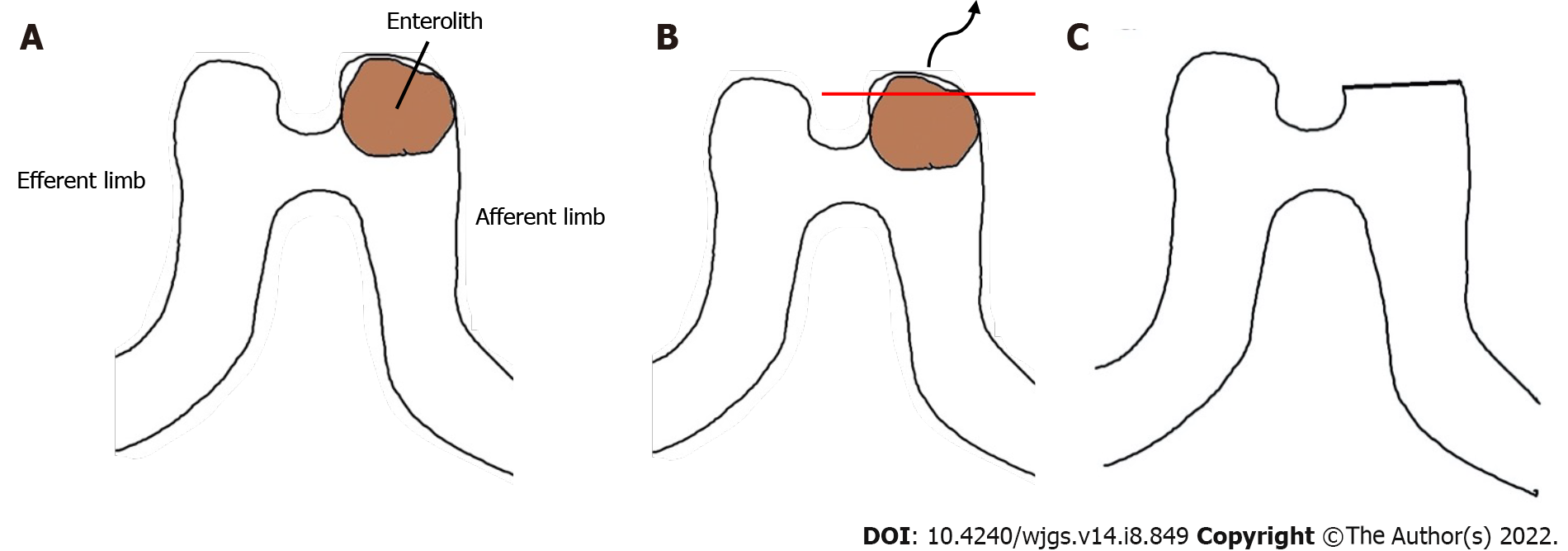
Figure 3 Animated depiction of intraoperative management of recurrent enterolith impaction.
A: Enterolith impaction in blind-ended pouch of previous side-to-side stapled anastomosis; B: Enterotomy at blind-ended pouch with enterolith extraction; C: Final configuration following closure of enterotomy with a linear stapler.
- Citation: Lee C, Menezes G. Recurrent small bowel obstruction secondary to jejunal diverticular enterolith: A case report. World J Gastrointest Surg 2022; 14(8): 849-854
- URL: https://www.wjgnet.com/1948-9366/full/v14/i8/849.htm
- DOI: https://dx.doi.org/10.4240/wjgs.v14.i8.849


