©The Author(s) 2025.
World J Diabetes. Sep 15, 2025; 16(9): 109597
Published online Sep 15, 2025. doi: 10.4239/wjd.v16.i9.109597
Published online Sep 15, 2025. doi: 10.4239/wjd.v16.i9.109597
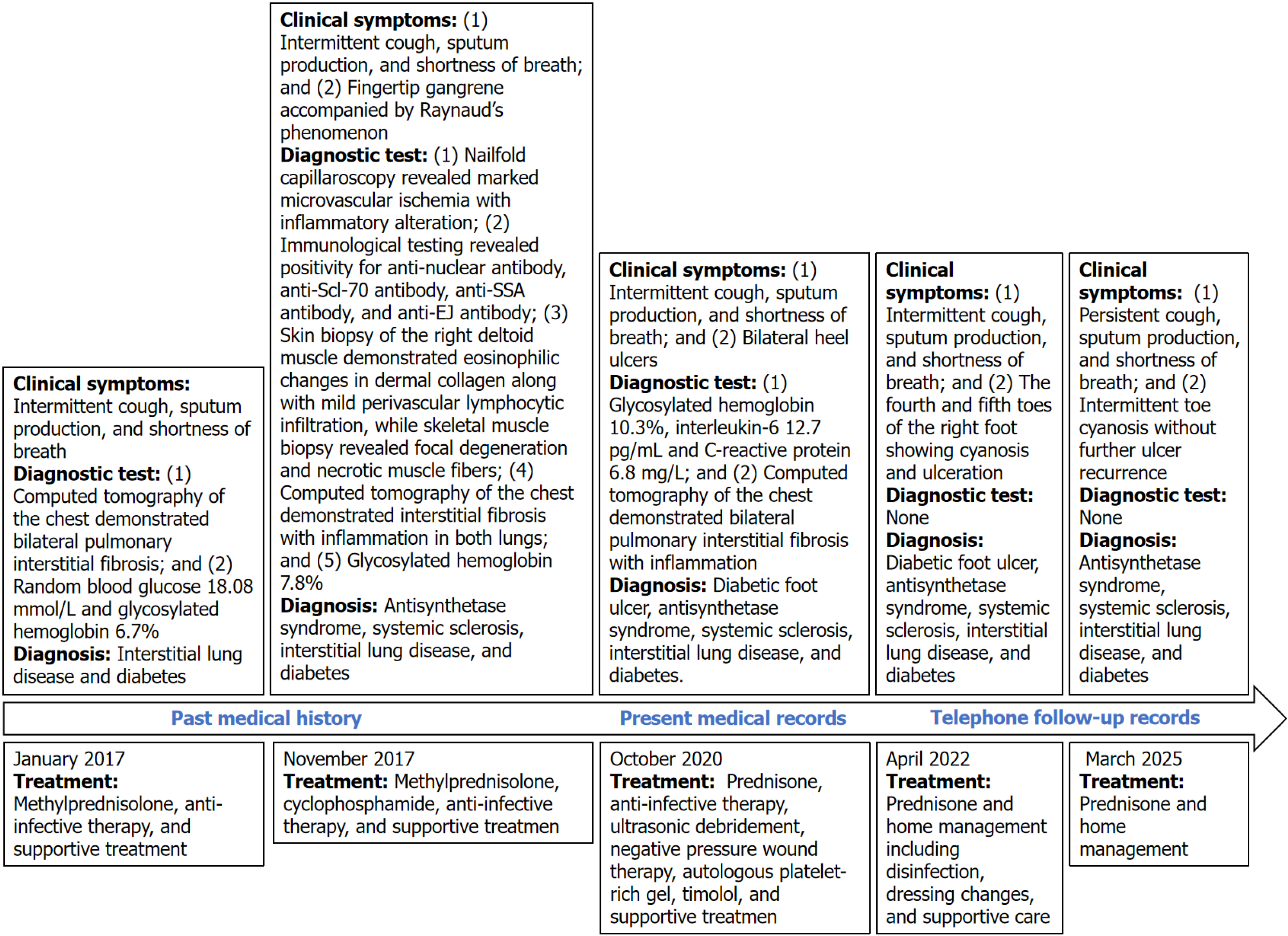
Figure 1 The timeline diagram of patient diagnosis and treatment.
anti-Scl-70 antibody: Anti-scleroderma-70 antibody; anti-SSA antibody: Anti-Sjogren’s-syndrome-related antigen A antibody; anti-EJ antibody: Anti-glycyl-tRNA synthetase.
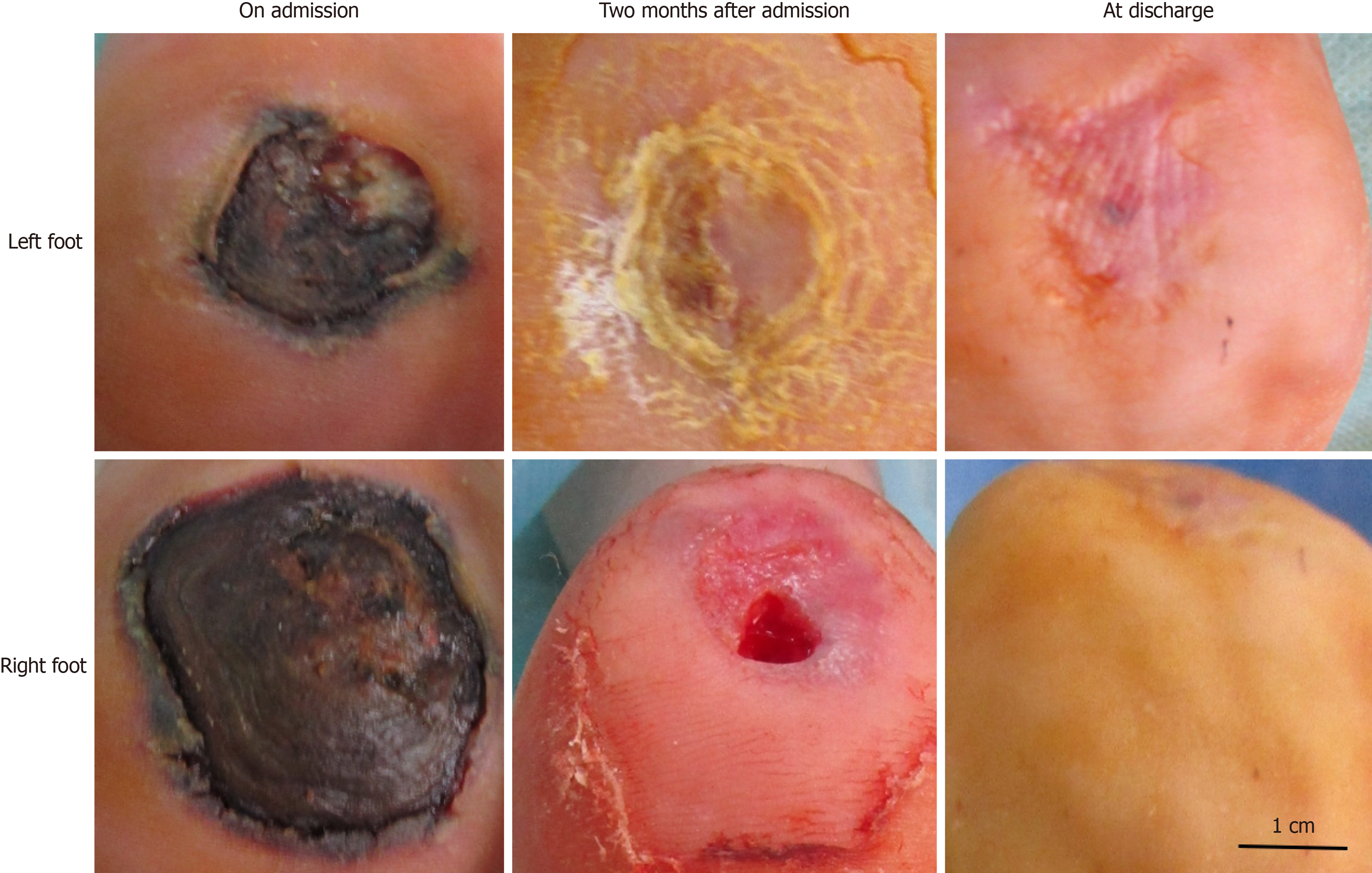
Figure 2 The condition of the patient’s foot ulcers.
Figure 3 Imageological examinations.
A: Computed tomography of the chest; B: Magnetic resonance imaging of the right foot (T1-weighted and sagittal image); C: Magnetic resonance imaging of the left foot (T1-weighted and sagittal image).
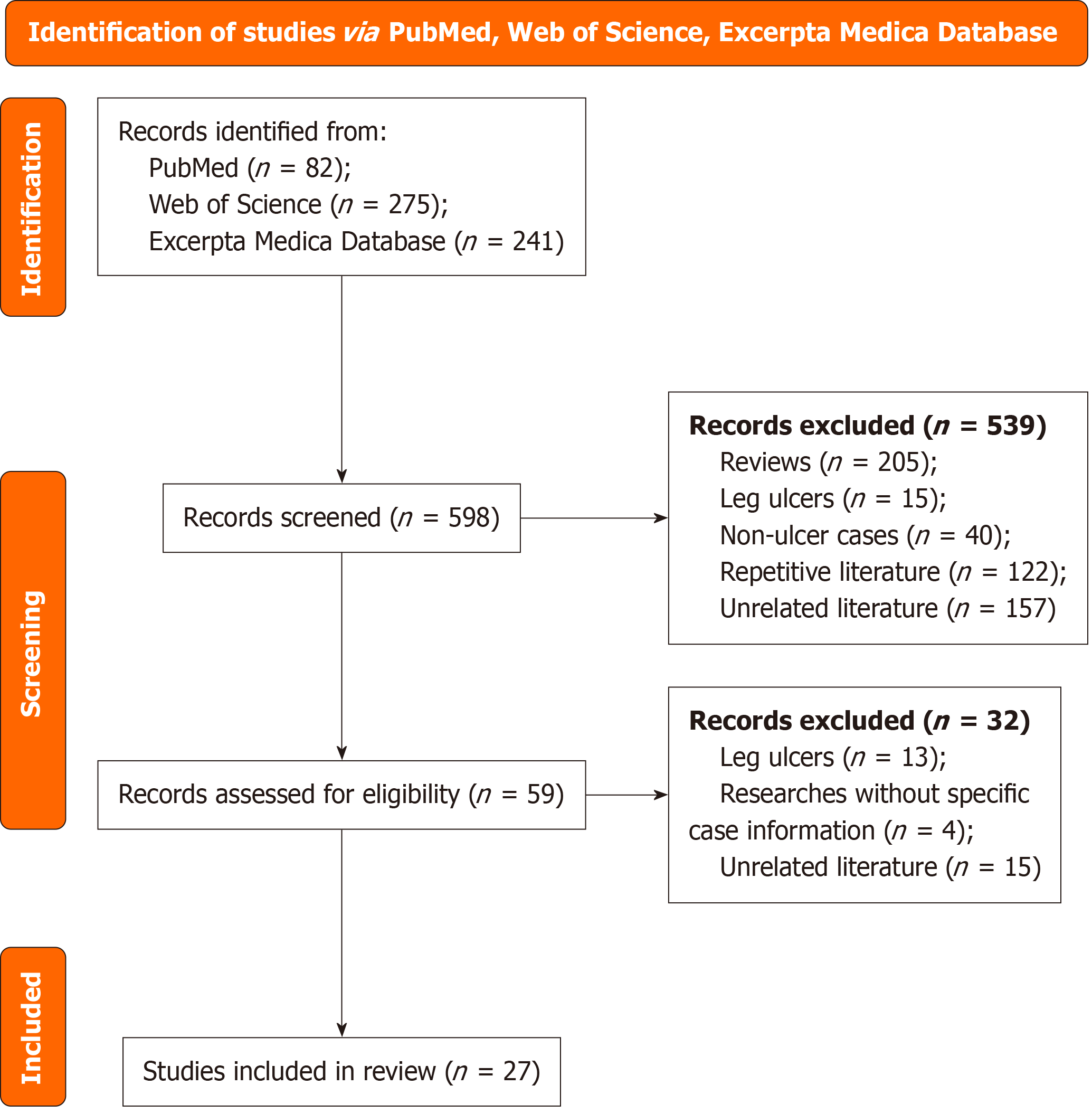
Figure 4 The preferred reporting items for systematic reviews and meta-analyses flow diagram of literature screening.
- Citation: Sun SY, Chen DW, Wu J, Li Y, Ran XW. Diabetic foot ulcer with overlap syndrome: A case report and review of literature. World J Diabetes 2025; 16(9): 109597
- URL: https://www.wjgnet.com/1948-9358/full/v16/i9/109597.htm
- DOI: https://dx.doi.org/10.4239/wjd.v16.i9.109597


