©The Author(s) 2025.
World J Diabetes. Oct 15, 2025; 16(10): 107640
Published online Oct 15, 2025. doi: 10.4239/wjd.v16.i10.107640
Published online Oct 15, 2025. doi: 10.4239/wjd.v16.i10.107640
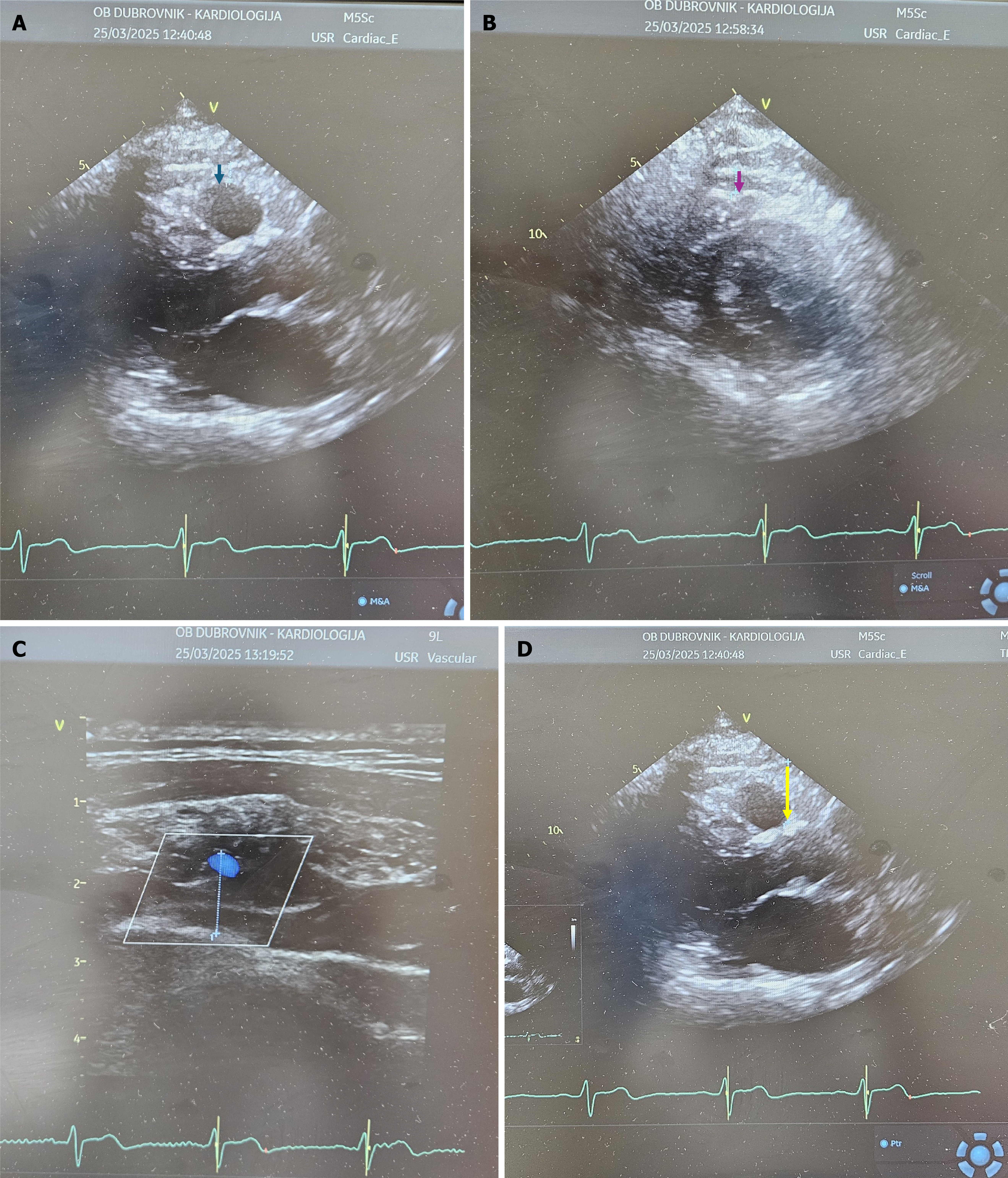
Figure 1 Epicardial adipose tissue thickness.
A: Measured from parasternal long-axis view (blue arrow); B: Measured from parasternal short-axis view (purple arrow); C: Measured from modified three-chamber view with a linear probe (dotted line); D: Measured from parasternal long-axis view at Rindfleisch fold (yellow arrow).
- Citation: Đuzel Čokljat A, Grubić Rotkvić P, Čokljat D, Ferri Certić J, Babić Z. Epicardial adipose tissue in diabetic myocardial disorder: Role of echocardiography. World J Diabetes 2025; 16(10): 107640
- URL: https://www.wjgnet.com/1948-9358/full/v16/i10/107640.htm
- DOI: https://dx.doi.org/10.4239/wjd.v16.i10.107640


