©The Author(s) 2024.
World J Diabetes. Jun 15, 2024; 15(6): 1299-1316
Published online Jun 15, 2024. doi: 10.4239/wjd.v15.i6.1299
Published online Jun 15, 2024. doi: 10.4239/wjd.v15.i6.1299
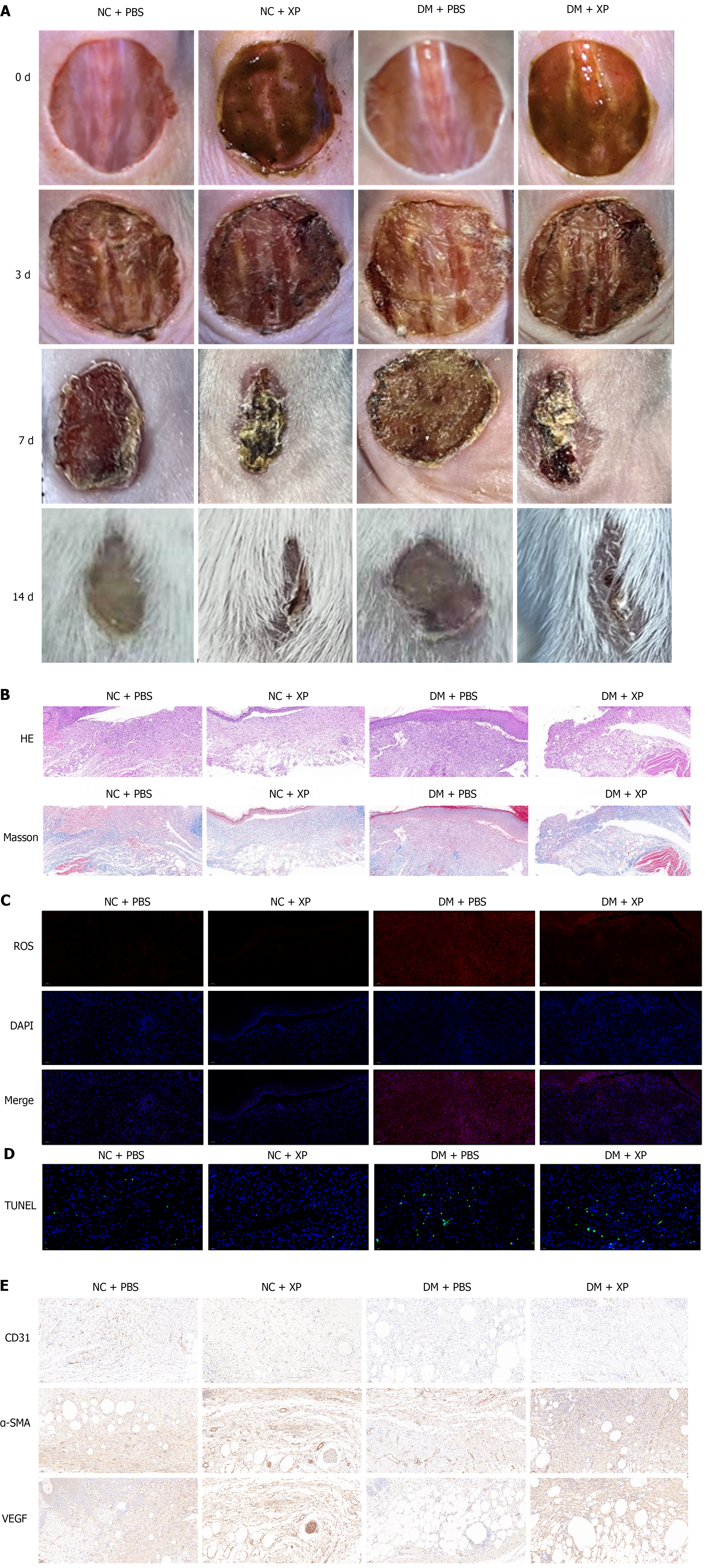
Figure 1 X-Paste promotes wound healing in diabetic foot ulcers mice.
A: Images of mice grouped in either normal control (NC), NC plus X-Paste (XP), diabetic wound plus phosphate-buffered saline or diabetic wound plus XP were captured at days 0, 3, 7 and 14; B: Wound tissue sections were stained with Hematoxylin and Eosin and Masson in different groups; C: The representative images of reactive oxygen species production in wound tissue sections from different groups; D: The representative images of TUNEL in wound tissue sections from different groups; E: The representative images of CD31, alpha-smooth muscle actin, and VEGFA from different groups. NC: Normal control; PBS: Phosphate-buffered saline; DM: Diabetic wound; XP: X-Paste; α-SMA: Alpha-smooth muscle actin.
Figure 2 Screening and analysis of effective substances in X-Paste.
A: High-performance liquid chromatography analyzed 10 batches of the X-Paste (XP); B: The volcano plot shows deferentially expressed genes (DEGs) between the XP treatment group and the diabetic foot ulcer group; C: GO enrichment analysis of DEGs; D: The KEGG enrichment analysis of DEGs; E: The molecular docking between Nrf2 and Andro. BP: Biological process; MF: Molecular function; CC: Cell component.
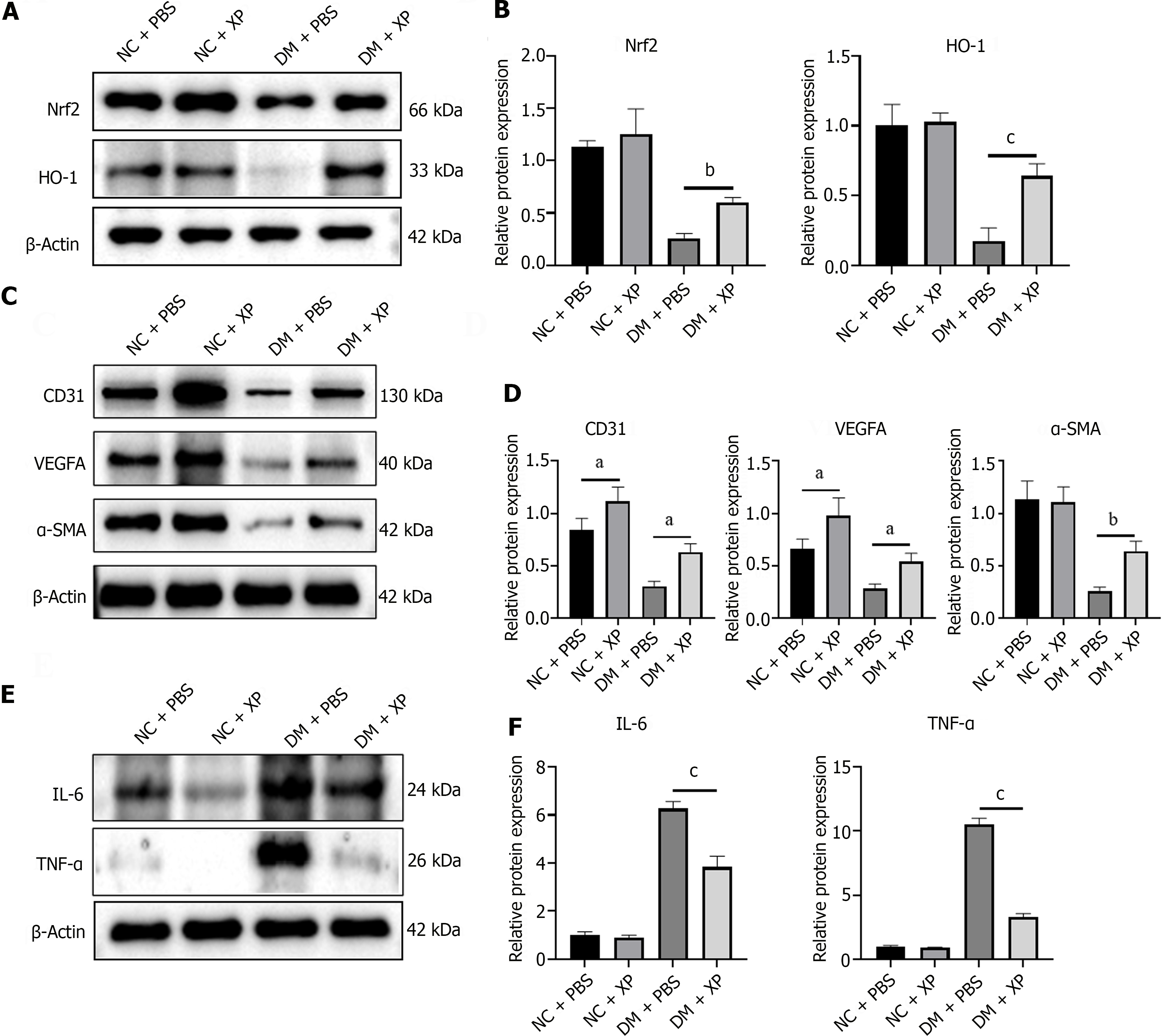
Figure 3 X-Paste promotes diabetic foot ulcers wound healing through the NF-E2-related factor-2/HO-1 pathway.
A: The expression of NF-E2-related factor-2 (Nrf2) and HO-1 were measured by Western blot from skin wound tissues; B: Quantification of Western blot results; C: The protein of CD31, VEGFA, and alpha-smooth muscle actin were measured by Western blot from skin wound tissues; D: Quantification of Western blot results; E: Western blot analysis of interleukin-6 and tumour necrosis factor alpha protein expression; F: Quantification of Western blot results. Statistical significance is expressed as aP < 0.05, bP < 0.01, cP < 0.001, P > 0.05 was not denoted. NC: Normal control; PBS: Phosphate-buffered saline; DM: Diabetic wound; XP: X-Paste; α-SMA: Alpha-smooth muscle actin; IL: Interleukin; TNF-α: Tumour necrosis factor alpha; Nrf2: NF-E2-related factor-2.
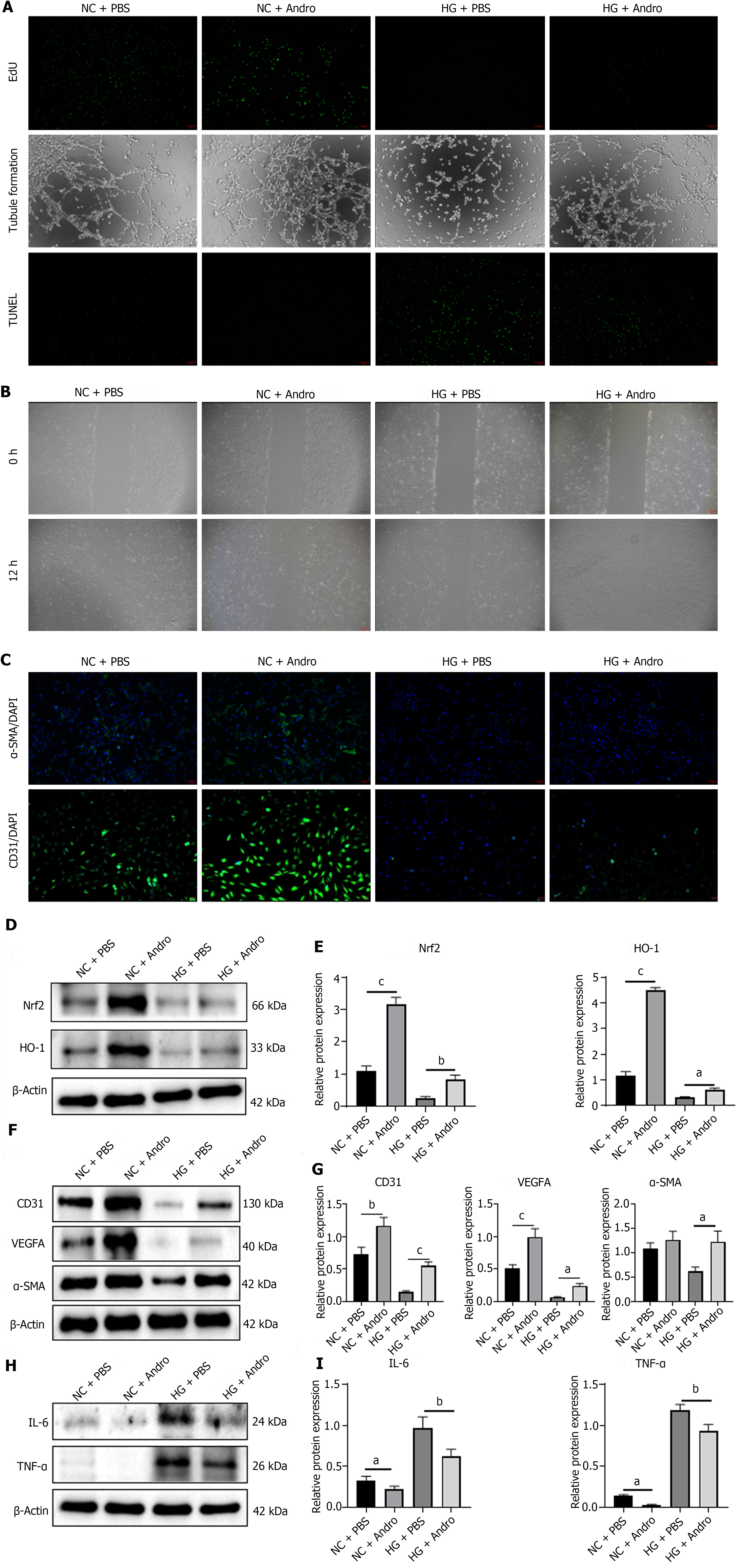
Figure 4 Effect of Andro on proliferation and metastasis of human umbilical vein endothelial cells.
A: The representative images of 5-ethynyl-2’-deoxyuridine, tubule formation and TUNEL in wound tissue sections from different groups; B: The wound healing assay in 0 h and 24 h from different groups; C: Immunofluorescence staining of CD31 and alpha-smooth muscle actin (α-SMA) in different groups; D and E: Western blot analysis of NF-E2-related factor-2 and HO-1 expression in each group and quantification of Western blot results; F and G: Western blot analysis of CD31, VEGFA, and α-SMA expression in each group and quantification of Western blot results; H and I: Western blot analysis of interleukin-6 and TNF-α expression in each group and quantification of Western blot results. Statistical significance is expressed as aP < 0.05, bP < 0.01, cP < 0.001, P > 0.05 was not denoted. HG: High glucose; NC: Normal control; PBS: Phosphate-buffered saline; IL: Interleukin; TNF-α: Tumour necrosis factor alpha; Nrf2: NF-E2-related factor-2; Andro: Andrographolide.
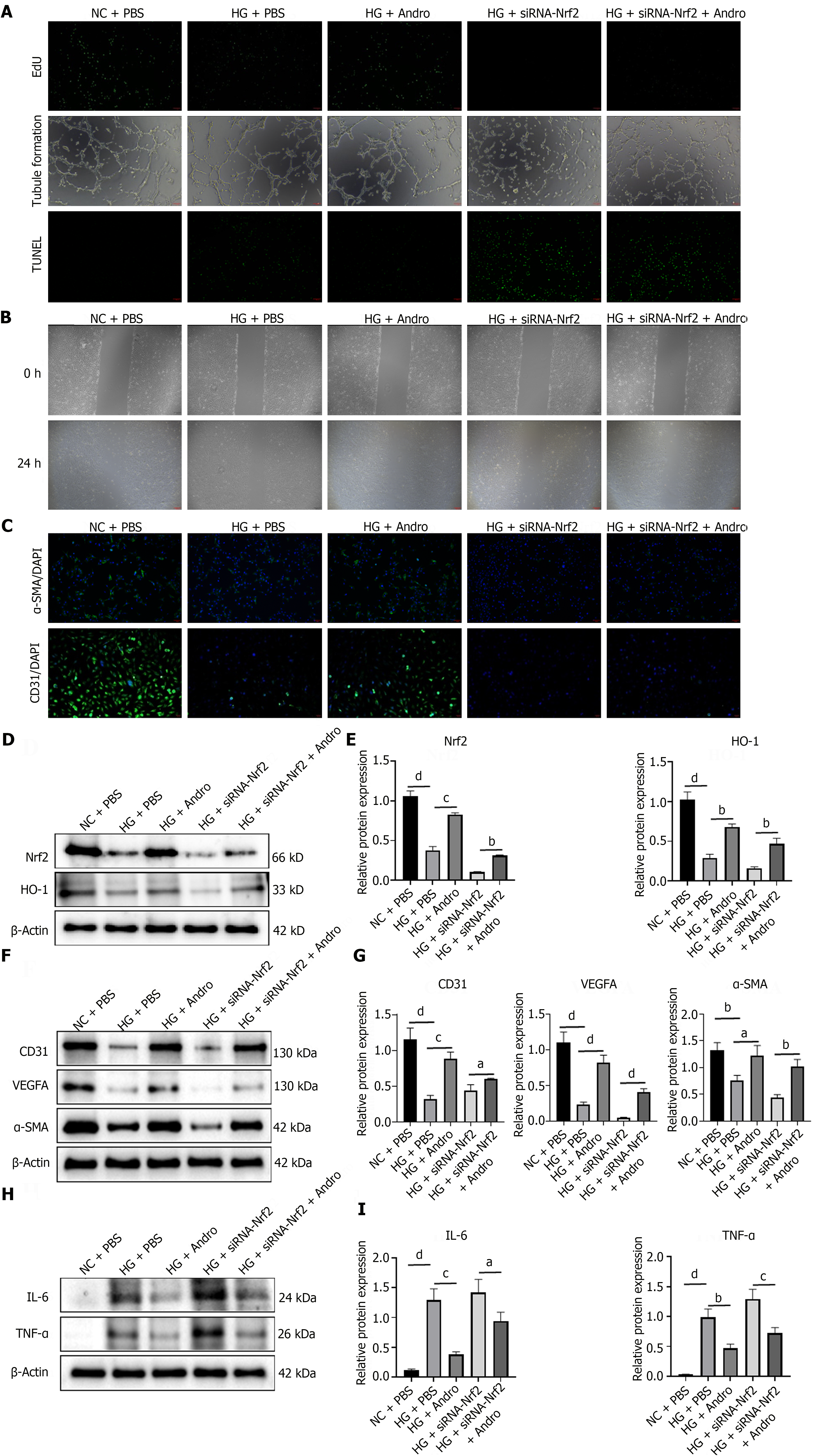
Figure 5 Andro accelerates diabetic foot ulcers wound healing by activating the NF-E2-related factor-2/HO-1 pathway.
A: The representative images of 5-ethynyl-2’-deoxyuridine, tubule formation and TUNEL in wound tissue sections from different groups; B: The wound healing assay in 0 h and 24 h from different groups; C: Immunofluorescence staining of CD31 and alpha-smooth muscle actin (α-SMA); D and E: Western blot of NF-E2-related factor-2 and HO-1 expression in each group and quantification of Western blot results; F and G: CD31, VEGFA, and α-SMA expression in each group and quantification of Western blot results; H and I: Western blot analysis of interleukin-6 and tumour necrosis factor alpha expression and quantification of Western blot results. Statistical significance is expressed as aP < 0.05, bP < 0.01, cP < 0.001, dP < 0.0001, P > 0.05 was not denoted.
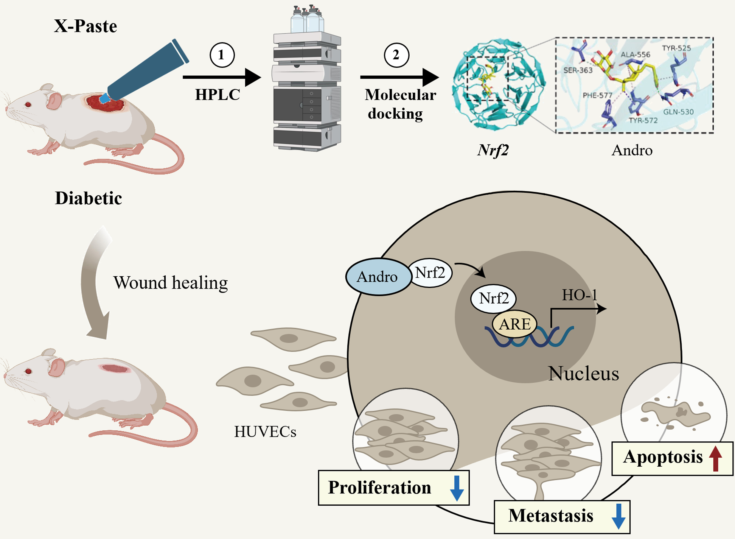
Figure 6 Effects on the wound healing in diabetic mice by the X-Paste through the NF-E2-related factor-2/HO-1 signaling pathway.
X-Paste (XP) obviously promotes the healing of skin wounds in diabetic mice, resulting in an accelerated healing process and shortened healing time. High-performance liquid chromatography was used to analyze the 21 main components of XP, among which Andro exhibited a strong binding ability with NF-E2-related factor-2 (Nrf2). At the cellular level, the addition of Andro alleviated high glucose (HG)-induced proliferation, migration, vascular injury, and inflammatory product inhibition in human umbilical vein endothelial cells (HUVECs). Mechanically, Andro relieved HG-induced damage to HUVECs by activating the Nrf2/HO-1 signaling pathway. HPLC: High-performance liquid chromatography; Nrf2: NF-E2-related factor-2; Andro: Andrographolide; HUVECs: Human umbilical vein endothelial cells.
- Citation: Du MW, Zhu XL, Zhang DX, Chen XZ, Yang LH, Xiao JZ, Fang WJ, Xue XC, Pan WH, Liao WQ, Yang T. X-Paste improves wound healing in diabetes via NF-E2-related factor/HO-1 signaling pathway. World J Diabetes 2024; 15(6): 1299-1316
- URL: https://www.wjgnet.com/1948-9358/full/v15/i6/1299.htm
- DOI: https://dx.doi.org/10.4239/wjd.v15.i6.1299


