©The Author(s) 2021.
World J Diabetes. May 15, 2021; 12(5): 556-568
Published online May 15, 2021. doi: 10.4239/wjd.v12.i5.556
Published online May 15, 2021. doi: 10.4239/wjd.v12.i5.556
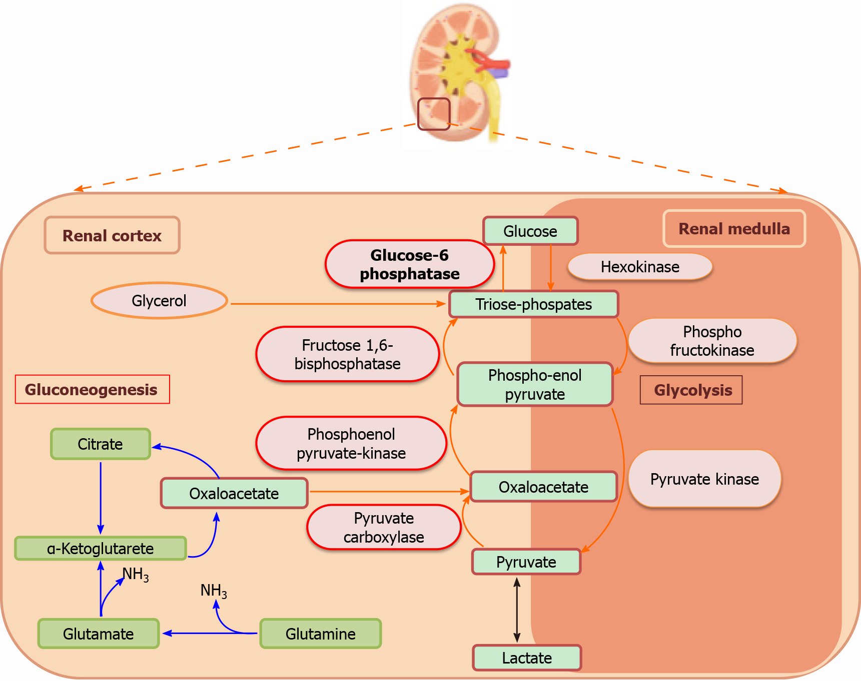
Figure 1 Schematic overview of renal gluconeogenesis and glycolysis pathway and enzyme localization.
The key enzymes of gluconeogenesis (1) pyruvate carboxylase; (2) phosphoenolpyruvate carboxykinase; (3) fructose-1,6-biphosphatase; and (4) glucose 6-phosphatase are predominantly localized in the renal cortical cells whereas, the glycolytic key enzymes (1) hexokinase; (2) phosphofructokinase; and (3) pyruvate kinase are found in the renal medulla.
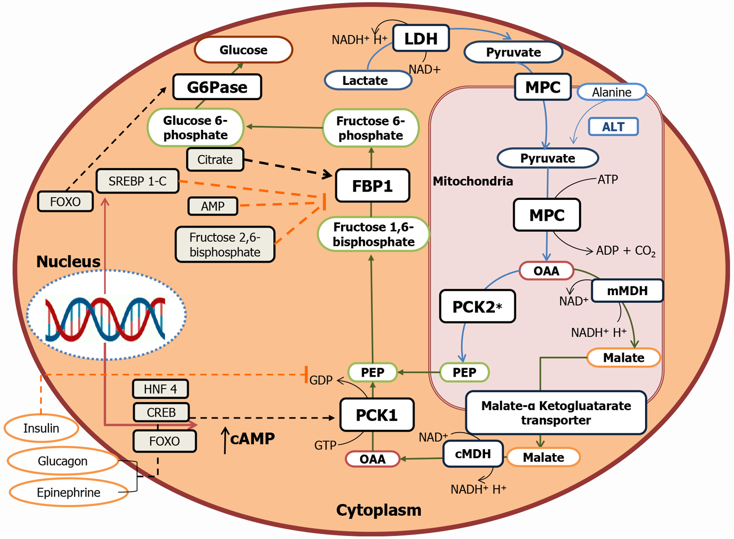
Figure 2 Gluconeogenesis Pathway and cellular compartmentalization of the gluconeogenic enzymes.
Pyruvate from lactate enters mitochondria by mitochondrial pyruvate transporter. Pyruvate provided by alanine transamination or lactate dehydrogenation is converted to oxaloacetate (OAA) by mitochondrial pyruvate carboxylase. OAA is either reduced to malate and exported out in the cytoplasm by malate ketoglutarate transporter or directly converted to phosphoenolpyruvate (PEP) by phosphoenolpyruvate carboxykinase (PCK) 2 (mitochondrial isoform) and exported out in the cytoplasm. In the cytoplasm, malate is first oxidized to OAA and then converted to PEP by PCK1 (cytoplasmic isoform). Fructose-1,6-bisphosphate (FBP) is then converted to fructose-6-phosphate by cytoplasmic FBP1. Glucose-6-phosphatase in the cytoplasm ultimately dephosphorylates glucose-6-phosphate to release glucose. G6Pase: Glucose-6-phosphatase; LDH: Lactate dehydrogenase; MPC: Mitochondrial pyruvate carrier; ALT: Alanine aminotransferase; FBP: Fructose-1,6-bisphosphate; OAA: Oxaloacetate; PCK: Phosphoenolpyruvate carboxykinase; PEP: Phosphoenolpyruvate; mMDH: Malate dehydrogenase; cAMP: Cyclic adenosine monophosphate.
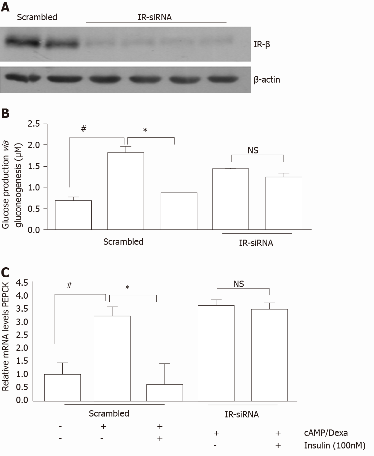
Figure 3 siRNA mediated knockdown of insulin receptor in the human proximal tubule cells increased glucose production via gluconeogenesis stimulation.
A: Western blot showing reduced insulin receptor insulin receptor (IR) expression in IR-siRNA treated human proximal tubule (hPT) cells relative to scrambled; B:cAMP/Dexa induced gluconeogenesis/glucose production in the hPT cell culture media; and C: Relative phosphoenolpyruvate carboxykinase mRNA transcript levels in scrambled and IR-siRNA treated hPT cells with or without insulin treatment. “Citation: Pandey G, Shankar K, Makhija E, Gaikwad A, Ecelbarger C, Mandhani A, Srivastava A, Tiwari S. Reduced Insulin Receptor Expression Enhances Proximal Tubule Gluconeogenesis. J Cell Biochem 2017; 118: 276-285 [PMID: 27322100 DOI: 10.1002/jcb.25632] Copyright © The Author(s) 2017. Published by John/Wiley & Sons, Inc[65]”
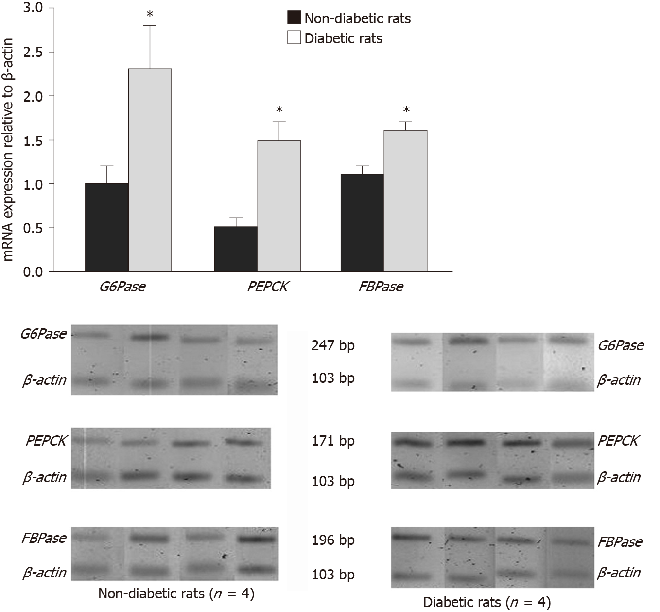
Figure 4 mRNA and protein levels of glucose-6-phosphatase, phosphoenolpyruvate carboxykinase, and fructose-1,6-bisphosphatase in diabetic rats and their non-diabetic controls.
“Citation: Eid A, Bodin S, Ferrier B, Delage H, Boghossian M, Martin M, Baverel G, Conjard A. Intrinsic gluconeogenesis is enhanced in renal proximal tubules of Zucker diabetic fatty rats. J Am SocNephrol 2006; 17: 398-405 [PMID: 16396963 DOI: 10.1681/asn.2005070742] Copyright © The Author(s) 2006. Published by the American Society of Nephrology Inc[12]”
- Citation: Sharma R, Tiwari S. Renal gluconeogenesis in insulin resistance: A culprit for hyperglycemia in diabetes. World J Diabetes 2021; 12(5): 556-568
- URL: https://www.wjgnet.com/1948-9358/full/v12/i5/556.htm
- DOI: https://dx.doi.org/10.4239/wjd.v12.i5.556


