©The Author(s) 2020.
World J Diabetes. Jan 15, 2020; 11(1): 1-12
Published online Jan 15, 2020. doi: 10.4239/wjd.v11.i1.1
Published online Jan 15, 2020. doi: 10.4239/wjd.v11.i1.1
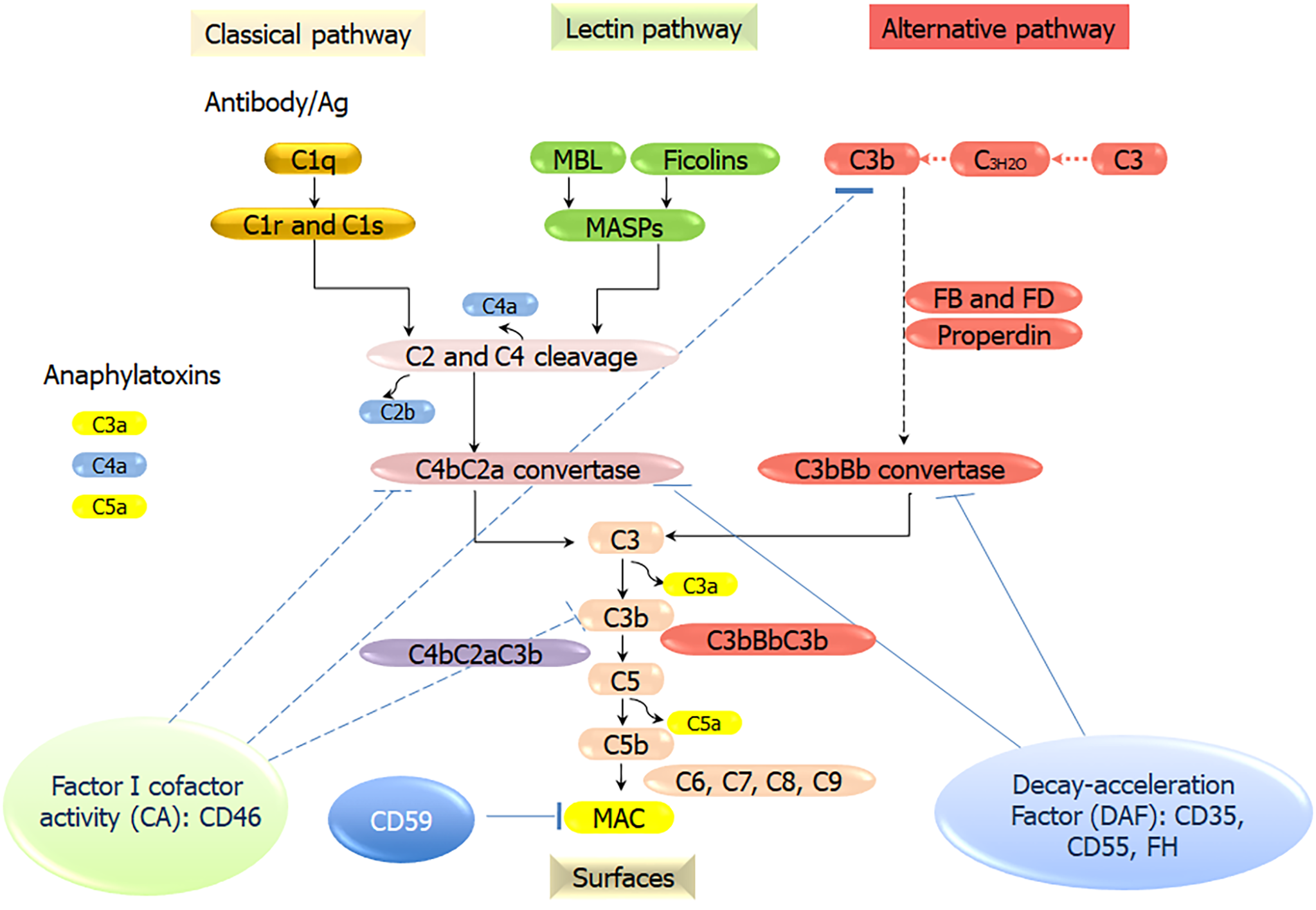
Figure 1 Overview of the activation and regulation of the complement pathways.
Three different recognition and activation pathways, including classical pathway (CP), lectin pathway (LP), and alternative pathway (AP). The CP is triggered by the formation of antigen-antibody immune complexes that bind to the C1 complex (C1q, C1r2, C1s2). The LP is activated by the binding of mannan-binding lectin (MBL), ficolins, or collectin 11 to carbohydrates/mannan/other pathogen-associated molecular patterns. MBL-associated serine proteases (MASP)-1, MASP-2, and MASP-3 are activated. Serine protease C1s/C1r (CP) or MASPs (LP) sequentially cleave C4 and C2 to form C3 convertase C4bC2b. The AP C3 convertase (C3bBb) is generated through a chain of reactions involving factor B, factor D and properdin. C3b then binds with the C3 convertase to form C5 convertases (C4bC2bC3b or C3bBbC3b), which cleaves C5 into C5a and C5b. C5b binds C6, C7, C8, and C9 molecules to form the membrane attack complex (MAC), the lytic machinery of the complement system. MBL: Mannan-binding lectin; MASP: Mannan-binding lectin-associated serine proteases; FB: Factor B; FD: Factor D; MAC: Membrane attack complex.
- Citation: Shim K, Begum R, Yang C, Wang H. Complement activation in obesity, insulin resistance, and type 2 diabetes mellitus. World J Diabetes 2020; 11(1): 1-12
- URL: https://www.wjgnet.com/1948-9358/full/v11/i1/1.htm
- DOI: https://dx.doi.org/10.4239/wjd.v11.i1.1


