Copyright
©2011 Baishideng Publishing Group Co.
World J Gastrointest Oncol. Jun 15, 2011; 3(6): 99-102
Published online Jun 15, 2011. doi: 10.4251/wjgo.v3.i6.99
Published online Jun 15, 2011. doi: 10.4251/wjgo.v3.i6.99
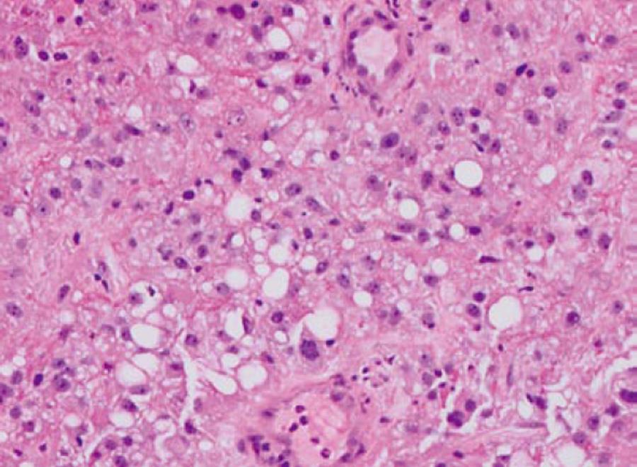
Figure 1 Histopathology of the nephrectomy specimen showing clear cell type renal cancer.
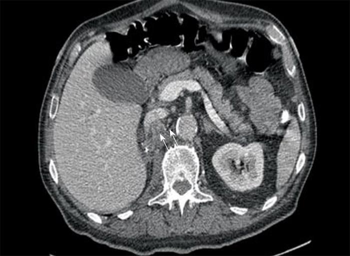
Figure 2 Computed tomography scan of abdomen showing heterogeneous soft tissue mass extending into the duodenum from right nephrectomy bed.
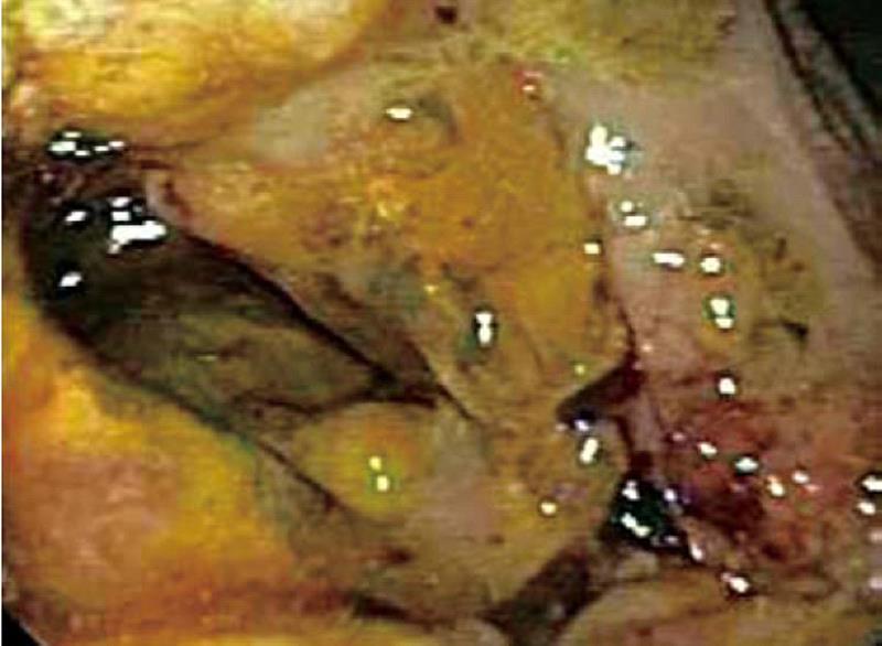
Figure 3 Endoscopic picture of fungating mass in the duodenum.
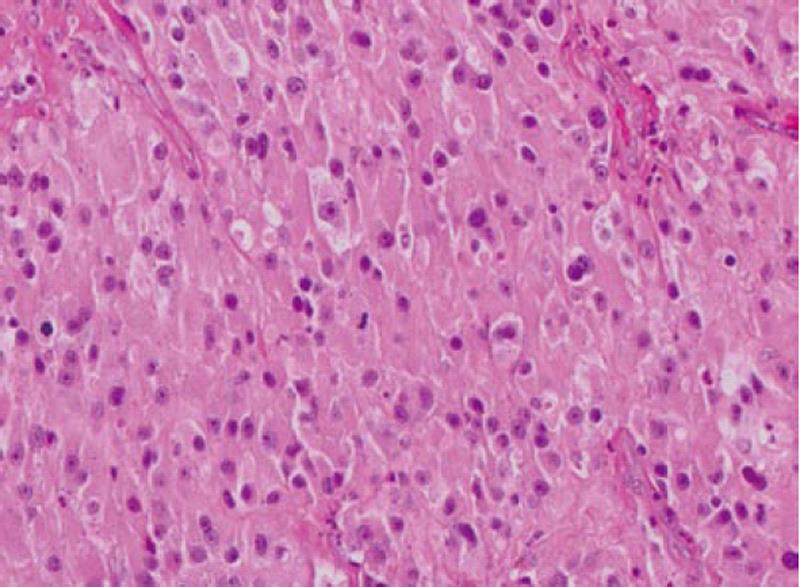
Figure 4 Duodenal biopsy showing large cell malignant epithelial neoplasm with abundant eosinophilic cytoplasm and occasional mitotic figures.
- Citation: Cherian SV, Das S, Garcha AS, Gopaluni S, Wright J, Landas SK. Recurrent renal cell cancer presenting as gastrointestinal bleed. World J Gastrointest Oncol 2011; 3(6): 99-102
- URL: https://www.wjgnet.com/1948-5204/full/v3/i6/99.htm
- DOI: https://dx.doi.org/10.4251/wjgo.v3.i6.99


