Case Report Open Access
Copyright ©2011 Baishideng Publishing Group Co., Limited. All rights reserved.
World J Gastrointest Oncol. Jun 15, 2011; 3(6): 103-106
Published online Jun 15, 2011. doi: 10.4251/wjgo.v3.i6.103
Adult intussusception secondary to an ileum hamartoma
Carlos M Nuño-Guzmán, José Arróniz-Jáuregui, José Ignacio Gómez-Ontiveros, Arturo Vargas-Gerónimo, Jesús Valle-González, Department of General Surgery, Antiguo Hospital Civil de Guadalajara “Fray Antonio Alcalde”, Calle Hospital No. 278, Sector Hidalgo. C.P. 44280, Guadalajara, Jalisco, Mexico
Ismael Espejo, Department of Histopathology, Antiguo Hospital Civil de Guadalajara “Fray Antonio Alcalde”, Calle Hospital No. 278, Sector Hidalgo. C.P. 44280 Guadalajara, Jalisco, Mexico
Josué Solís-Ugalde, Department of Radiology, Antiguo Hospital Civil de Guadalajara “Fray Antonio Alcalde”, Calle Hospital No. 278, Sector Hidalgo. C.P. 44280, Guadalajara, Jalisco, Mexico
Author contributions: Nuño-Guzmán CM, Arróniz-Jáuregui J, Espejo I and Solís-Ugalde J supplemented the case report data; Nuño-Guzmán CM, Gómez-Ontiveros JI and Vargas-Gerónimo A analyzed the case data; Nuño-Guzmán CM and Espejo I wrote the document; Nuño-Guzmán CM, Arróniz-Jáuregui J, Gómez-Ontiveros JI, Vargas-Gerónimo A and Valle-González J participated in surgery on the patient.
Correspondence to: Carlos M Nuño-Guzmán, MD, MSc, Department of General Surgery, Antiguo Hospital Civil de Guadalajara “Fray Antonio Alcalde”, Calle Hospital No. 278. Sector Hidalgo. C.P. 44280, Guadalajara, Jalisco, Mexico. carlosnunoguzman@hotmail.com
Telephone: +1-5233-36145501 Fax: +1-5233-36690229
Received: October 15, 2010
Revised: March 12, 2011
Accepted: March 18, 2011
Published online: June 15, 2011

Abstract

Intussusception is a rare condition in the adult population. However, in contrast to its presentation in children, an identifiable etiology is found in the majority of cases. Clinical manifestations of adult intussusception are non-specific and patients may present with acute, intermittent or chronic symptoms, predominantly those of intestinal obstruction. A 27-year-old male patient with recurrent abdominal pain secondary to intussusception is herein reported. The clinical presentation and ultrasonographic findings led to the diagnosis. At laparotomy, an ileal hamartoma was found as the lead point of the intussusception. Surgical management and histopathologic studies are described. A recurrent intestinal obstruction and classic ultrasound findings may lead to the diagnosis of intussusception but surgical exploration remains essential. The principle of resection without reduction is well established.

Key Words: Adult intussusception; Ileum hamartoma; Intestinal obstruction


INTRODUCTION

Intussusception accounts for 1%-5% of all cases of intestinal obstruction in adults[1]. In the majority of adult patients, a cause is identified. However, clinical presentation is not specific, manifesting as chronic intestinal obstruction symptoms[2]. Although radiographic findings at abdominal ultrasonography and computed tomography may be indicative, a preoperative diagnosis is made less frequently in adult patients than in children[2,3].

CASE REPORT

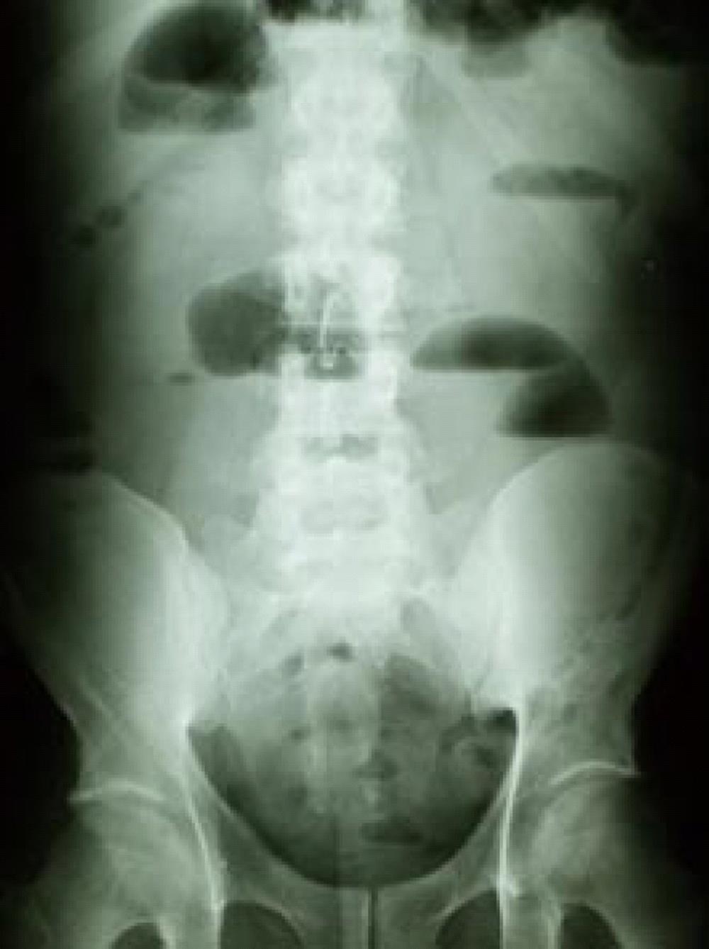
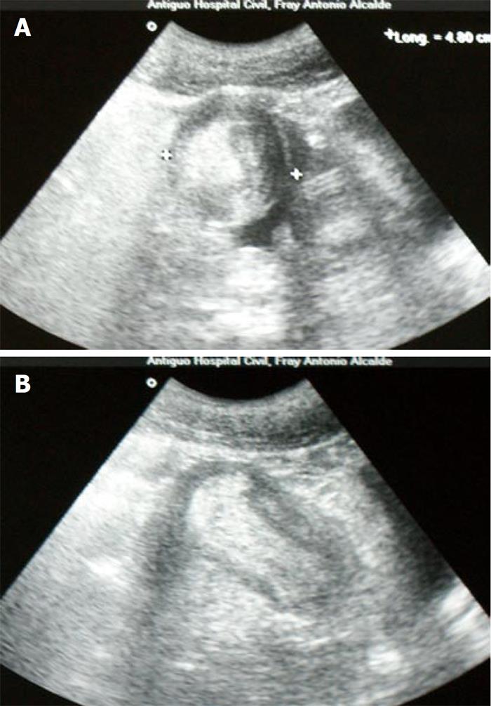
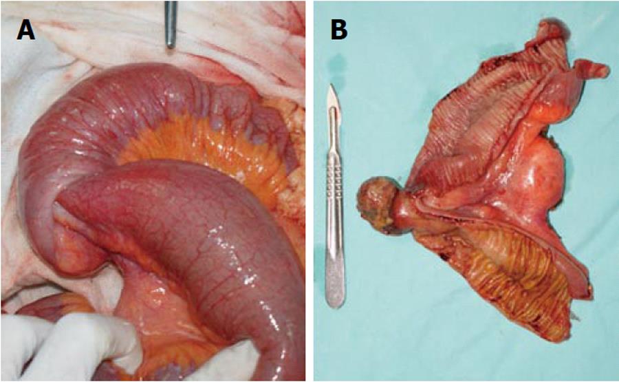
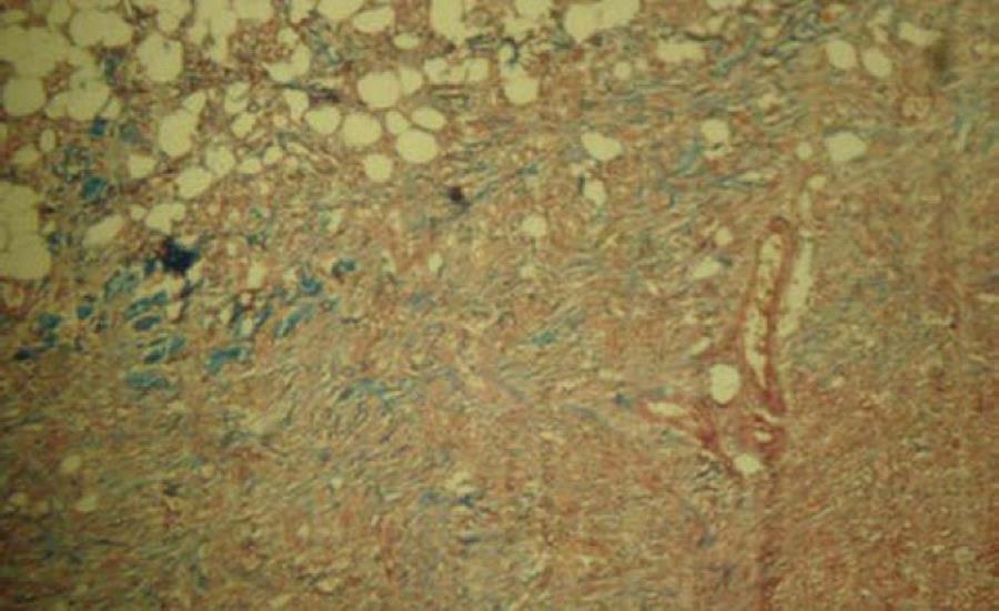
A 27-year-old male patient presented at the emergency department complaining of a 6-wk history of recurrent cramping abdominal pain. He had previously been admitted at three hospitals. Increased abdominal pain, nausea and seven episodes of vomiting occurred during the 24 h prior to admission at our institution. Physical examination revealed signs of dehydration, a temperature of 36.8°C, a pulse of 94 beats per minute, a respiratory rate of 18 per minute and blood pressure 100/60 mmHg. Bowel sounds were hyperactive, his abdomen was distended, with tenderness, but no guarding or rebound. White blood cell count was 8090 per cubic millimeter, with 76% neutrophils. Other tests were unremarkable. Plain abdominal film showed dilated bowel loops and air-fluid levels (Figure 1). At the second day after admission, after fluid resuscitation, the patient recovered transit of feces and gases, and vital signs normalized; white blood cell count was 6400 per cubic millimeter, with 80% neutrophils. Transabdominal ultrasound, gave images suggestive of an intussusception (Figure 2A and B). On an elective basis, the patient underwent a laparotomy. Dilated jejunum and ileum proximal to an intussusception which was 30 cm proximal to the ileocecal valve were found (Figure 3A). Resection and anastomosis were performed around the intussusception, whose lead point was a 3 cm pedunculated-type polypoid tumor (Figure 3B). No other palpable tumor was found during exploration of the bowel. Histopathology revealed a solid mass with vascular congestion and superficial necrosis, formed by well-differentiated adult-type adipose cells, separated by fibrous tissue, fibrocytes, fibroblasts and mesenchymal cells, which showed no pleomorphic or mitotic features, although irregular vessel distribution was observed. These findings are compatible with the diagnosis of an ileal hamartoma (Figure 4). After an uneventful recovery, the patient was discharged on the fifth postoperative day. After 18 mo, the patient is in good health.

Figure 1
Figure 1 Plain abdominal film showed dilated bowel loops and air-fluid levels.
Figure 2
Figure 2 Ultrasonographic feature of a “target” sign on a transverse view (A), and a “sausage-shaped image” in a longitudinal view (B).
Figure 3
Figure 3 Distended bowel proximal to the intussusception (A), open surgical specimen showing a 3 cm pedunculated-type polypoid tumor (B).
Figure 4
Figure 4 Histopathologic appearance of the ileal tumor showing an active mesenchymal lesion, with no malignant transformation (Masson’s trichrome stain, × 10).
DISCUSSION

Intussusception refers to the telescoping displacement of a proximal segment of bowel (intussusceptum) into the lumen of the adjacent distal segment (intussuscipiens). It accounts for 1%-5% of all cases of intestinal obstruction in adults[1]. In contrast to children, in whom 95% of intussusceptions take place, an etiology is found in 70%-90% of adult patients[2]. However, preoperative diagnosis in adult cases is infrequent, due to its varying presentation, which most often is consistent with intestinal obstruction, but may manifest with acute, intermittent or chronic symptoms[2,3]. In up to 90% of adult cases, a well defined lesion serves as a lead point for the adjacent bowel segment to telescope into the lumen of the distal segment, causing mesentery compromise. The bowel edema and subsequent compression of vessels in the mesentery may cause ischemic necrosis of the bowel wall[4]. Clinical presentation of intussusception is nonspecific. The predominant symptoms are those of partial intestinal obstruction, where the most important characteristic of abdominal pain is its periodic, intermittent and cramping nature[2,4,5]. Other signs and symptoms such as nausea, vomiting, constipation, fever, intestinal bleeding, diarrhea, and a palpable abdominal mass are less frequent. Although there are acute presentations, the mean duration of symptoms exceeds 7 d, while clinical manifestations have been present from two weeks to several months in most of the cases, sometimes reaching one to five years[1,2,4-9].

A correct preoperative diagnosis ranging from 30% to 70% has been reported, mainly due to the varying and nonspecific clinical presentation[1,2,4-7,9]. Since obstructive symptoms predominate in most cases, plain abdominal films are the first diagnostic modality. Signs of intestinal obstruction such as dilated loops and air fluid level may be seen, and information about the site of obstruction may be obtained[1,4,7-10]. An upper gastrointestinal series may reveal a small bowel intussusception; a proximal dilated bowel and a beaklike change in the caliber at the obstruction. The classic “stacked coin” or “coiled spring” signs are characteristic in upper gastrointestinal series[1,2,4,5,10,11]. Ultrasonography is a useful diagnostic modality; the classic imaging features are the “target” or “doughnut” signs in the transverse view and the “pseudo-kidney” sign in the longitudinal view. However, obesity, the presence of air in the distended bowel loops and operator skill may limit the study accuracy[9,10,12]. Abdominal Computed Tomography scan is considered the most useful imaging modality, with a diagnostic accuracy of 58% to 100%. It is particularly useful when a mass is found on physical examination. The characteristic features correspond to an early target mass with enveloped, eccentrically located areas of low density, which may appear as “target sign”, “sausage shaped mass” or “reniform mass”. A CT scan may define the location, nature of the mass, its relationship to surrounding tissues, and staging in the case of suspected malignancy[1,2,8,10].

In adult intussusception, surgical exploration remains essential. Nevertheless, controversy persists concerning the optimal surgical management strategy. The principle of resection without reduction is well established[11]. Several considerations have been highlighted: the frequency of an underlying etiology, the prevalence of associated malignancy, the anatomic site and extent of intussusception, and the degree of inflammation and ischemia in the affected bowel segment[5]. The high likelihood of malignancy in colonic intussusception justifies resection without reduction. In small bowel intussusception, a more selective approach seems feasible, although resection is advocated unless a benign lesion has been previously confirmed. Nevertheless, in the majority of cases, the inability to differentiate benign from malignant etiologies, signs of bowel ischemia and the possibility of perforation should be considered[1,4,6]. The overall incidence of malignancy in adult intussusception lesions is approximately 40%; an overall malignancy incidence of up to 40% in small bowel intussusception has been reported, whereas in colonic intussusception it has been as high as 65%[1,2,4,5,10,11].

In 1940, Clarke used the term “Myoepithelial hamartoma” to describe gastrointestinal submucosal tumors comprising glandular elements, lined by epithelial cells and smooth muscle[13]. The predominant tissue in these tumors may be either connective tissue derivatives such as lamina propia, smooth muscle, vasoformative tissue, or nerve elements, or epithelial elements[14]. Only a few cases of intussusception secondary to a solitary hamartoma have been reported, most of them in the pediatric population[15-22]. In adult patients, reported cases are also scarce, both among series and in case reports[1,4,23-27].

In the adult patient herein reported, a 6-wk history of recurrent cramping abdominal pain, requiring three previous hospital admissions, as well as the increased pain, nausea and vomits that occurred during the 24 h, were consistent with intermittent intestinal obstruction. Dilated intestinal loops with air-fluid levels seen on the plain abdominal film were accordingly indicative. The classic ultrasound findings were consistent with an intussusception as the cause of the intestinal obstruction, making the CT scan non- essential. The apparent transient resolution manifested by the regaining of transit of gas and feces allowed a non-urgent laparotomy. Reduction was avoided for reasons already described, and resection of an intestinal segment proximal and distal to the intussusception was performed.

In conclusion, we have reported an unusual cause of obstruction in an adult patient, secondary to a hamartoma as the lead point of an ileal intussusception.

Footnotes

Peer reviewer: Goran Stanojevic, MD, PhD, Proffesor, Department of Surgery, Clinical Centre Nis, Bul Zorana Djindjica 48, 18000 Nis, Serbia

S- Editor Wang JL L- Editor Hughes D E- Editor Ma WH

References
1.  Eisen LK, Cunningham JD, Aufses AH Jr. Intussusception in adults: institutional review. J Am Coll Surg. 1999;188:390-395.  [PubMed]  [DOI]  [Cited in This Article: ]
2.  Azar T, Berger DL. Adult intussusception. Ann Surg. 1997;226:134-138.  [PubMed]  [DOI]  [Cited in This Article: ]
3.  Felix EL, Cohen MH, Bernstein AD, Schwartz JH. Adult intussusception; case report of recurrent intussusception and review of the literature. Am J Surg. 1976;131:758-761.  [PubMed]  [DOI]  [Cited in This Article: ]
4.  Begos DG, Sandor A, Modlin IM. The diagnosis and management of adult intussusception. Am J Surg. 1997;173:88-94.  [PubMed]  [DOI]  [Cited in This Article: ]
5.  Nagorney DM, Sarr MG, McIlrath DC. Surgical management of intussusception in the adult. Ann Surg. 1981;193:230-236.  [PubMed]  [DOI]  [Cited in This Article: ]
6.  Barussaud M, Regenet N, Briennon X, de Kerviler B, Pessaux P, Kohneh-Sharhi N, Lehur PA, Hamy A, Leborgne J, le Neel JC. Clinical spectrum and surgical approach of adult intussusceptions: a multicentric study. Int J Colorectal Dis. 2006;21:834-839.  [PubMed]  [DOI]  [Cited in This Article: ]  [Cited by in Crossref: 137]  [Cited by in F6Publishing: 156]  [Article Influence: 8.7]  [Reference Citation Analysis (0)]
7.  Erkan N, Haciyanli M, Yildirim M, Sayhan H, Vardar E, Polat AF. Intussusception in adults: an unusual and challenging condition for surgeons. Int J Colorectal Dis. 2005;20:452-456.  [PubMed]  [DOI]  [Cited in This Article: ]  [Cited by in Crossref: 106]  [Cited by in F6Publishing: 111]  [Article Influence: 5.8]  [Reference Citation Analysis (0)]
8.  Tabrizian P, Nguyen SQ, Greenstein A, Rajhbeharrysingh U, Argiriadi P, Barlow M, Chao TE, Divino CM. Significant parameters for surgery in adult intussusception. Surgery. 2010;147:227-232.  [PubMed]  [DOI]  [Cited in This Article: ]  [Cited by in Crossref: 10]  [Cited by in F6Publishing: 11]  [Article Influence: 0.7]  [Reference Citation Analysis (0)]
9.  Yakan S, Caliskan C, Makay O, Denecli AG, Korkut MA. Intussusception in adults: clinical characteristics, diagnosis and operative strategies. World J Gastroenterol. 2009;15:1985-1989.  [PubMed]  [DOI]  [Cited in This Article: ]  [Cited by in CrossRef: 111]  [Cited by in F6Publishing: 127]  [Article Influence: 8.5]  [Reference Citation Analysis (0)]
10.  Marinis A, Yiallourou A, Samanides L, Dafnios N, Anastasopoulos G, Vassiliou I, Theodosopoulos T. Intussusception of the bowel in adults: a review. World J Gastroenterol. 2009;15:407-411.  [PubMed]  [DOI]  [Cited in This Article: ]  [Cited by in CrossRef: 428]  [Cited by in F6Publishing: 474]  [Article Influence: 31.6]  [Reference Citation Analysis (2)]
11.  Weilbaecher D, Bolin JA, Hearn D, Ogden W. Intussusception in adults. Review of 160 cases. Am J Surg. 1971;121:531-535.  [PubMed]  [DOI]  [Cited in This Article: ]
12.  Nylund K, Ødegaard S, Hausken T, Folvik G, Lied GA, Viola I, Hauser H, Gilja OH. Sonography of the small intestine. World J Gastroenterol. 2009;15:1319-1330.  [PubMed]  [DOI]  [Cited in This Article: ]  [Cited by in CrossRef: 59]  [Cited by in F6Publishing: 58]  [Article Influence: 3.9]  [Reference Citation Analysis (0)]
13.  Ikegami R, Watanabe Y, Tainaka T. Myoepithelial hamartoma causing small-bowel intussusception: a case report and literature review. Pediatr Surg Int. 2006;22:387-389.  [PubMed]  [DOI]  [Cited in This Article: ]  [Cited by in Crossref: 18]  [Cited by in F6Publishing: 19]  [Article Influence: 1.1]  [Reference Citation Analysis (0)]
14.  Dawson I. Hamartomas in the alimentary tract. Gut. 1969;10:691-694.  [PubMed]  [DOI]  [Cited in This Article: ]  [Cited by in Crossref: 27]  [Cited by in F6Publishing: 28]  [Article Influence: 0.5]  [Reference Citation Analysis (0)]
15.  Cox ME, Parker EF. MYO-EPITHELIAL HAMARTOMA OF THE ILEUM WITH INTUSSUSCEPTION. Ann Surg. 1942;116:355-359.  [PubMed]  [DOI]  [Cited in This Article: ]
16.  Estevão-Costa J, Soares-Oliveira M, Campos M, Carvalho JL. [Intestinal invagination secondary to myoepithelial hamartoma in children]. Rev Esp Enferm Dig. 2001;93:485-486.  [PubMed]  [DOI]  [Cited in This Article: ]
17.  Gonzálvez J, Marco A, Andújar M, Iñiguez L. Myoepithelial hamartoma of the ileum: a rare cause of intestinal intussusception in children. Eur J Pediatr Surg. 1995;5:303-304.  [PubMed]  [DOI]  [Cited in This Article: ]
18.  Goodwin DP. Intussusception due to hamartoma. Br Med J. 1967;4:681-682.  [PubMed]  [DOI]  [Cited in This Article: ]  [Cited by in Crossref: 5]  [Cited by in F6Publishing: 6]  [Article Influence: 0.1]  [Reference Citation Analysis (0)]
19.  Lamki N, Woo CL, Watson AB, Kim HS. Adenomyomatous hamartoma causing ileoileal intussusception in a young child. Clin Imaging. 1993;17:183-185.  [PubMed]  [DOI]  [Cited in This Article: ]
20.  Lo Bello Gemma G, Corradino R, Cavuoto F, Motta M. Myoepithelial jejunal hamartoma causing small bowel intussusception and volvolus. Radiol Med. 2003;105:246-249.  [PubMed]  [DOI]  [Cited in This Article: ]
21.  Schwartz SI, Radwin HM. Myoepithelial hamartoma of the ileum causing intussusception. AMA Arch Surg. 1958;77:102-104.  [PubMed]  [DOI]  [Cited in This Article: ]
22.  Yamagami T, Tokiwa K, Iwai N. Myoepithelial hamartoma of the ileum causing intussusception in an infant. Pediatr Surg Int. 1997;12:206-207.  [PubMed]  [DOI]  [Cited in This Article: ]  [Cited by in Crossref: 18]  [Cited by in F6Publishing: 19]  [Article Influence: 0.7]  [Reference Citation Analysis (0)]
23.  Ahn JH, Choi SC, Lee KJ, Jung YS. A clinical overview of a retrospective study about adult intussusceptions: focusing on discrepancies among previous studies. Dig Dis Sci. 2009;54:2643-2649.  [PubMed]  [DOI]  [Cited in This Article: ]  [Cited by in F6Publishing: 1]  [Reference Citation Analysis (0)]
24.  Chang CC, Chen YY, Chen YF, Lin CN, Yen HH, Lou HY. Adult intussusception in Asians: clinical presentations, diagnosis, and treatment. J Gastroenterol Hepatol. 2007;22:1767-1771.  [PubMed]  [DOI]  [Cited in This Article: ]  [Cited by in Crossref: 55]  [Cited by in F6Publishing: 54]  [Article Influence: 3.2]  [Reference Citation Analysis (0)]
25.  Gal R, Kolkow Z, Nobel M. Adenomyomatous hamartoma of the small intestine: a rare cause of intussusception in an adult. Am J Gastroenterol. 1986;81:1209-1211.  [PubMed]  [DOI]  [Cited in This Article: ]
26.  Lee JS, Kim HS, Jung JJ, Kim YB. Adenomyoma of the small intestine in an adult: a rare cause of intussusception. J Gastroenterol. 2002;37:556-559.  [PubMed]  [DOI]  [Cited in This Article: ]
27.  Tung SP, Chern CH, Chen JD, How CK, Shih HC, Wang LM, Huang CI, Lee CH. Epigastraglia with tarry stools in a middle-aged female caused by jejunal intussusception due to a hamartoma. Emerg Radiol. 2005;11:298-300.  [PubMed]  [DOI]  [Cited in This Article: ]  [Cited by in Crossref: 2]  [Cited by in F6Publishing: 2]  [Article Influence: 0.1]  [Reference Citation Analysis (0)]