Copyright
©2010 Baishideng.
World J Gastrointest Oncol. Jul 15, 2010; 2(7): 307-310
Published online Jul 15, 2010. doi: 10.4251/wjgo.v2.i7.307
Published online Jul 15, 2010. doi: 10.4251/wjgo.v2.i7.307
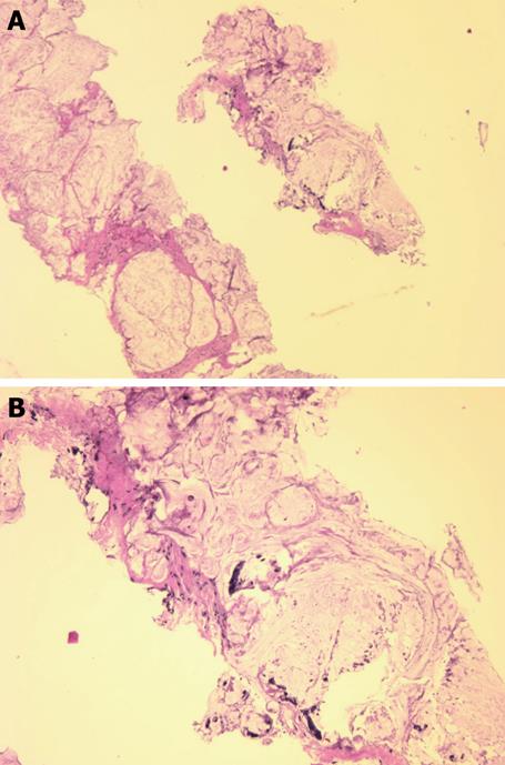
Figure 1 Needle biopsy fragments showing mucinous adenocarcinoma (HE stain).
A: Low power photomicrograph above shows fragments of infiltrating cancer; B: High power photomicrograph below shows individual cancer cells or clusters of cancer cells within mucus.
Figure 2 Magnetic resonance images of the patient.
A and B show the axial T2-weighted fat suppressed MR images of the pelvis. A heterogeneous but predominantly high signal intensity tumor mass (T) is seen arising from the right lateral wall (arrow) of the rectum (R). This mass extends anteriorly to invade both seminal vesicles (S) and abuts and displaces the prostate gland (P). A high signal focus in the left acetabulum represents a bone metastasis (M). The bladder is noted as B. C is the MR image about 5 wk later, now repeated with contrast. T1-weighted post-contrast fat saturation MR image correlating to image in Figure 2b shows rim enhancement of the mass with a large central low signal area consistent with central necrosis. There is now invasion of the tumor into the prostate gland (P) and multiple new osseous metastases (M).
- Citation: Freeman HJ, Perry T, Webber DL, Chang SD, Loh MY. Mucinous carcinoma in Crohn’s disease originating in a fistulous tract. World J Gastrointest Oncol 2010; 2(7): 307-310
- URL: https://www.wjgnet.com/1948-5204/full/v2/i7/307.htm
- DOI: https://dx.doi.org/10.4251/wjgo.v2.i7.307


