
















Copyright: ©Author(s) 2026.
World J Gastrointest Oncol. Aug 15, 2026; 18(8): 121799
Published online Aug 15, 2026. doi: 10.4251/wjgo.v18.i8.121799
Published online Aug 15, 2026. doi: 10.4251/wjgo.v18.i8.121799
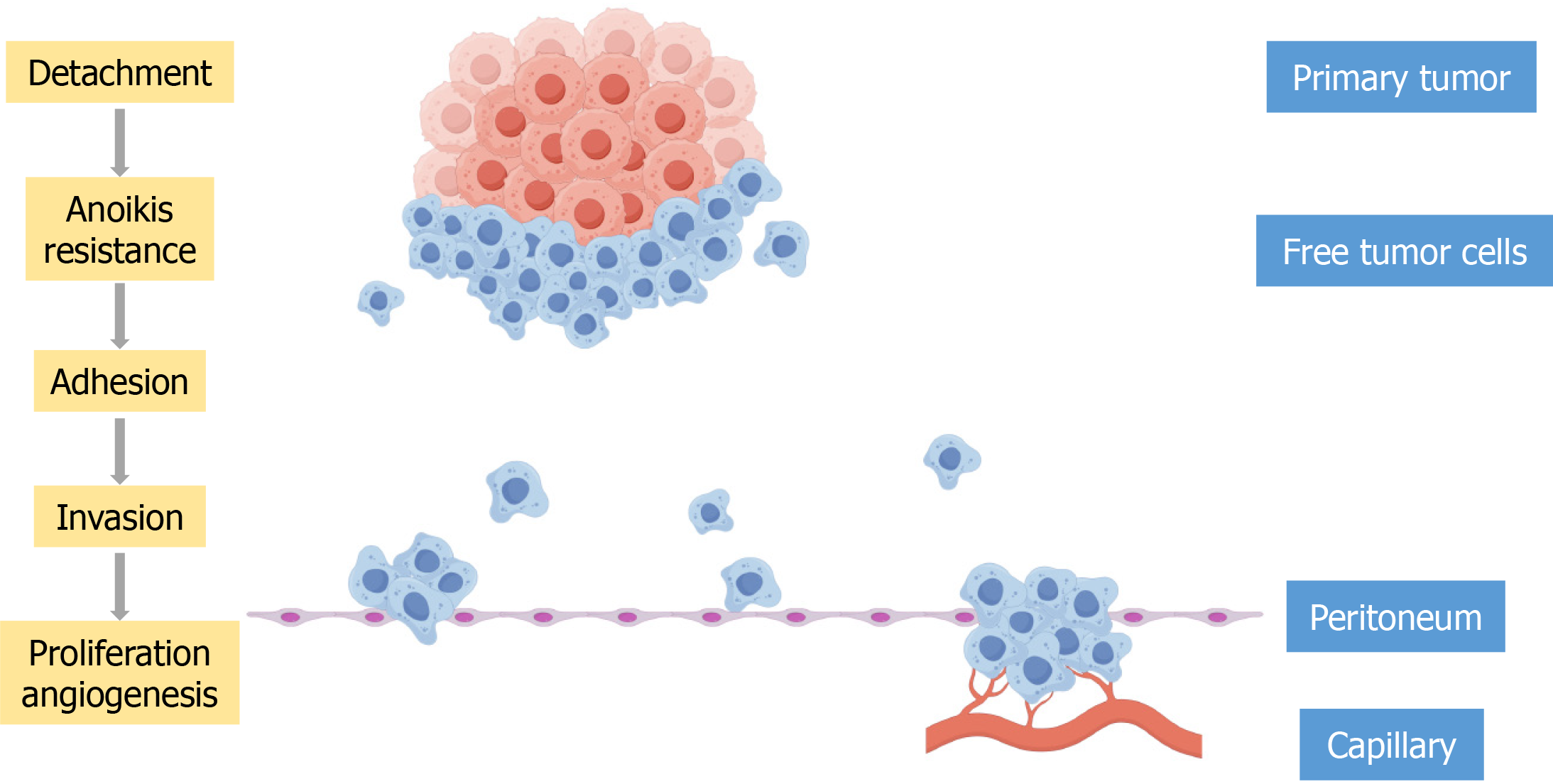
Figure 1 The stepwise process of gastric cancer peritoneal metastasis.
This schematic illustrates the sequential steps by which gastric cancer cells disseminate from the primary tumor and establish metastatic lesions in the peritoneum. The process involves six critical phases: (1) Shedding: Gastric cancer cells detach from the primary tumor by downregulating intercellular adhesion molecules; (2) Anoikis resistance: Detached cells survive in the peritoneal fluid by activating pro-survival pathways; (3) Adhesion: Free-floating cells adhere to the peritoneal mesothelial layer via adhesion molecules; (4) Invasion: Adhered cells secrete matrix metalloproteinases and cathepsins to degrade the extracellular matrix, invading the subperitoneal space; (5) Proliferation: Invaded cells proliferate, forming micrometastases; and (6) Angiogenesis: The growing metastatic foci secrete angiogenic factors (e.g., vascular endothelial growth factor), inducing new blood vessel formation to supply nutrients and oxygen.
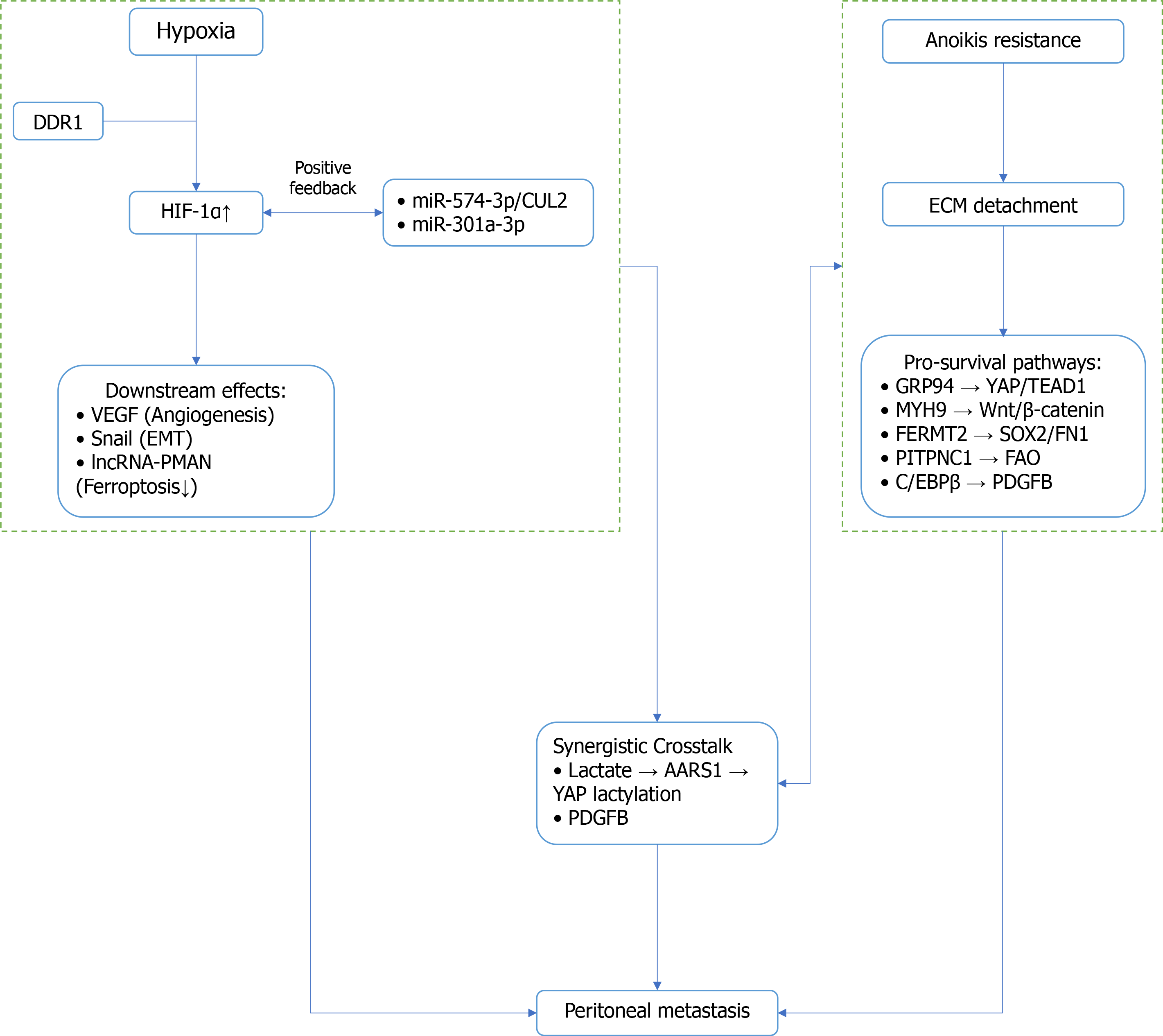
Figure 2 Schematic diagram of the synergistic interplay between hypoxia and anoikis resistance in gastric cancer peritoneal metastasis.
Under hypoxic conditions, Hypoxia-inducible factor 1alpha (HIF-1α) is stabilized and activated. It promotes angiogenesis, epithelial-mesenchymal transition, and inhibits ferroptosis. Positive feedback loops involving miR-574-3p/CUL2 and miR-301a-3p sustain HIF-1α activation. Following detachment from the extracellular matrix, gastric cancer cells activate multiple pro-survival pathways. Hypoxia and anoikis resistance are connected through shared nodes. YAP/TEA domain transcription factor 1 is activated by both hypoxia and Glucose-regulated protein 94. PDGFB is co-regulated by HIF-1α and C/EBPβ. Extracellular matrix detachment: Loss of contact between tumor cells and the extracellular matrix. DDR1: Discoidin domain receptor 1; HIF-1α: Hypoxia-inducible factor 1alpha; VEGF: Vascular endothelial growth factor; EMT: Epithelial-mesenchymal transition; lncRNA: Long non-coding RNA; ECM: Extracellular matrix; GRP94: Glucose-regulated protein 94; TEAD1: TEA domain transcription factor 1; MYH9: Myosin-9; SOX2: SRY box 2; FN1: Fibronectin 1.
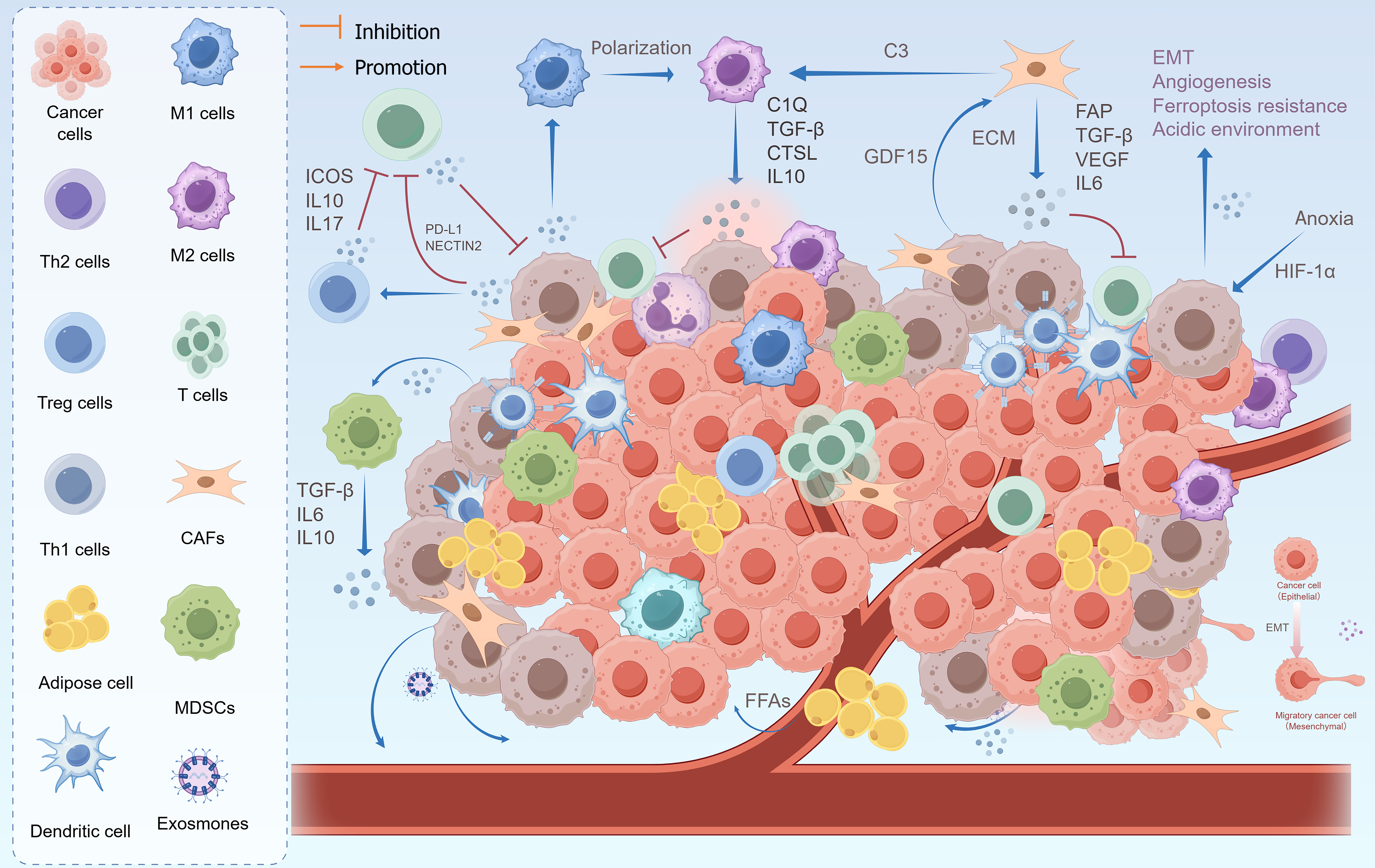
Figure 3 Outline of tumor microenvironment in peritoneal metastasis of gastric cancer.
M1 macrophages, dendritic cells, and T cells are innate immune cells that fight tumors. Gastric cancer cells induce effector T cells into an exhausted state by expressing relevant immune checkpoints, recruiting and promoting the polarization of M2 macrophages, and modifying regulatory T cell and myeloid-derived suppressor cells to suppress immune responses. Immune cells normally secrete cytokines to suppress tumor growth, however, suppressor T cells cannot produce these cytokines. In addition, gastric cancer cells induce the differentiation of cancer-associated fibroblasts and promote the maturation of preadipocytes, shaping the aggressive phenotype and immune tolerance of tumor cells. Anoxia is a core feature of peritoneal metastasis, which can drive epithelial-mesenchymal transition, angiogenesis, ferroptosis resistance, and acidic tumor microenvironment. EMT: Epithelial-mesenchymal transition; ICOS: Inducible T-cell co-stimulator; IL: Interleukin; PD-L1: Programmed death ligand 1; TGF: Transforming growth factor; CTSL: Cathepsin L; GDF15: Growth differentiation factor 15; ECM: Extracellular matrix; FAP: Fibronectin attachment protein; VEGF: Vascular endothelial growth factor; HIF-1α: Hypoxia-inducible factor 1alpha; CAFs: Cancer-associated fibroblasts; MDSC: Myeloid-derived suppressor cell; FFAs: Free fatty acids.
- Citation: Yan HQ, Lai QQ, Huang MW, Wang TA, Wu LC, Qin YZ. Advances in molecular mechanism of gastric cancer peritoneal metastasis. World J Gastrointest Oncol 2026; 18(8): 121799
- URL: https://www.wjgnet.com/1948-5204/full/v18/i8/121799.htm
- DOI: https://dx.doi.org/10.4251/wjgo.v18.i8.121799