
















Copyright: ©Author(s) 2026.
World J Gastrointest Oncol. Aug 15, 2026; 18(8): 119679
Published online Aug 15, 2026. doi: 10.4251/wjgo.119679
Published online Aug 15, 2026. doi: 10.4251/wjgo.119679
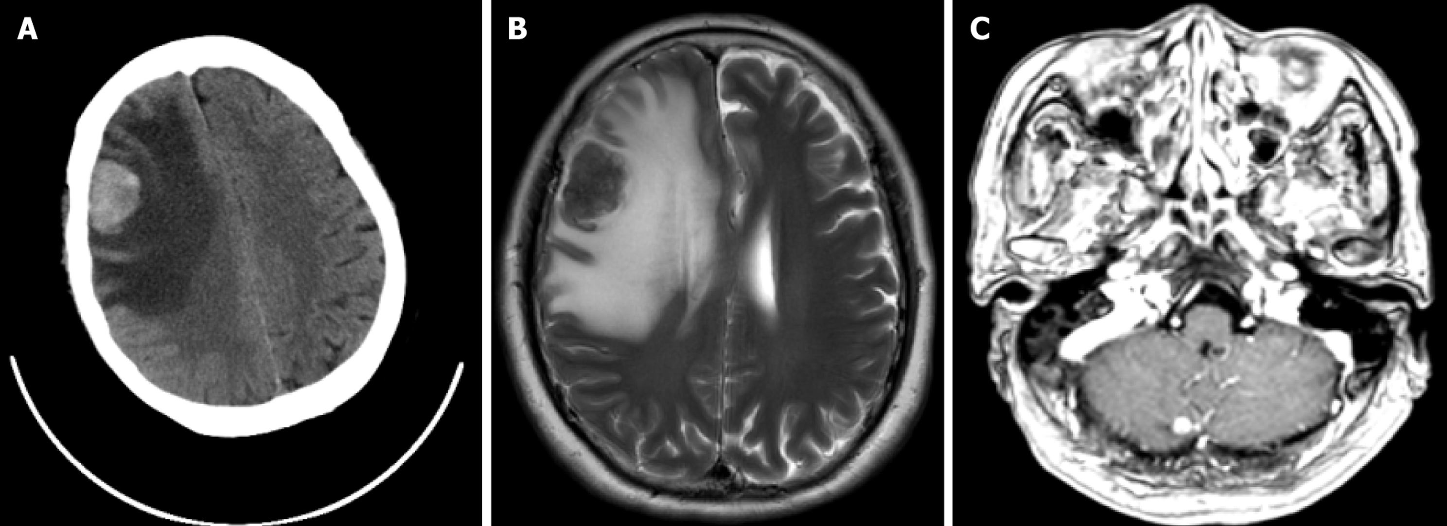
Figure 1 Head computed tomography and magnetic resonance imaging examinations of the patient.
A: Head computed tomography revealed an abnormal density focus in the right frontal lobe; B: Brain magnetic resonance imaging demonstrated a space-occupying lesion in the right frontal cortex, measuring approximately 3.1 cm × 2.4 cm; C: A follow-up brain magnetic resonance imaging scan prior to radiotherapy showed a newly developed small nodular lesion, approximately 0.6 cm in diameter, in the right cerebellar hemisphere.
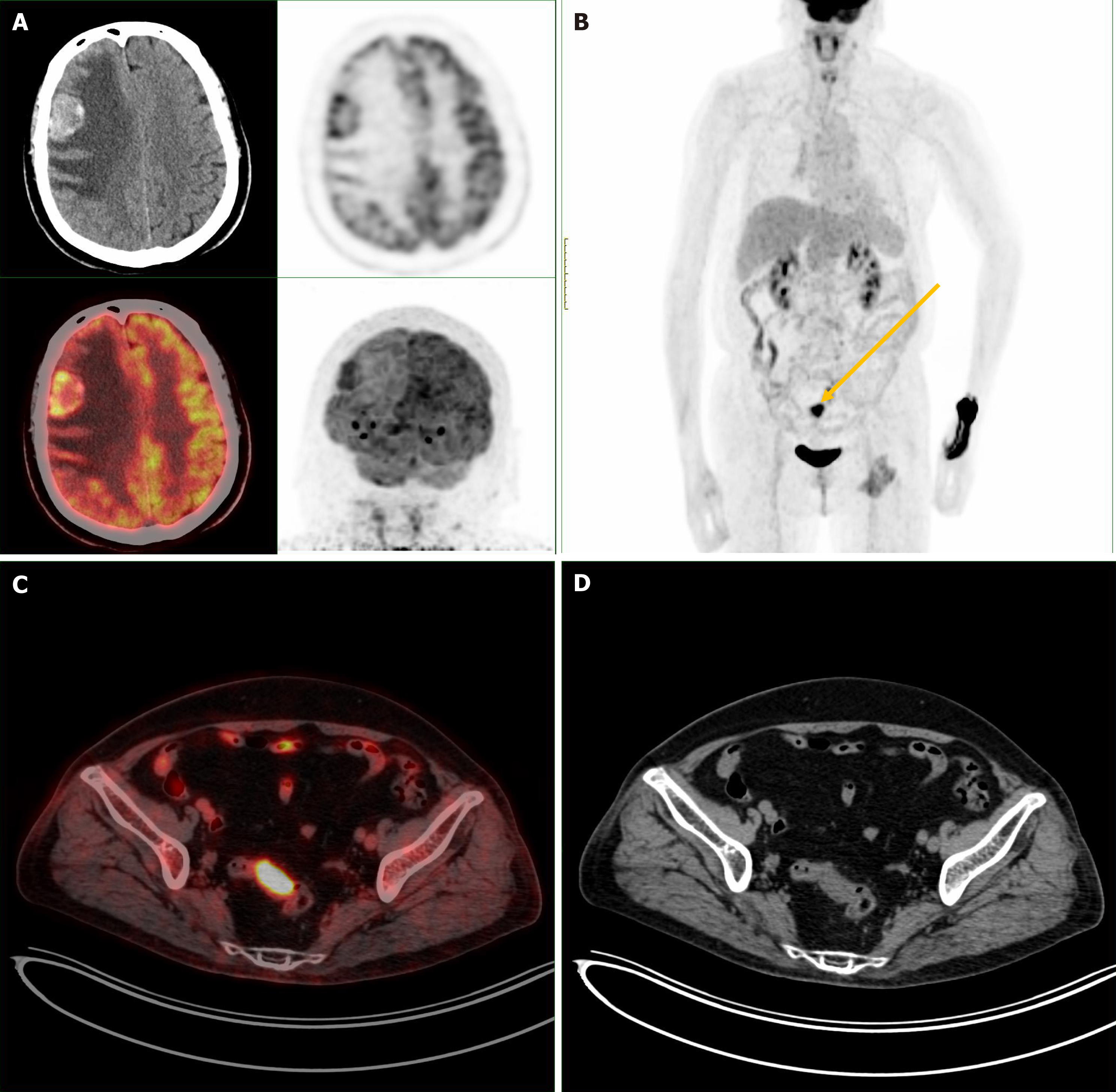
Figure 2 Whole-body positron emission tomography-computed tomography findings.
A: Positron emission tomography-computed tomography showed a hypermetabolic nodule with surrounding edema in the right frontal lobe, consistent with a brain metastasis; B: Whole-body positron emission tomography-computed tomography imaging. The arrow indicates the primary focus, with no evidence of extracranial organ metastasis. The bilateral renal and bladder activity represents physiologic tracer retention. The area of increased uptake in the medial aspect of the left proximal femur is due to muscular inflammation (yellow arrow: Primary tumor lesions in the intestine); C and D: Hypermetabolic primary rectal tumor.
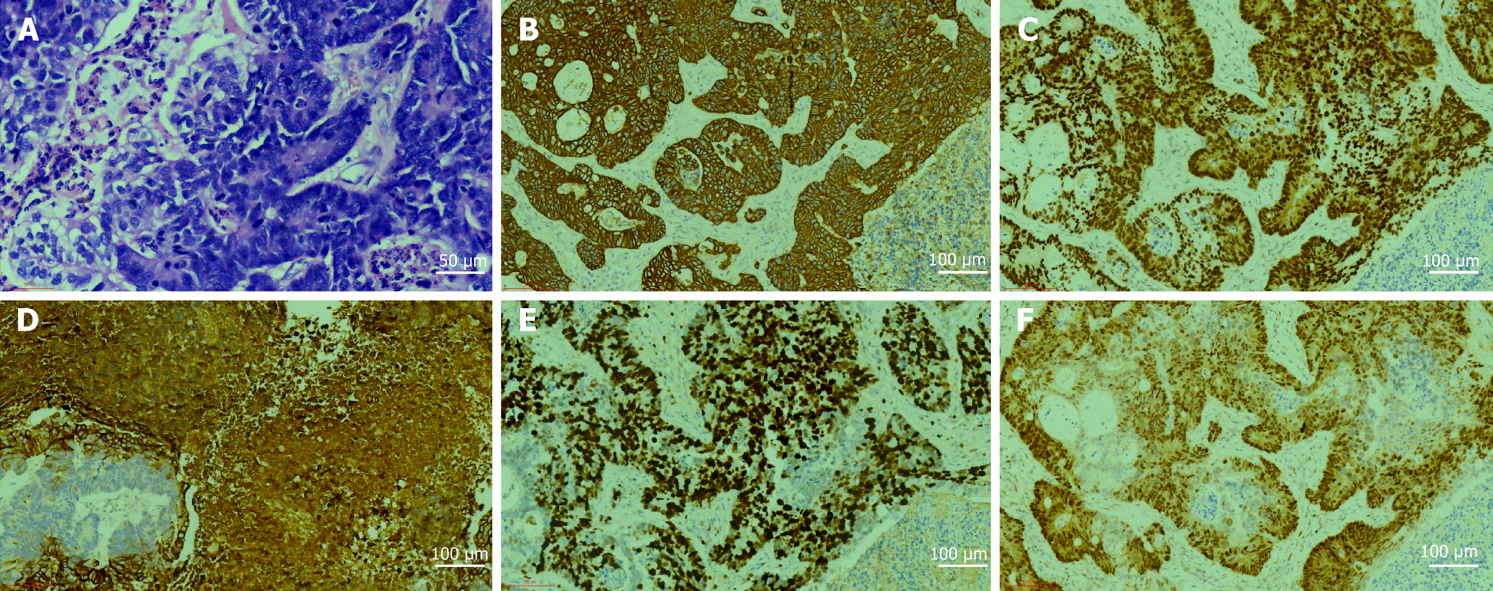
Figure 3 Postoperative pathology and immunohistochemistry of the brain metastasis.
A: Postoperative pathology of the brain metastasis (hematoxylin and eosin, × 400); B: Positive expression of AE1/AE3 (magnification, × 200); C: Positive expression of caudal-type homeobox transcription factor 2 (magnification, × 200); D: Positive expression of CK20 (magnification, × 200); E: The Ki-67 index was 80% (magnification, × 200); F: Positive expression of P53 (mutant type) (magnification, × 200).
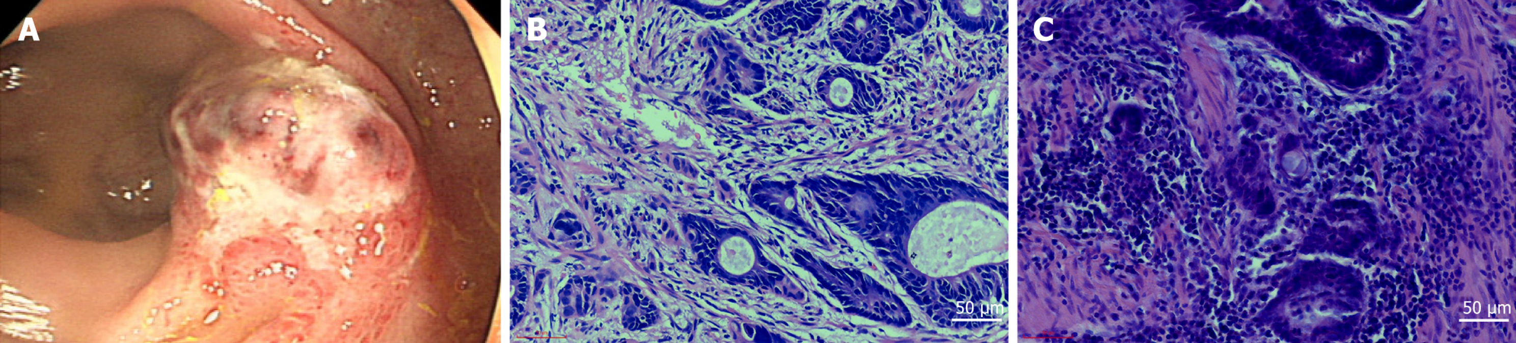
Figure 4 Colonoscopy and postoperative pathological analysis of the primary lesion.
A: Colonoscopy revealed a 4 cm × 3 cm ulcerated mass in the rectum; B: Endoscopic biopsy pathology indicates a moderately differentiated adenocarcinoma (hematoxylin and eosin, × 400); C: Postoperative pathology of the primary rectal cancer lesion shows an invasive, moderately to poorly differentiated adenocarcinoma (hematoxylin and eosin, × 400).
- Citation: Xu JL, Ge ZW, Wu CW, Wang YH, Mao JD. Multidisciplinary care for colorectal cancer brain metastases with headache as initial symptom: A case report and literature review. World J Gastrointest Oncol 2026; 18(8): 119679
- URL: https://www.wjgnet.com/1948-5204/full/v18/i8/119679.htm
- DOI: https://dx.doi.org/10.4251/wjgo.119679