
















Copyright: ©Author(s) 2026.
World J Gastrointest Oncol. Jul 15, 2026; 18(7): 118522
Published online Jul 15, 2026. doi: 10.4251/wjgo.v18.i7.118522
Published online Jul 15, 2026. doi: 10.4251/wjgo.v18.i7.118522
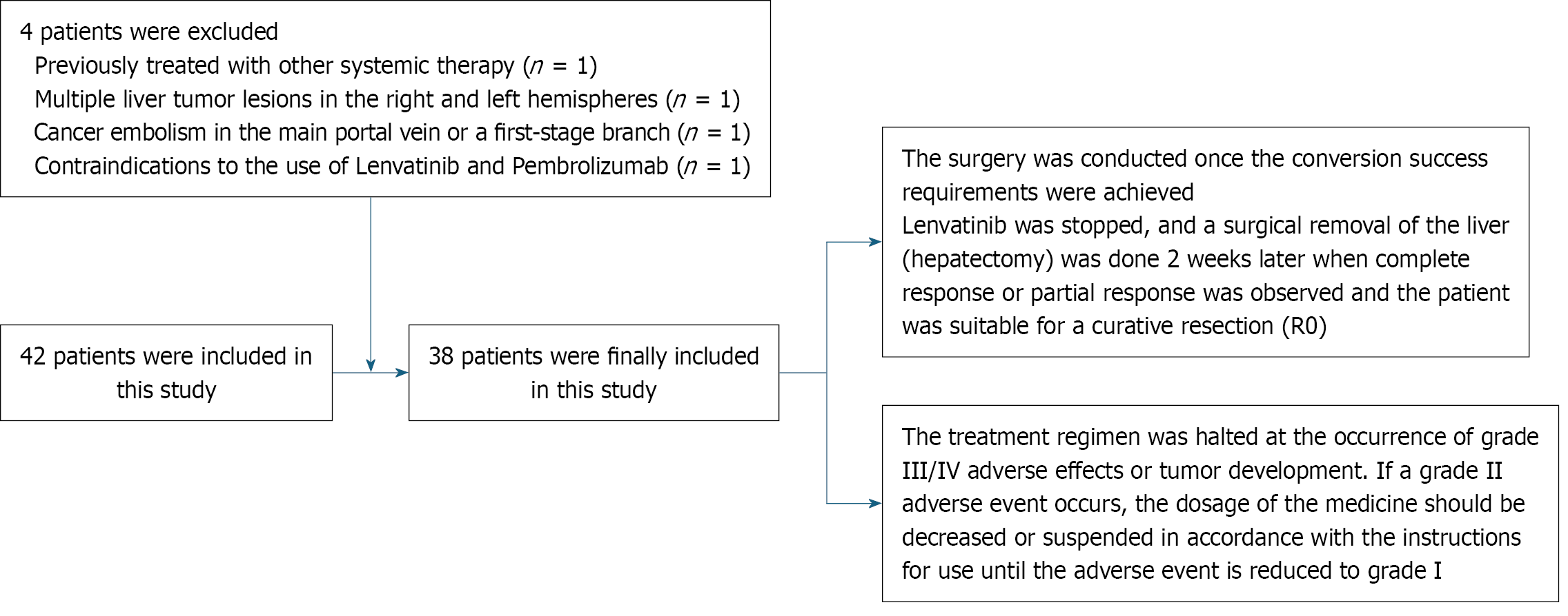
Figure 1
Flow diagram of study.
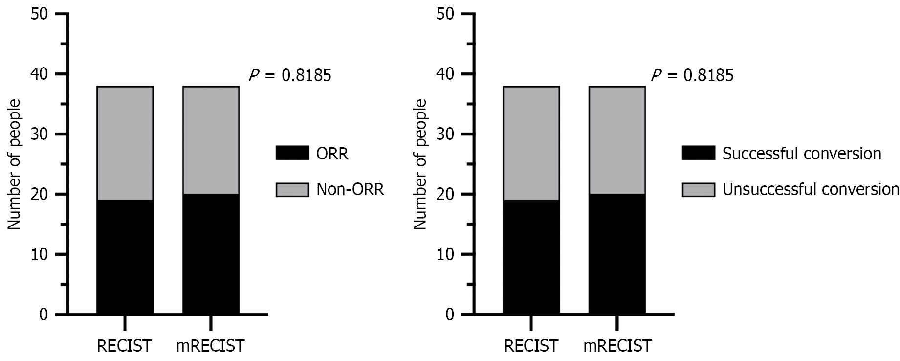
Figure 2 Comparative analysis of objective response rate and conversion success rates using Response Evaluation Criteria in Solid Tumors and modified Response Evaluation Criteria in Solid Tumors assessment criteria.
mRECIST: Modified Response Evaluation Criteria in Solid Tumors; ORR: Objective response rate; RECIST: Response Evaluation Criteria in Solid Tumors.
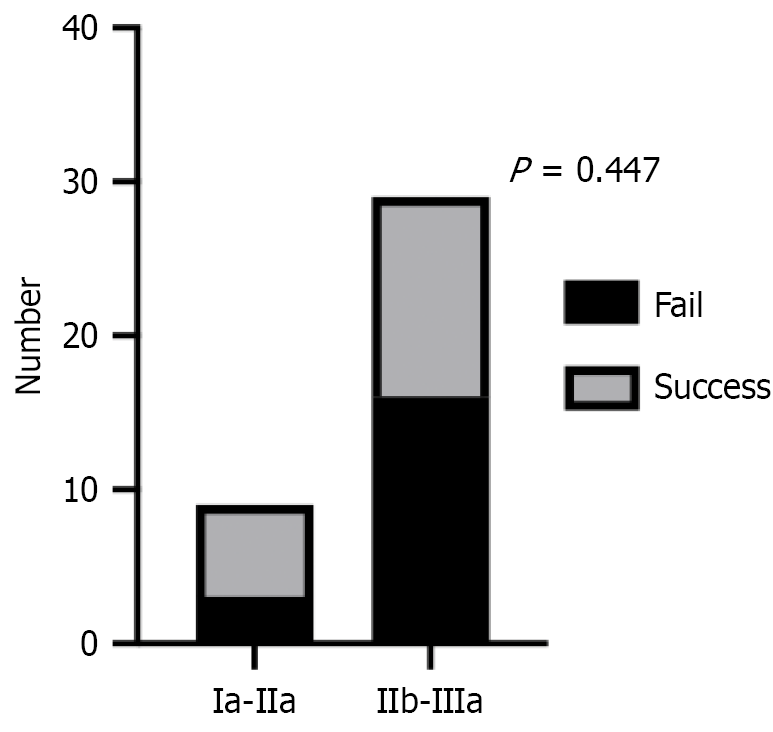
Figure 3
Comparative analysis of the success rate of transformation in early-stage and advanced-stage liver cancer.
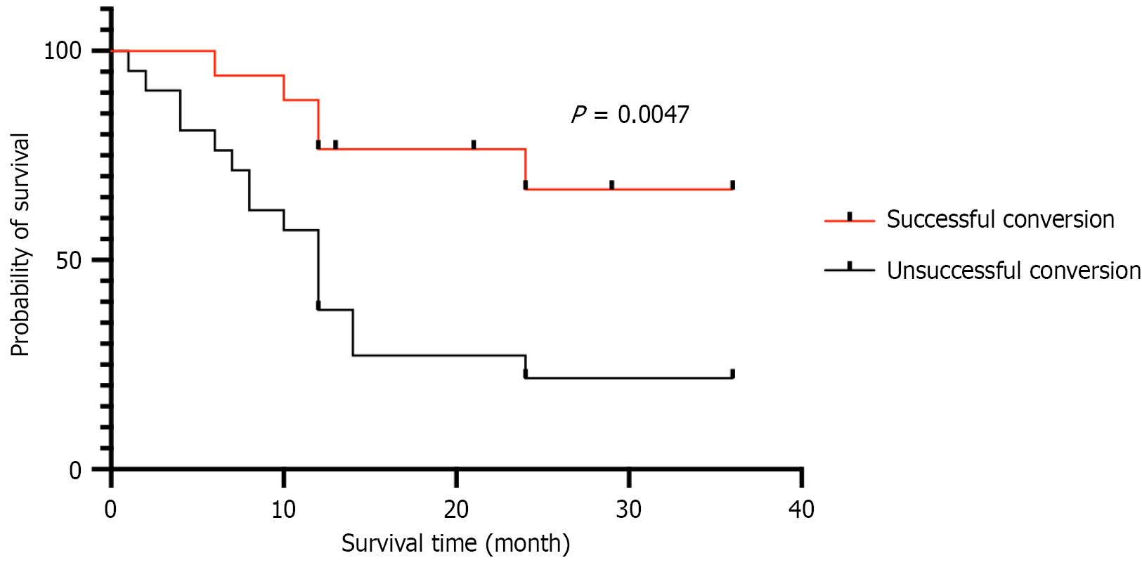
Figure 4
Kaplan-Meier overall survival curves for the successful and unsuccessful transformation groups.
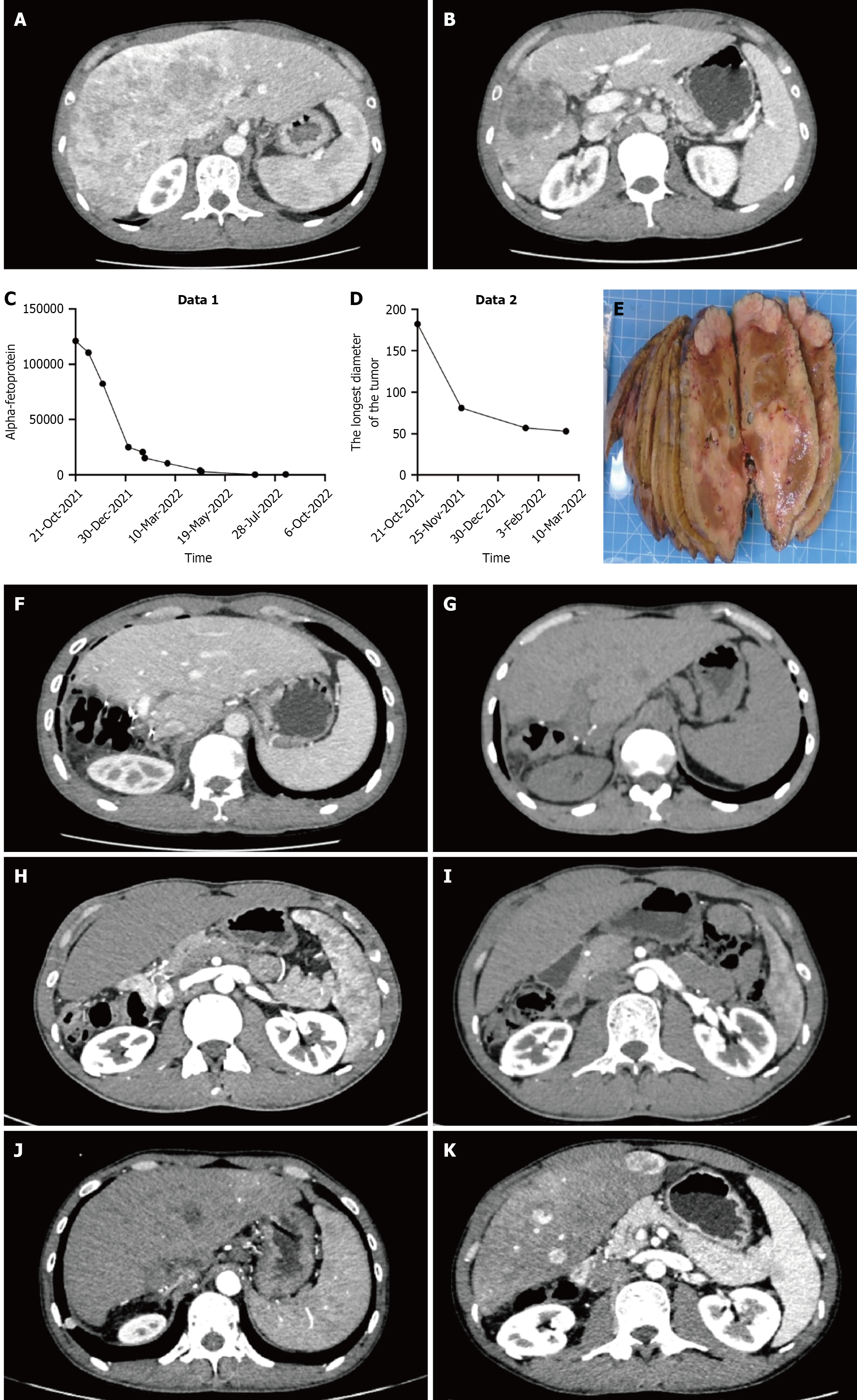
Figure 5 Enhanced computed tomography findings before transformation (primary treatment).
A and B: Postoperative computed tomography (CT) findings showed multiple nodules in the right liver lobe, measuring 46 mm × 44 mm × 53 mm in total. The intrahepatic mass and nodules were reduced in size compared to the previous scan, indicating ongoing tumor activity. The right portal vein and hepatic vein showed minor constriction due to external pressure. Imaging assessment indicated a partial response; C: Alpha-fetoprotein levels; D: Variation curve of the maximum diameter of the tumor; E: Gross specimen after surgery. A liver tissue sample (12.5 cm × 12 cm × 6.5 cm) was cut, revealing multiple nodules ranging from 0.6 cm to 6.5 cm in size. These nodules were greyish-yellow, solid, soft, and had distinct borders. They were located near the periosteum and did not reach the cautery margin; F: Postoperatively, the right liver lobe showed changes with a remaining cavity in the operated area, accompanied by minor fluid and gas around the liver and cavity. The area was being drained; G: One month after surgery, the right liver lobe was completely absent, with a partial wedge-shaped absence in hepatic S4. There was swelling near the S4 margin and a small amount of fluid around the liver; H: Three months after surgery, the right liver lobe and S2 showed postoperative changes, including a residual cavity at the surgical site. The gallbladder was absent, and fluid around the liver had been absorbed. A new abnormal growth in liver S2 suggested metastasis, warranting magnetic resonance imaging with liver-specific contrast (Prometrium) for further diagnosis. Additionally, multiple lung nodules had grown, indicating metastatic tumors; I: Six months after surgery, the right liver lobe and S4 showed postoperative changes with a residual cavity, and the gallbladder remained absent. A mildly enhancing nodule in liver S2 had grown slightly, suggesting a high likelihood of metastatic tumor. The spleen remained enlarged, and cranial CT showed no clear abnormalities; J: One year after surgery, the right liver lobe and S4 showed postoperative changes with a residual cavity, and the gallbladder was still absent. A mildly enhanced nodule in liver S2 had grown and showed less enhancement, suggesting a metastatic tumor. Other nodules did not show clear findings. The spleen remained enlarged, and cranial CT revealed no evident abnormalities; K: Two years after surgery, the right liver lobe and S4 showed postoperative changes with a residual cavity, and the gallbladder remained absent. The liver had several metastases; most of which had invaded the middle and right hepatic veins. Both lungs had multiple cancerous nodules, with most increasing in size. Lymph node metastases in the mediastinum and right hilum had also enlarged, with invasion of the single vein and compression of the right upper pulmonary artery. The spleen was enlarged, the main portal vein widened, the splenic vein thickened, and varicose veins developed in the lower esophagus and stomach fundus.
- Citation: Chen X, Liao H, Chen J, Peng LH, Zhang JH, Xuyun XX, Chen YJ, Chen T. Lenvatinib and pembrolizumab plus FOLFOX-hepatic arterial infusion chemotherapy in locally advanced, potentially resectable hepatocellular carcinoma. World J Gastrointest Oncol 2026; 18(7): 118522
- URL: https://www.wjgnet.com/1948-5204/full/v18/i7/118522.htm
- DOI: https://dx.doi.org/10.4251/wjgo.v18.i7.118522