©The Author(s) 2025.
World J Gastrointest Oncol. Nov 15, 2025; 17(11): 113262
Published online Nov 15, 2025. doi: 10.4251/wjgo.v17.i11.113262
Published online Nov 15, 2025. doi: 10.4251/wjgo.v17.i11.113262
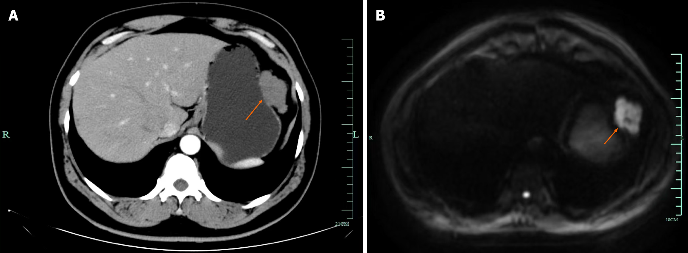
Figure 1 Imaging examinations.
A: Computed tomography scan indicates a mass on the greater curvature side of the stomach; B: Magnetic resonance indicates a mass on the greater curvature side of the stomach.
Figure 2 Computed tomography shows a solid mass lesion in the small intestine within the abdominal cavity.
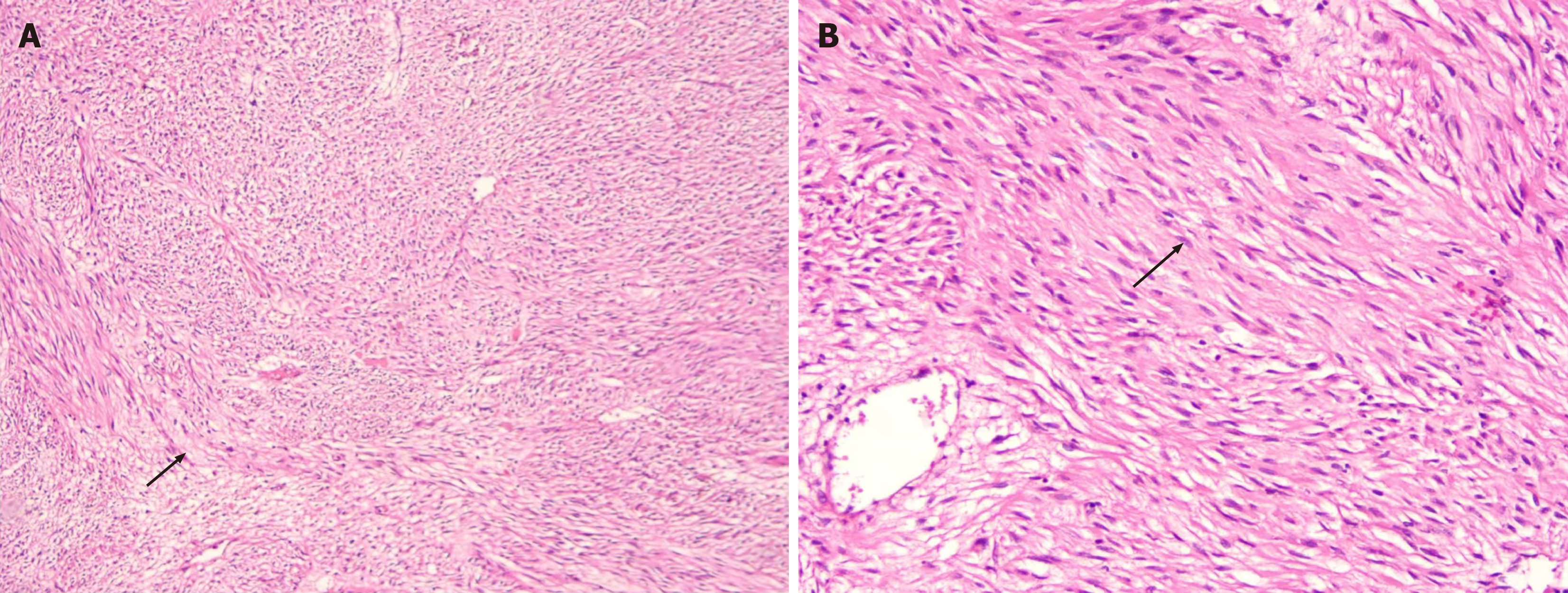
Figure 3 Microscopic examination.
A: Confirmed that this was a spindle cell type gastrointestinal stromal tumors; B: Indicates a composition of spindle-shaped cells.
- Citation: Hong YY, Shou CH, Yang WL, Wang XD, Zhang Q, Liu XS, Yu JR. FGFR2 fusions as novel oncogenic drivers in gastrointestinal stromal tumors: Two case reports and review of literature. World J Gastrointest Oncol 2025; 17(11): 113262
- URL: https://www.wjgnet.com/1948-5204/full/v17/i11/113262.htm
- DOI: https://dx.doi.org/10.4251/wjgo.v17.i11.113262


