©The Author(s) 2025.
World J Gastrointest Oncol. Oct 15, 2025; 17(10): 109450
Published online Oct 15, 2025. doi: 10.4251/wjgo.v17.i10.109450
Published online Oct 15, 2025. doi: 10.4251/wjgo.v17.i10.109450
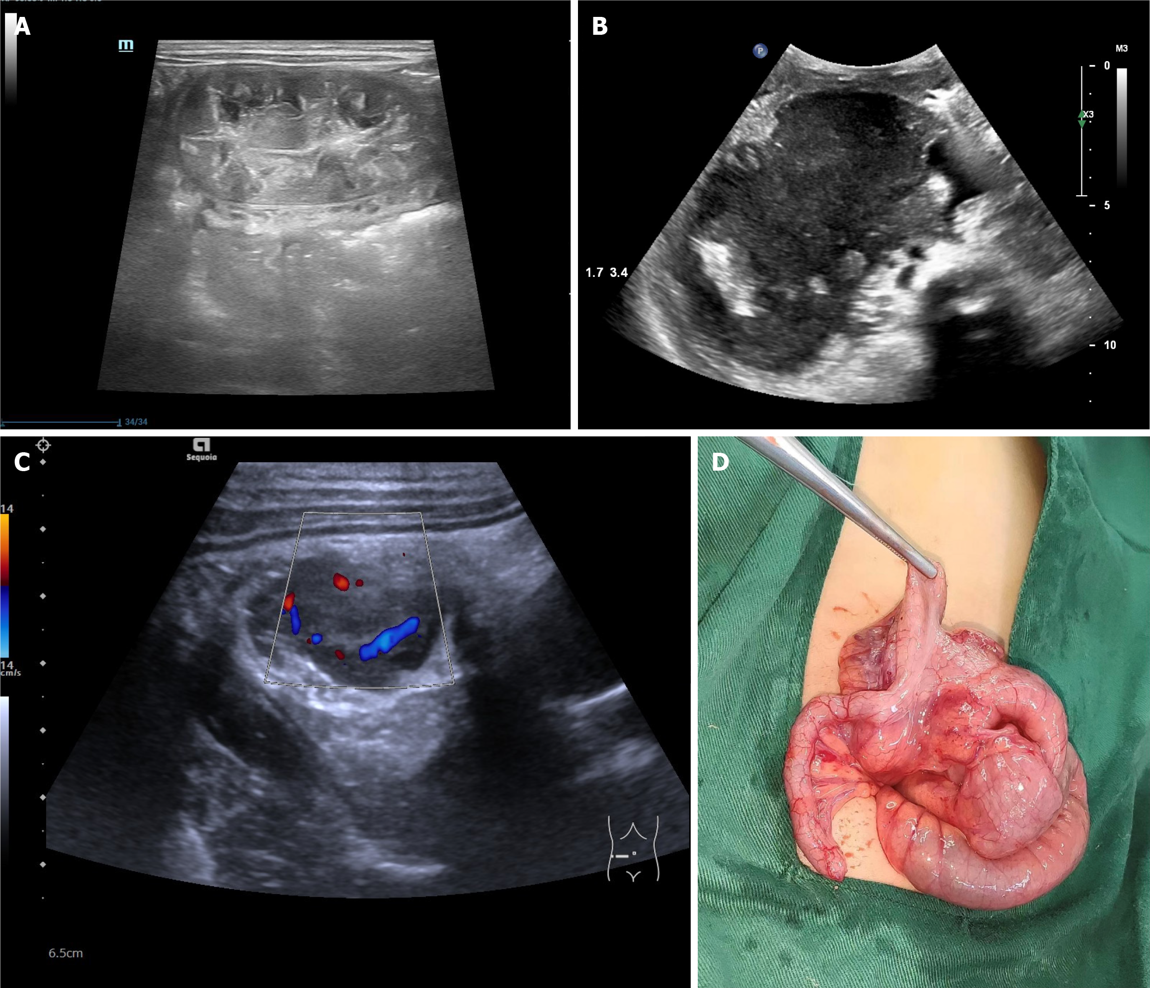
Figure 1 Ultrasound manifestations of the intestinal wall in intestinal lymphoma.
A: Focal segmental concentric annular thickening of the intestinal wall; B: Focal segmental the intestinal wall was eccentrically tumor-like thickened; C: Focal segmental solid masses of the intestinal wall to intussusception; D: Surgical photo.
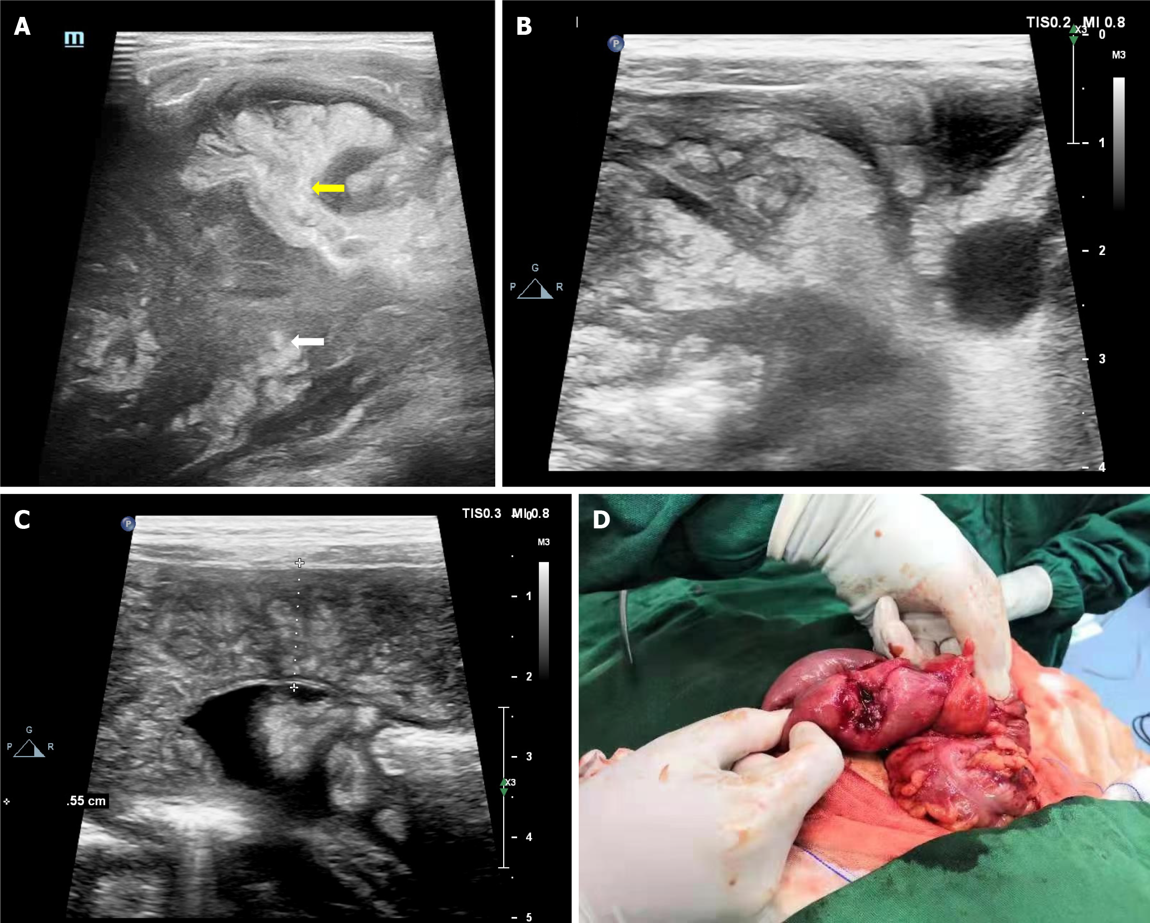
Figure 2 Changes in intestinal accessory structures in intestinal lymphoma.
A-C: Eccentric tumor-like thickening of the intestinal wall, eccentric intestinal mucosa (white arrow), and narrowed intestinal lumen, the mesentery was thickened and radially pulled the intestinal tract (yellow arrow), the adjacent adipose tissue was nodular (A and B), and the peritoneum was thickened and nodular (C); D: During the operation, tumor-like thickening of the intestinal wall and hyperplasia of the surrounding adipose tissue were observed.
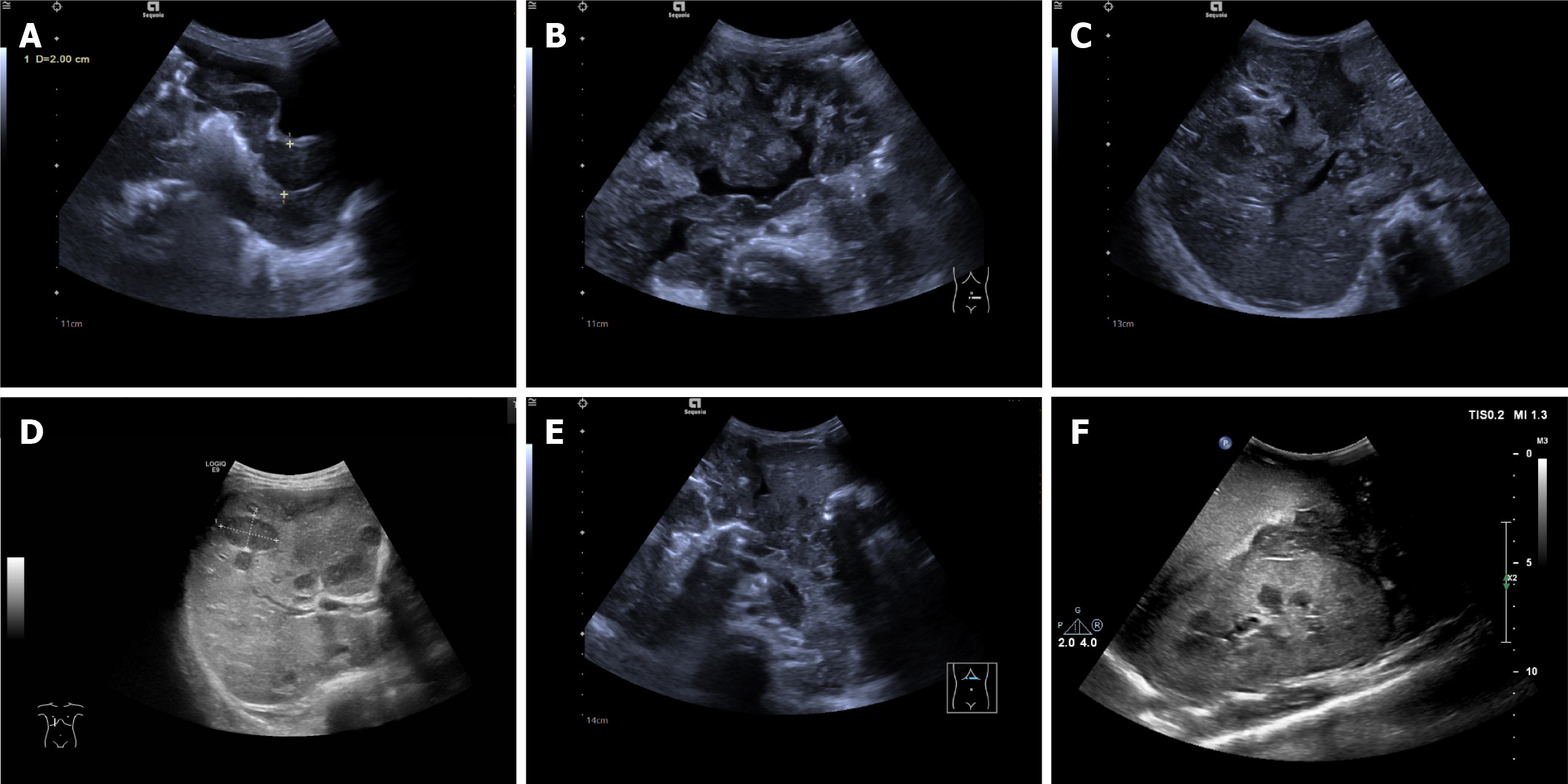
Figure 3 Manifestations of abdominal cavity and organ invasion in intestinal lymphoma.
A: The intestinal wall (sigmoid colon and rectum) of the primary part was circular thickening thickened, with decreased echo and small intestinal lumen; B: In the remaining abdominal cavities, the intestinal wall was widely thickened, and the thickening was not as obvious as that of the primary lesion, with interintestinal effusion; C and D: Extremely hypoechoic distributed along the Greenland sheath in the liver and multiple hypoechoic metastatic foci in the liver; E: A widely distributed low-echo area was observed around the pancreas; F: Scattered low-echo areas in the kidney.
- Citation: Huang SF, Yang F, Chen WJ, Zhang XH. Ultrasound features of primary intestinal lymphoma in children and their correlation with prognosis: A two-center experiment. World J Gastrointest Oncol 2025; 17(10): 109450
- URL: https://www.wjgnet.com/1948-5204/full/v17/i10/109450.htm
- DOI: https://dx.doi.org/10.4251/wjgo.v17.i10.109450


