©The Author(s) 2024.
World J Gastrointest Oncol. Jun 15, 2024; 16(6): 2487-2503
Published online Jun 15, 2024. doi: 10.4251/wjgo.v16.i6.2487
Published online Jun 15, 2024. doi: 10.4251/wjgo.v16.i6.2487
Figure 1 Pathological images of a case of gastric cancer with Helicobacterpylori infection with a low level of tumor-infiltrating lymphocytes.
A: Hematoxylin and eosin (HE) staining showing the presence of Helicobacter pylori (yellow arrow) at 100 × magnification; B: HE staining of the intratumoral compartment with a low level of tumor-infiltrating lymphocytes at 20 ×.
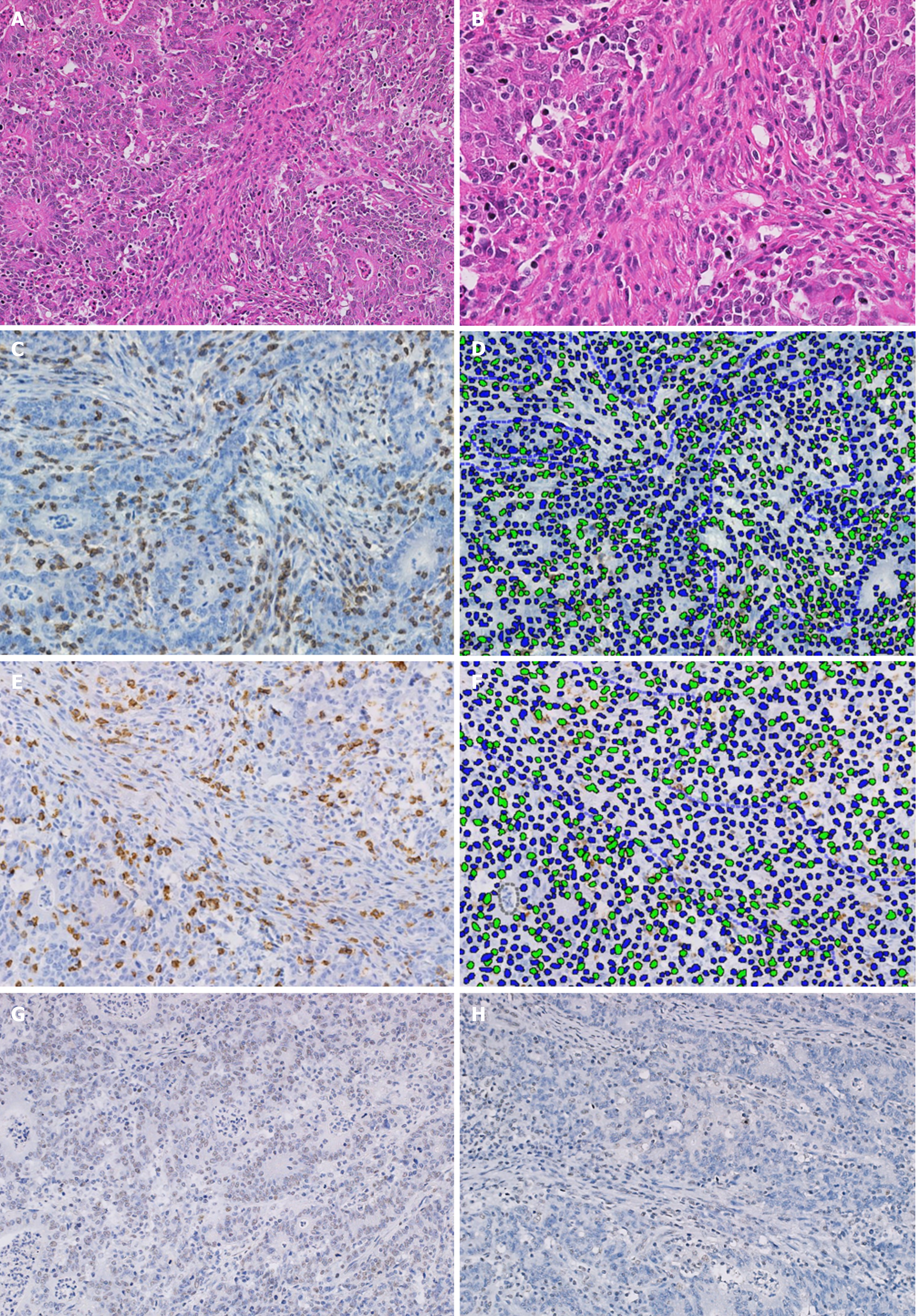
Figure 2 Pathological images of a case of gastric cancer with deficient mismatch-repair and a high level of tumor-infiltrating lymphocytes.
A: Hematoxylin and eosin (HE) staining of stromal compartment with high level of tumor-infiltrating lymphocytes (TILs) at 20 × magnification; B: Field magnification of HE with high TIL stain at 40 ×; C: CD3 immunohistochemistry (IHC) staining at 40 ×; D: Identification of CD3 density by machine learning-based image processing showing positive (green) and negative (blue) cells; E: CD8 IHC staining; F: Digital identification of CD8 density; G: IHC staining showing absence of MSH6 expression and; H: Absence of PMS2 expression.
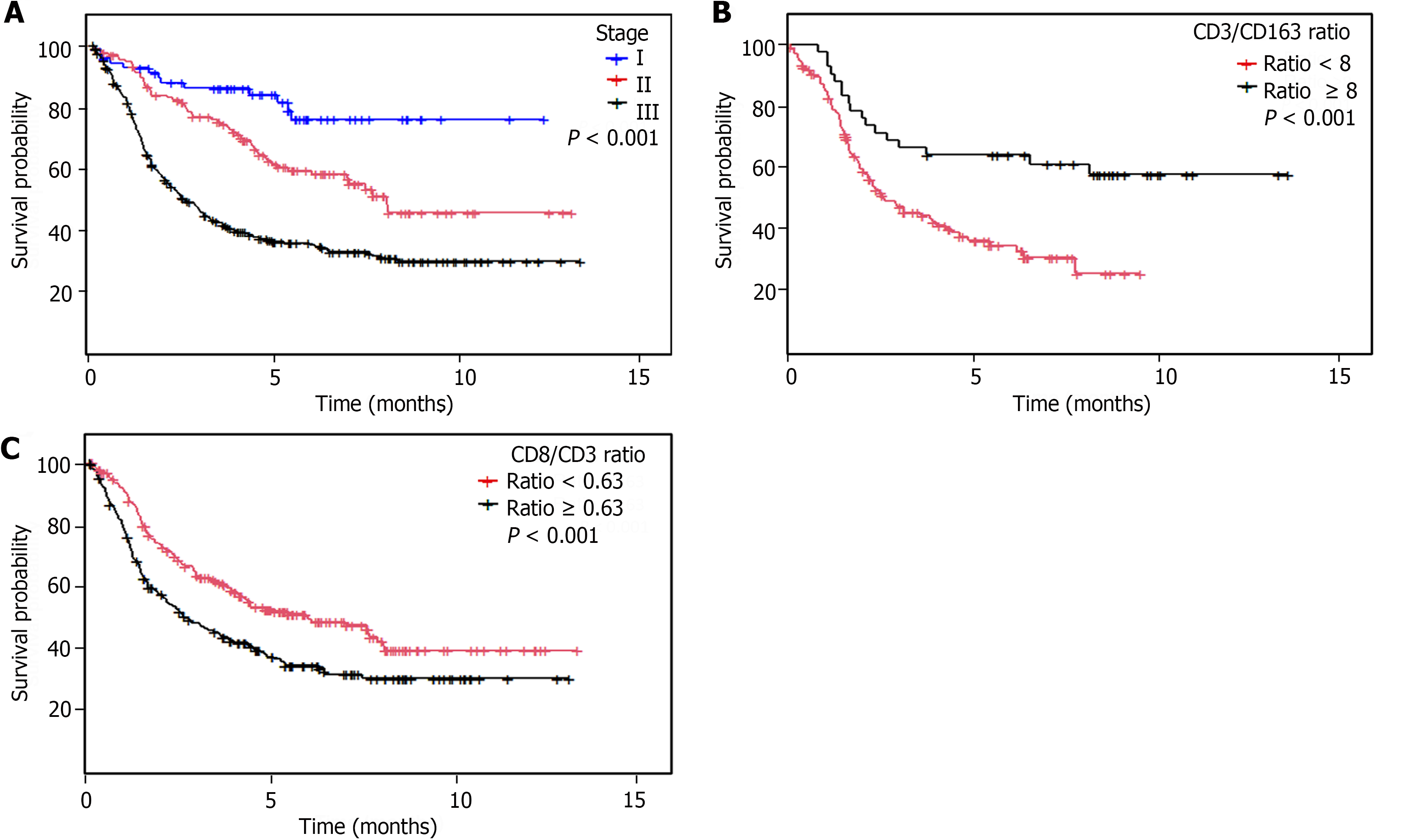
Figure 3 Overall survival analyses.
A: Kaplan-Meier overall survival curve according to clinical stage; B: Intratumoral CD3/163 ratio; C: Intratumoral CD3/CD8 ratio.
- Citation: Castaneda CA, Castillo M, Bernabe LA, Sanchez J, Fassan M, Tello K, Wistuba II, Chavez Passiuri I, Ruiz E, Sanchez J, Barreda F, Valdivia D, Bazan Y, Abad-Licham M, Mengoa C, Fuentes H, Montenegro P, Poquioma E, Alatrista R, Flores CJ, Taxa L. Association between Helicobacter pylori infection, mismatch repair, HER2 and tumor-infiltrating lymphocytes in gastric cancer. World J Gastrointest Oncol 2024; 16(6): 2487-2503
- URL: https://www.wjgnet.com/1948-5204/full/v16/i6/2487.htm
- DOI: https://dx.doi.org/10.4251/wjgo.v16.i6.2487


