©The Author(s) 2024.
World J Gastrointest Oncol. Dec 15, 2024; 16(12): 4738-4745
Published online Dec 15, 2024. doi: 10.4251/wjgo.v16.i12.4738
Published online Dec 15, 2024. doi: 10.4251/wjgo.v16.i12.4738
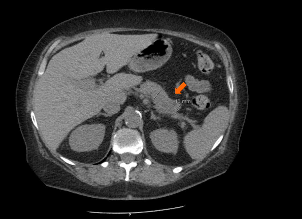
Figure 1 Computed tomography imaging of abdomen showing a 4.
0 cm pancreatic tail mass (arrow).
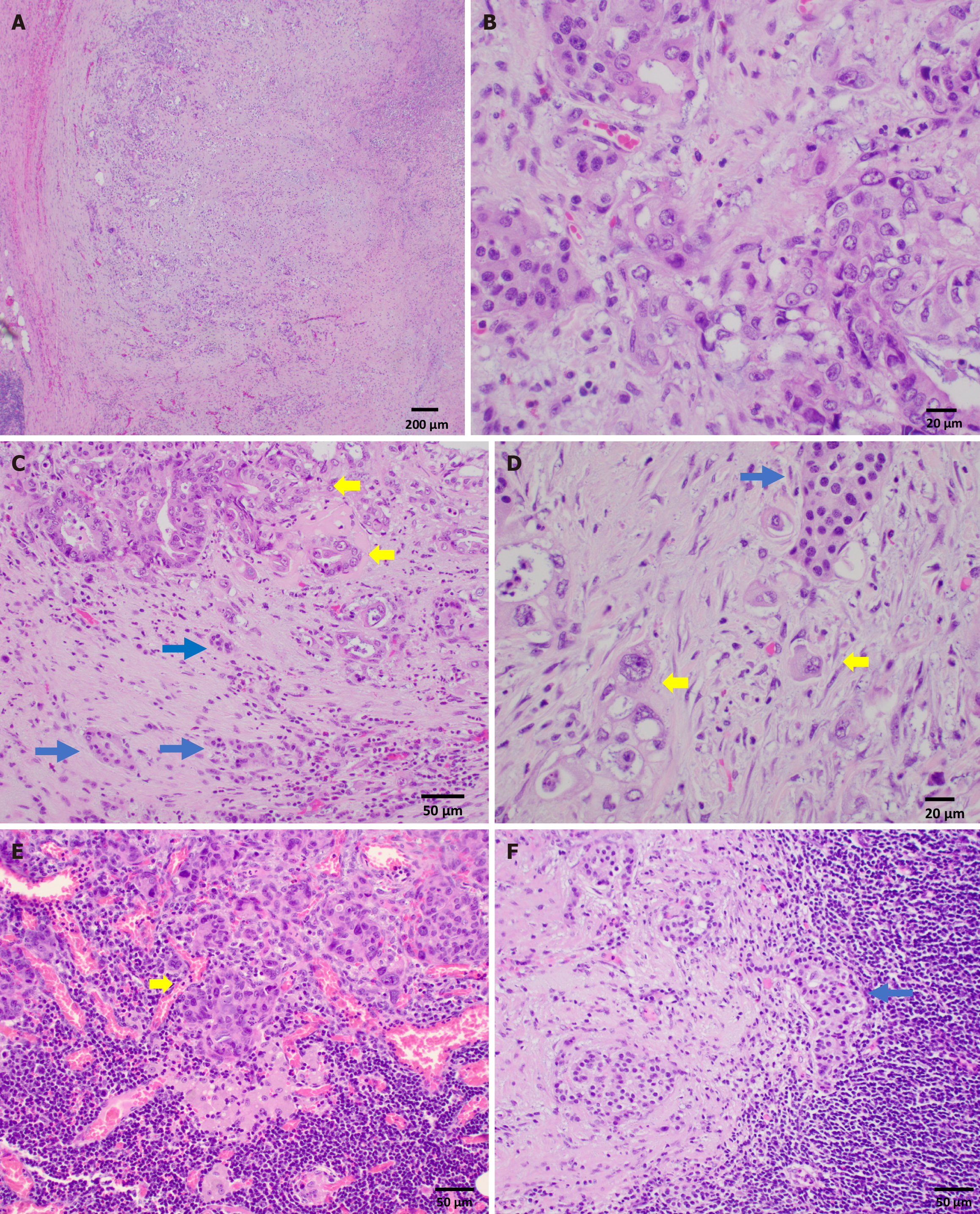
Figure 2 Pancreatic ductal adenocarcinoma and low grade neuroendocrine tumor components.
A: Pancreatic ductal adenocarcinoma component in low power (40 ×); B: Pancreatic ductal adenocarcinoma component in high power (400 ×); C and D: Intermingled pancreatic ductal adenocarcinoma (yellow arrow) and well-differentiated neuroendocrine tumor (blue arrow), hematoxylin and eosin (H&E, 200 × and 400 ×); E: Lymph node with metastatic ductal adenocarcinoma (H&E, 200 ×, yellow arrow); F: Lymph node with well-differentiated neuroendocrine tumor (H&E, 200 ×, blue arrow).
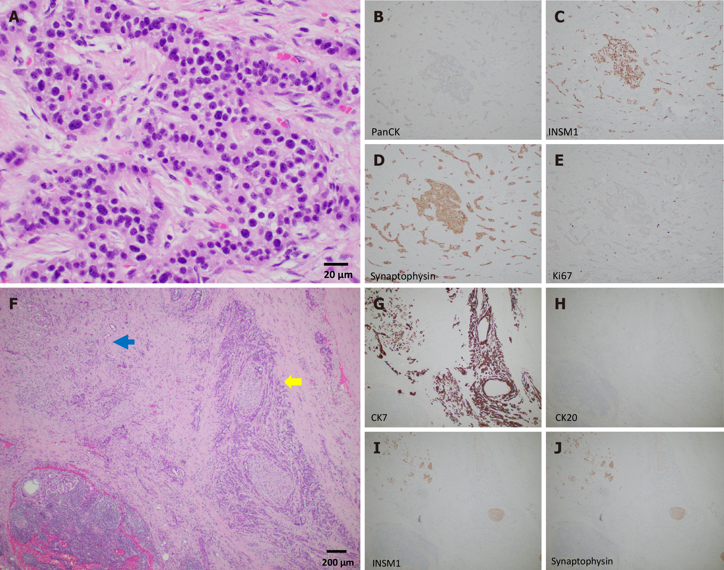
Figure 3 Neuroendocrine tumor component.
A-E: Well-differentiated neuroendocrine tumor component. Hematoxylin and eosin (H&E), 400 ×. The tumor is weakly positive for PanCK AE1/AE3 (B), strongly positive for INSM1 and synaptophysin (C and D), proliferative index Ki67 is very low (< 3%, E); F-J: Intermingled pancreatic ductal adenocarcinoma (yellow arrow) and well-differentiated neuroendocrine tumor (blue arrow). Hematoxylin and eosin, 40 ×. Perineural invasion is evident (F); Immunohistochemical stain for CK7 is positive for both component (G); CK20 is negative in both (H); INSM1 and synaptophysin are positive in well-differentiated neuroendocrine tumor (and neurons) (I and J).
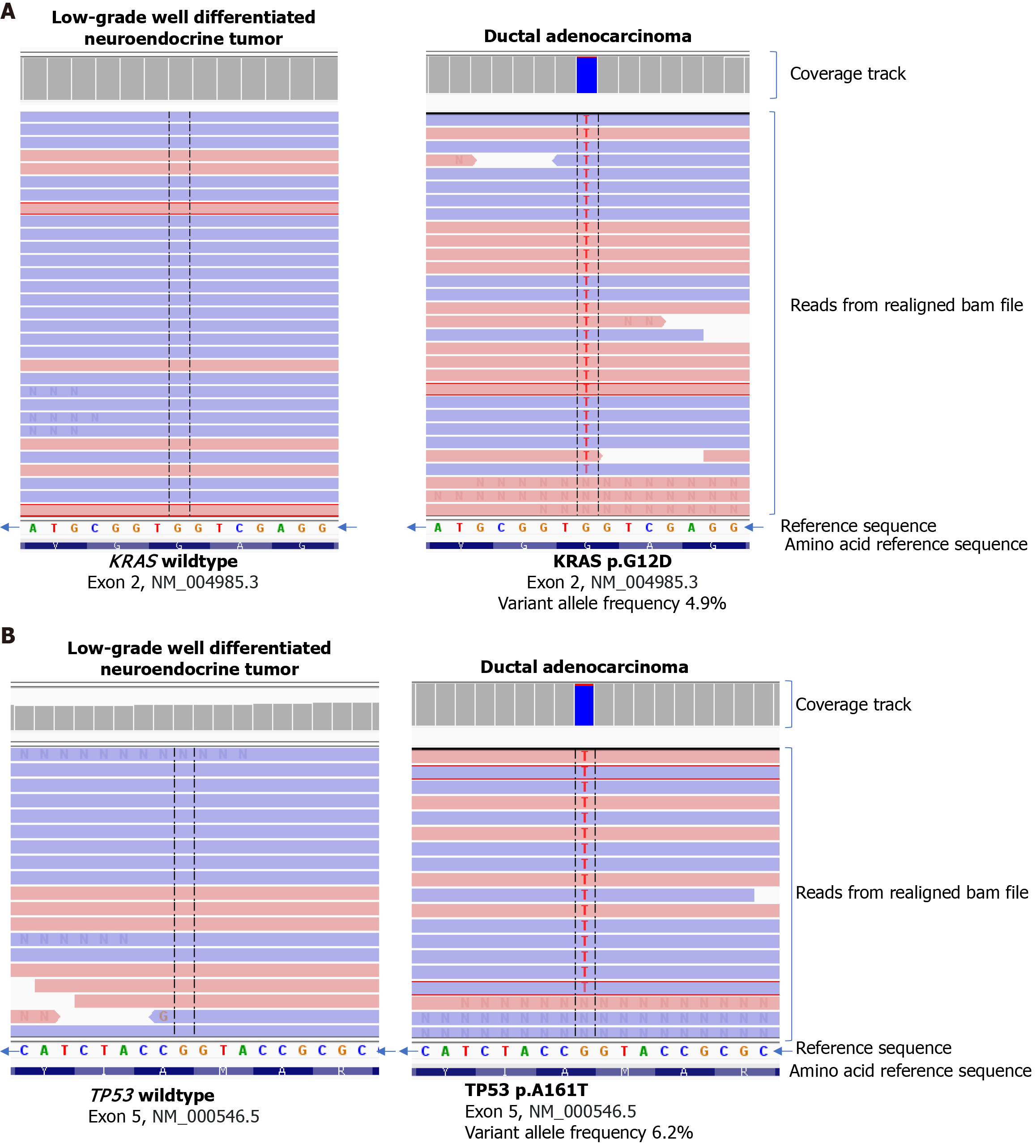
Figure 4 Next generation sequencing.
A and B: Next generation sequencing of the two different histologic components. KRAS exon 2 p.G12D (variant frequency 4.9%) (A) and TP53 exon 5 p.A161T (variant frequency 6.2%) (B) were identified in the adenocarcinoma component, but not in the well-differentiated neuroendocrine tumor component.
- Citation: Zhao X, Bocker Edmonston T, Miick R, Joneja U. Mixed pancreatic ductal adenocarcinoma and well-differentiated neuroendocrine tumor: A case report. World J Gastrointest Oncol 2024; 16(12): 4738-4745
- URL: https://www.wjgnet.com/1948-5204/full/v16/i12/4738.htm
- DOI: https://dx.doi.org/10.4251/wjgo.v16.i12.4738


