Copyright
©The Author(s) 2022.
World J Gastrointest Oncol. Mar 15, 2022; 14(3): 587-606
Published online Mar 15, 2022. doi: 10.4251/wjgo.v14.i3.587
Published online Mar 15, 2022. doi: 10.4251/wjgo.v14.i3.587
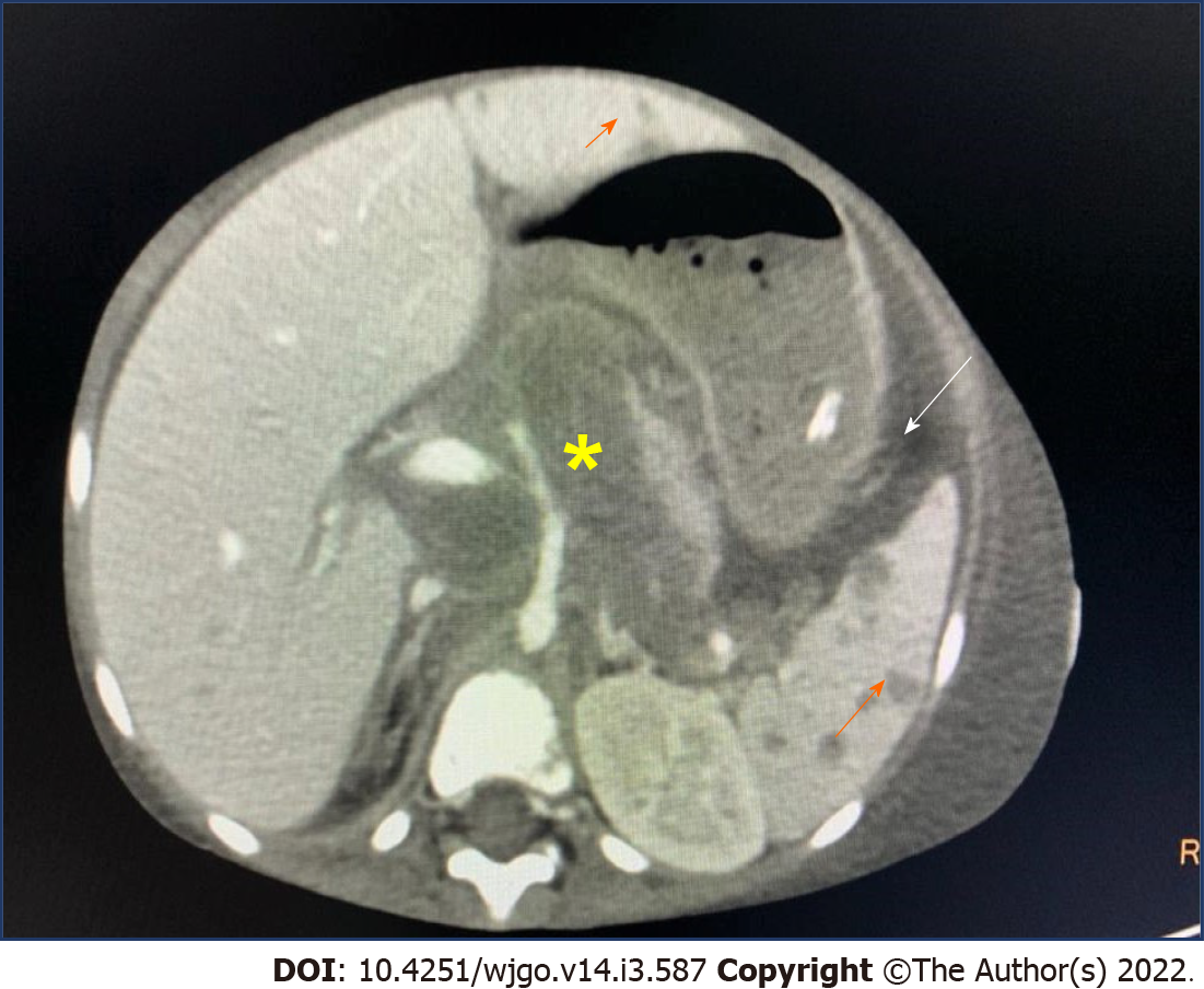
Figure 1 Axial post contrast computed tomography image showing retroperitoneal lymphadenopathy with encasement of celiac artery and portal vein (yellow asterisk).
There are multiple hypoenhancing lesions in liver, spleen (orange arrow) and presence of chylous ascites (white arrow).
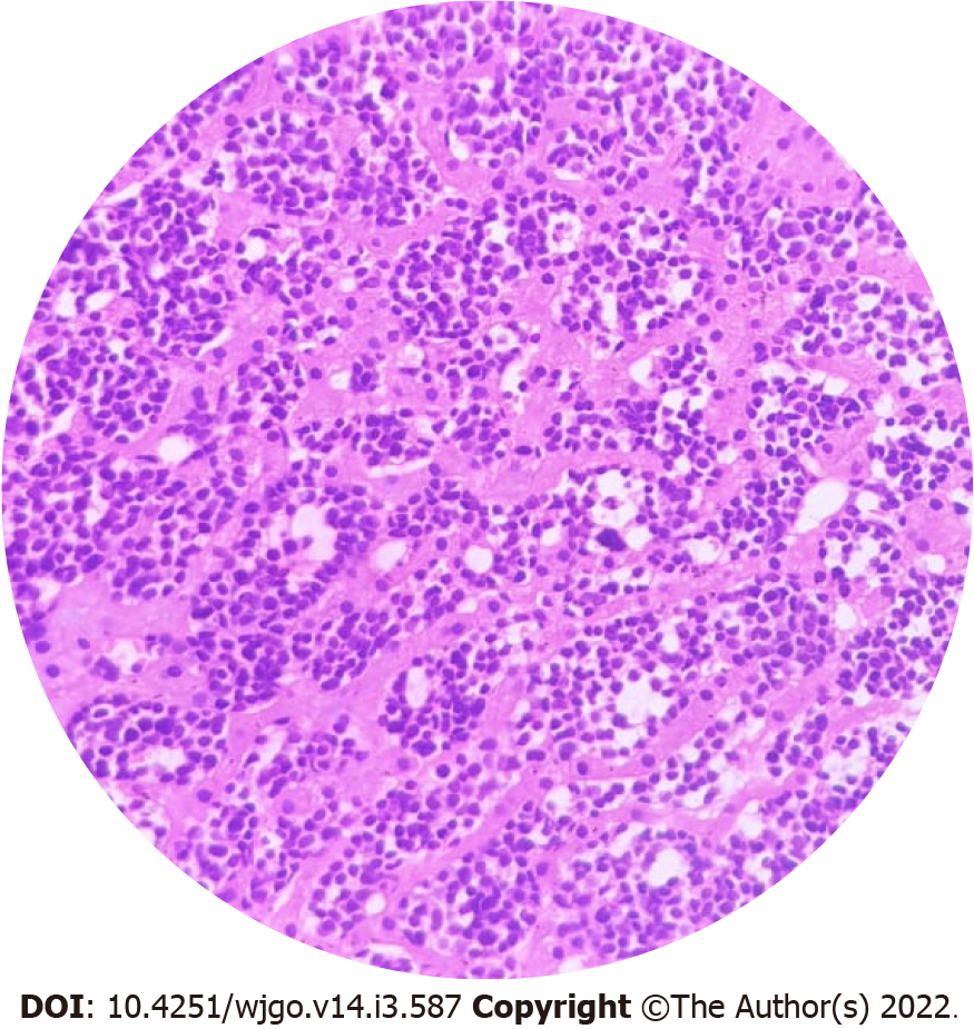
Figure 2 Liver biopsy tissue showing normal hepatocytes with the sinusoids infiltrated by monomorphic round cell with darkly staining nuclei, small inconspicuous nucleoli and a narrow rim of cytoplasm.
The infiltrating cells were positive for CD20, nuclear positivity for terminal deoxynucleotidyl transferase and high MIB1 index of 90%, suggestive of a B cell lymphoblastic lymphoma.
Figure 3 Primary gastrointestinal lymphoma.
A: Frequency of site involvement; B: Frequency of histological appearance. SEER: Surveillance, Epidemiology and End Results.
Figure 4 Primary gastrointestinal lymphoma.
Axial post contrast computed tomography abdomen with primarygastric lymphoma showing diffuse circumferential enhancing wall thickening (black arrow) and aneursymal dilatation of: A: Stomach; B: Ascending colon; C: Splenic flexure of colon.
Figure 5 In infants with initially localized disease, progression to a multisystemic involvement.
A: Lower common bile duct stricture (orange arrow) with dilated duct, beading and dilatation of intrahepatic ducts on magnetic resonance cholangiography; B: Punched out lytic lesion in skull (orange arrow) on skeletal survey; C: Honeycombing cystic lesions in lung parenchyma on high-resolution computed tomography chest.
- Citation: Devarapalli UV, Sarma MS, Mathiyazhagan G. Gut and liver involvement in pediatric hematolymphoid malignancies. World J Gastrointest Oncol 2022; 14(3): 587-606
- URL: https://www.wjgnet.com/1948-5204/full/v14/i3/587.htm
- DOI: https://dx.doi.org/10.4251/wjgo.v14.i3.587


