Copyright
©The Author(s) 2022.
World J Gastrointest Oncol. Oct 15, 2022; 14(10): 1918-1932
Published online Oct 15, 2022. doi: 10.4251/wjgo.v14.i10.1918
Published online Oct 15, 2022. doi: 10.4251/wjgo.v14.i10.1918
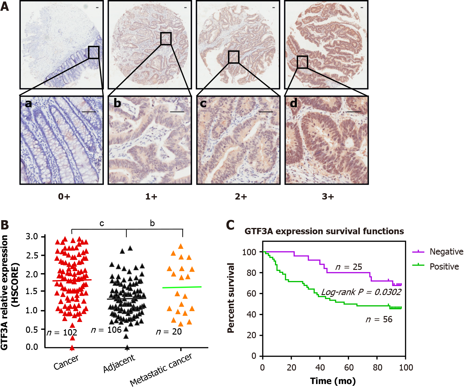
Figure 1 Transcriptional factor III A expression in colorectal cancer and survival analysis of patients with colorectal cancer.
A: Transcriptional factor III A (GTF3A) expression in adjacent normal, colorectal cancer (CRC), and metastatic tissues were detected using immunohistochemical staining. The grading standards were: (1) Negative, a, scored as 0; (2) Weakly positive, b, scored as 1; (3) Moderately positive, c, scored as 2; and (4) Strongly positive, d, scored as 3. Scale bar, 50 μm; B: GTF3A expression in adjacent normal, CRC, and metastatic tissues was quantified using semi-quantitative histological score. Scored dots expression groups were statistically analyzed using unpaired t test; C: Overall survival of patients with GTF3A negative (n = 25) or GTF3A positive (n = 56) expression was analyzed by log-rank (Mantel-Cox) test. bP < 0.01; cP < 0.001. GTF3A: Transcriptional factor III A; HSCORE: Semi-quantitative histological score.
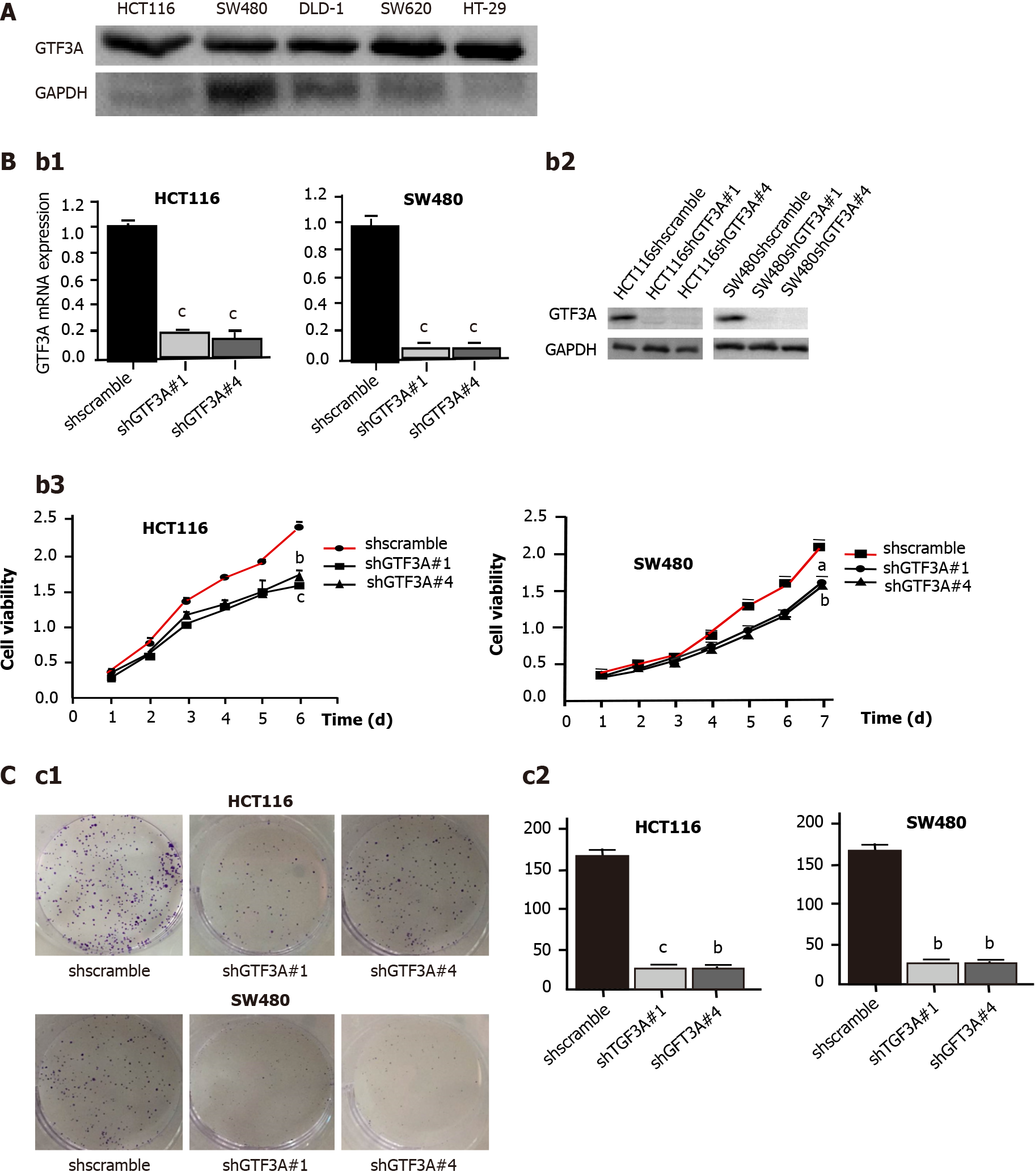
Figure 2 Knockdown of transcriptional factor III A gene inhibits colorectal cancer cell proliferation.
A: The expression of transcriptional factor III A (GTF3A) in HCT116, SW480, DLD-1, SW620, and HT-29 cells was detected by Western blot; B: HCT116 and SW480 cells were stably transfected with short hairpin (sh) shscramble and shGTF3A#1 and #4-GV112 virus, respectively. Gtf3a mRNA (b1) and GTF3A protein (b2) expression were detected using real-time quantitative polymerase chain reaction and Western blot, respectively. The cell viability of HCT116 and SW480-shscramble as well as shGTF3A#1 and #4 cells was detected using Cell Counting Kit assay (b3); C: Colony formation of the transfected HCT116 and SW480 cells was detected using colony formation assay (c1), and the colony number was counted (c2). Gtf3a mRNA expression, cell colony number, and cell viability are expressed as the mean ± SEM of three independent experiments. aP < 0.05; bP < 0.01; cP < 0.001. GTF3A: Transcriptional factor III A; GAPDH: Glyceraldehydes-3-phosphate dehydrogenase.
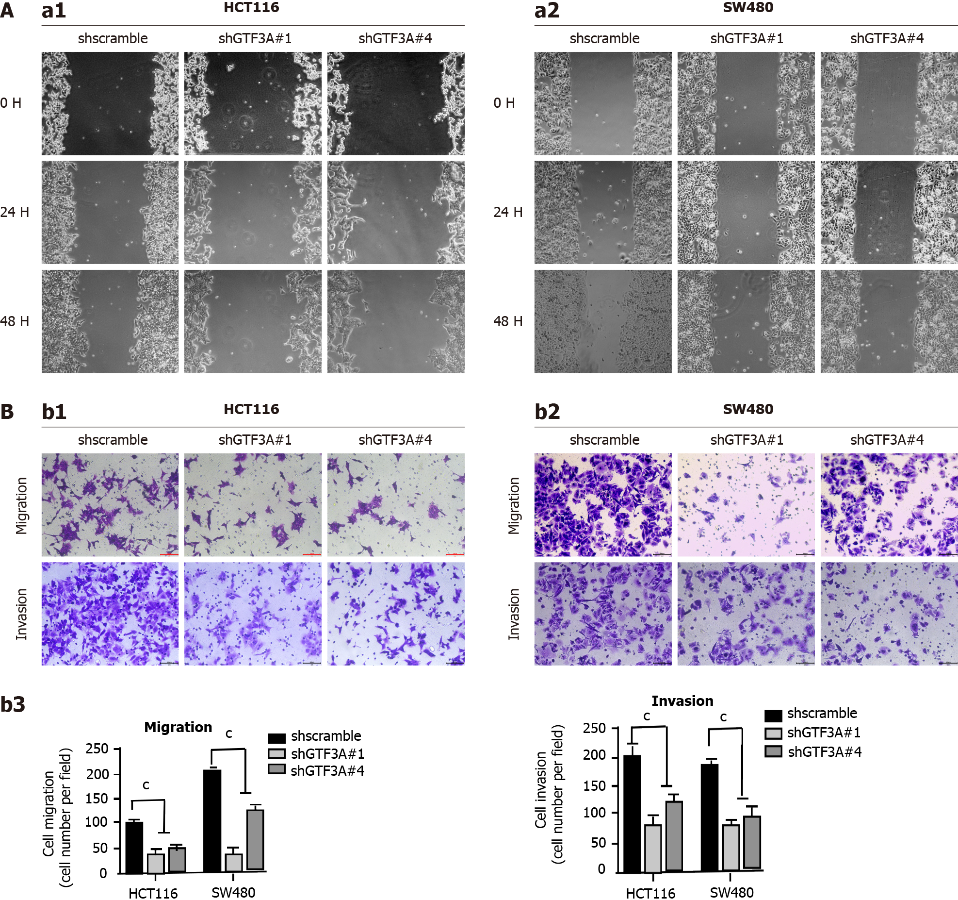
Figure 3 Knockdown of transcriptional factor III A gene inhibits the invasion and migration of colorectal cancer cell in vitro.
A: The migratory abilities of short hairpin (sh) GTF3A#1 and #4-HCT116 cells were detected using wound-healing assay (a1). The migratory abilities of shGTF3A#1 and #4-SW480 cells were detected as above (a2); B: The migratory and invasive capabilities of shGTF3A#1 and #4-HCT116 (b1) and shGTF3A#1 and #4-SW480 cells (b2) were detected using transwell assay. The migrated and invaded cells were counted (b3). Migratory and invasive capabilities are expressed as the mean ± SEM of three independent experiments. cP < 0.001.
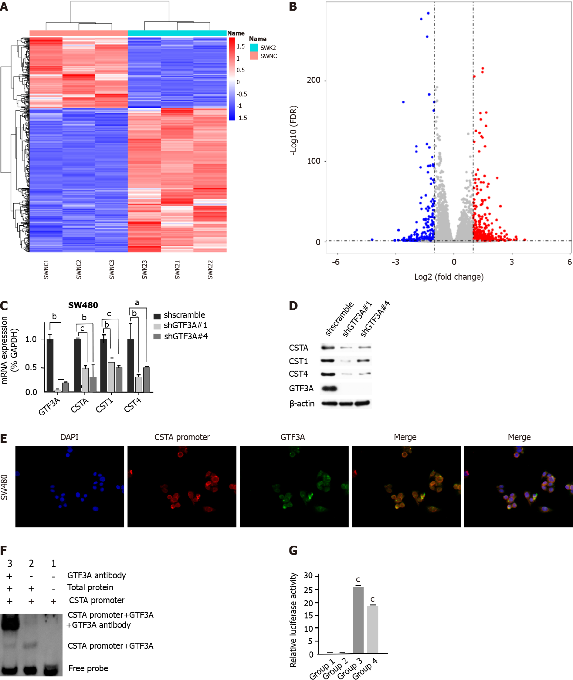
Figure 4 Transcriptional factor III A regulates cystatin A by binding to the promoter of cystatin A gene.
A: Heat map of RNA-sequencing (RNA-Seq) for transcriptional factor III A gene (Gtf3a)-knockdown and scramble control cells; B: Volcano plot of RNA-Seq for Gtf3a-knockdown and scramble control cells; C: mRNA expression of Gtf3a, cystatin A (Csta), cystatin SN gene (Cst1), and cystatin S genes (Cst4) in short hairpin (sh)scramble-SW480 and shGTF3A#1 and #4-SW480 cells were detected using real-time quantitative polymerase chain reaction; D: GTF3A, CSTA, CST1, and CST4 in shscramble-SW480 and shGTF3A#1 and #4-SW480 cells were detected using Western blot; E: RNA fluorescence in situ hybridization was performed to verify the locations of the Csta promoter probe (lncRNA) and GTF3A in SW480 cells; F: Electrophoretic mobility shift assay (EMSA) was used to test the interaction of the Csta promoter and GTF3A. The Csta promoter plus GTF3A antibody as the super shift in EMSA; G: Luciferase activity assay was used to detect the interaction of GTF3A with the Csta promoter and the transcription of Csta. Group 1 (Csta promoter-luc blank vector plus GTF3A blank plasmid) and group 2 (Csta promoter-luc blank vector plus GTF3A-plasmid) served as the control groups, and group 3 (Csta promoter-luc plus GTF3A) and group 4 (Csta promoter-luc plus GTF3A blank vector) as experimental groups. Csta expression is expressed as the mean ± SEM of three independent experiments. cP < 0.001. GTF3A: Transcriptional factor III A; GAPDH: Glyceraldehydes-3-phosphate dehydrogenase; CSTA: Cystatin A; CST1: Cystatin SN; CST4: Cystatin S.
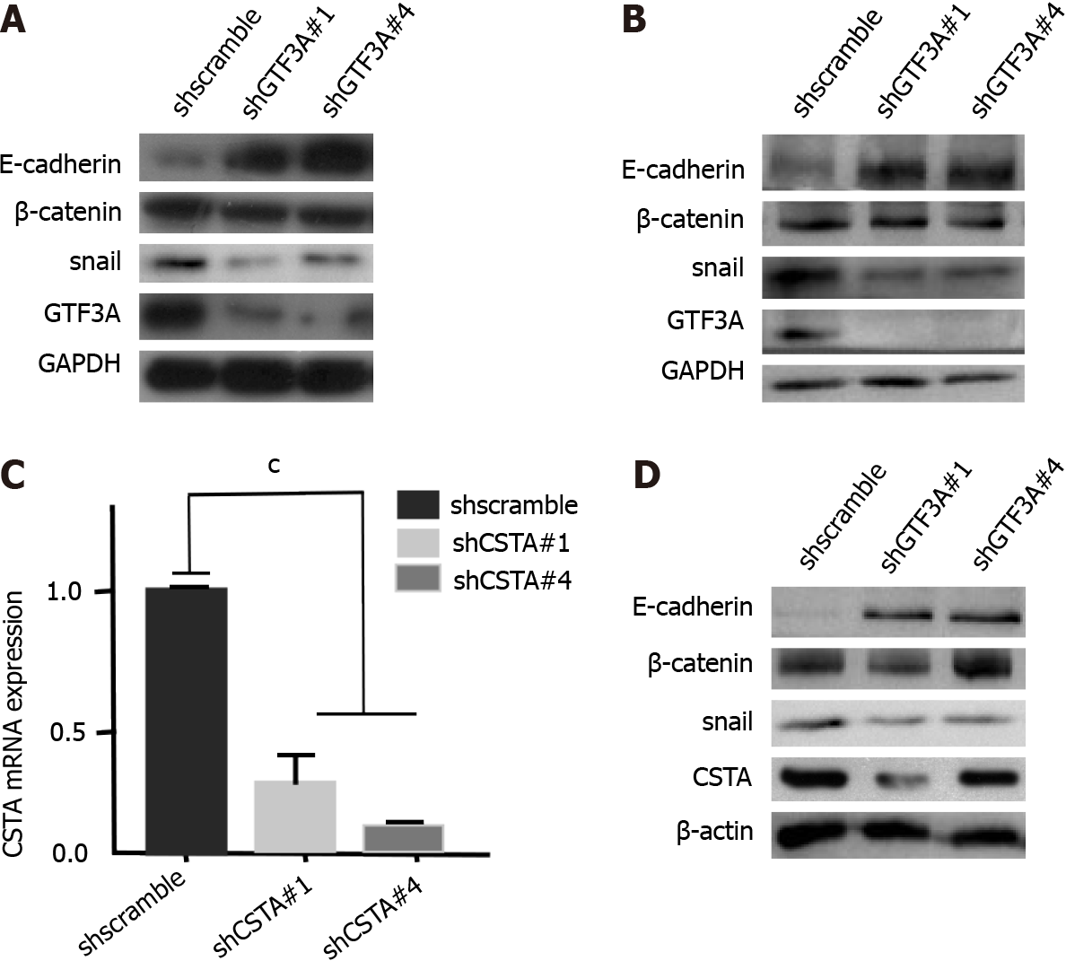
Figure 5 Transcriptional factor III A mediates epithelial-mesenchymal transition of colorectal cancer cells by regulating cystatin A.
A: Epithelial-mesenchymal transition (EMT) biomarkers E-cadherin, beta-catenin, and snail in short hairpin of transcriptional factor III A (shGTF3A) #1 and #4-HCT116 cells and the control group were detected by Western blot; B: EMT biomarkers were tested in shGTF3A#1 and #4-SW480 cells and the control group; C: mRNA expression of cystatin A (Csta) gene in shCSTA#1,2-SW480 and the control group was detected using real-time quantitative polymerase chain reaction; D: EMT biomarkers were analyzed in Csta-knockdown SW480 and the control cells. Cystatin A mRNA expression is expressed as the mean ± SEM of three independent experiments. There were statistically significant differences between shCSTA#1,2-SW480 and the control group. cP < 0.001. GTF3A: Transcriptional factor III A; GAPDH: Glyceraldehydes-3-phosphate dehydrogenase; CSTA: Cystatin A; shscramble: Short hairpin scramble; shCSTA: Short hairpin of transcriptional factor III A.
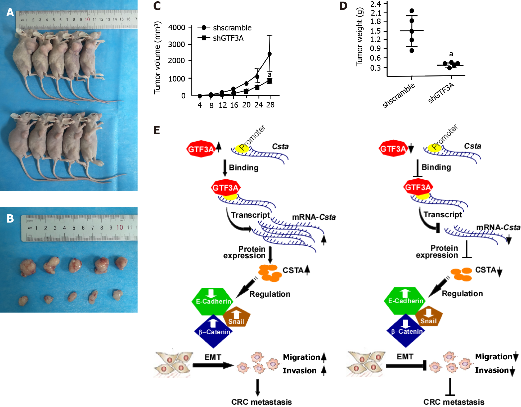
Figure 6 Transcriptional factor III A promotes colorectal cancer growth in vivo.
A: Short hairpin of transcriptional factor III A (shGTF3A)-HCT116 and short hairpin of scramble (shscramble)-HCT116 cells were subcutaneously injected into the right armpit of nude mice. After 28 d, the mice were euthanized, and the images of the representative nude mice are shown; B: Tumors were stripped from the nude mouse; C: The tumor growth curve of shGTF3A-HCT116 and shscramble-HCT116 cells was calculated by tumor volume; D: The removed tumors of shGTF3A-HCT116 and shscramble-HCT116 cells were weighed; E: Schematic diagram of GTF3A-promoting CRC metastasis. GTF3A bound to the promoter of cystatin A (Csta) gene to increase Csta gene transcription and protein expression, increased CSTA regulated the epithelial-mesenchymal transition (EMT) to promote invasion and metastasis of colorectal cancer (CRC) cells, while knockdown of Gtf3a decreased CSTA expression, inhibited the EMT, and reduced CRC cell invasion and metastasis. aP < 0.05. shscramble: Short hairpin scramble; shCSTA: Short hairpin of transcriptional factor III A; EMT: Epithelial-mesenchymal transition; CRC: Colorectal cancer; CSTA: Cystatin A.
- Citation: Wang J, Tan Y, Jia QY, Tang FQ. Transcriptional factor III A promotes colorectal cancer progression by upregulating cystatin A. World J Gastrointest Oncol 2022; 14(10): 1918-1932
- URL: https://www.wjgnet.com/1948-5204/full/v14/i10/1918.htm
- DOI: https://dx.doi.org/10.4251/wjgo.v14.i10.1918


