©The Author(s) 2025.
World J Gastrointest Endosc. Sep 16, 2025; 17(9): 108787
Published online Sep 16, 2025. doi: 10.4253/wjge.v17.i9.108787
Published online Sep 16, 2025. doi: 10.4253/wjge.v17.i9.108787
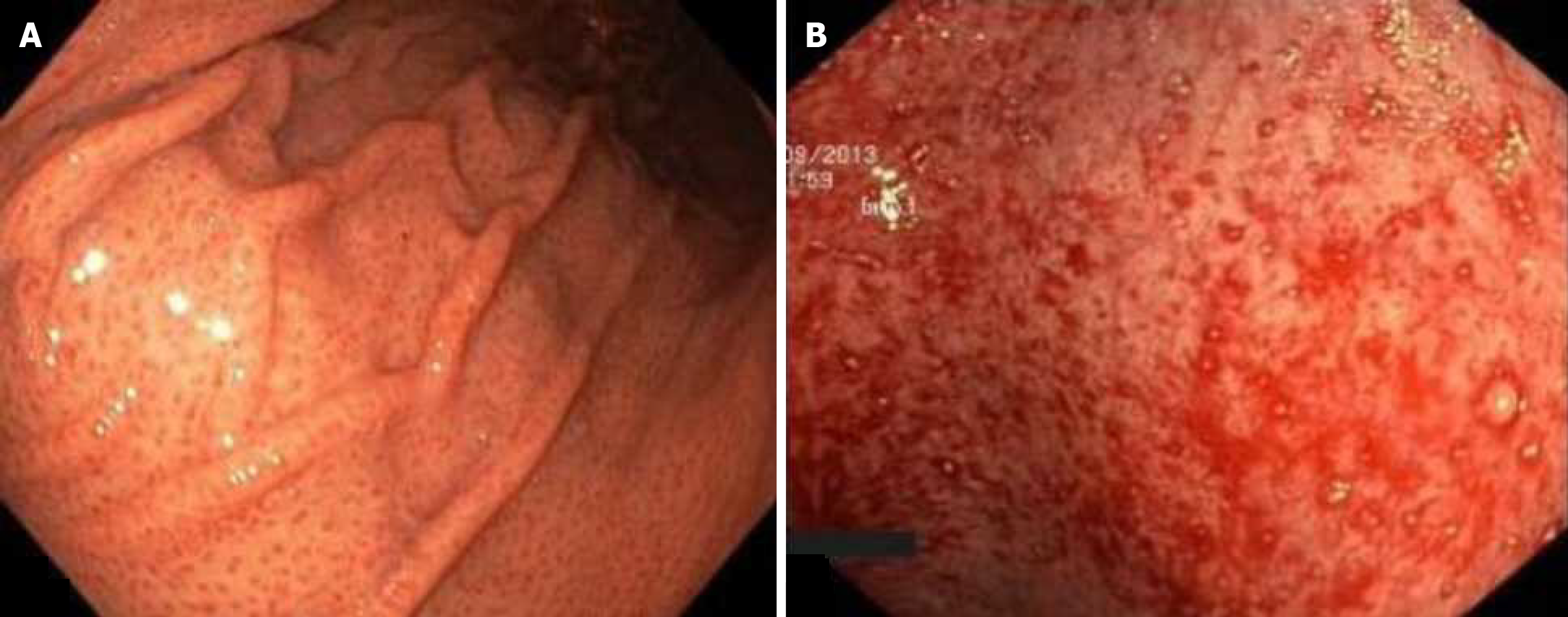
Figure 1 Portal hypertensive gastropathy.
A: Mild portal hypertensive gastropathy. A mosaic mucosal pattern of the body and fundus of the stomach; B: Severe portal hypertensive gastropathy. Hemorrhages on the background of erythema foci.
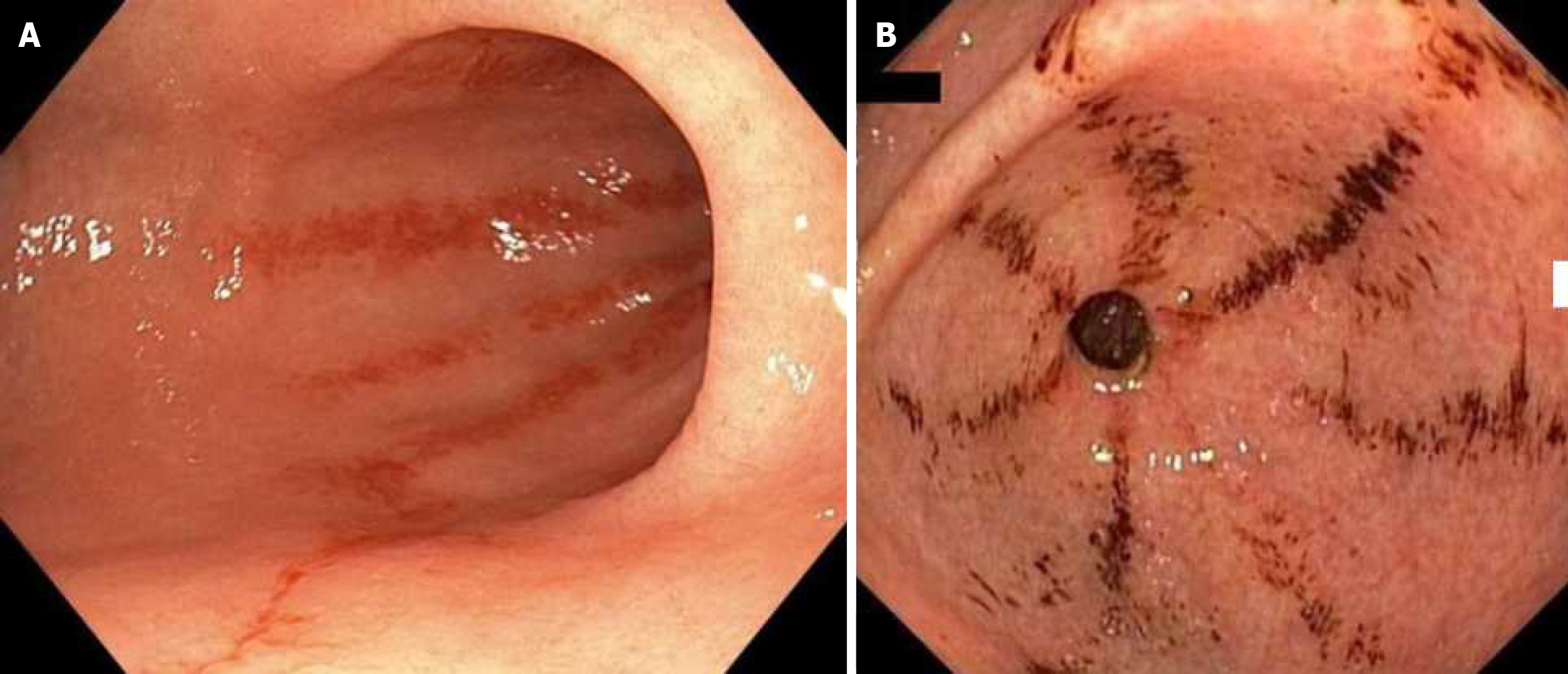
Figure 2 Gastric antral vascular ectasia.
A: Linear striped phenotype. Red vascular spots spiraling away from the pylorus; B: Linear striped phenotype. Hemorrhages on the background of red vascular spots spiraling away from the pylorus.
Figure 3 Gastric antral vascular ectasia.
A: Diffuse phenotype. Multiple red spots spread out in a diffuse pattern along the gastric antrum; B: Diffuse phenotype in white light endoscopy; C: Diffuse phenotype in endoscopy with narrow-band imaging.
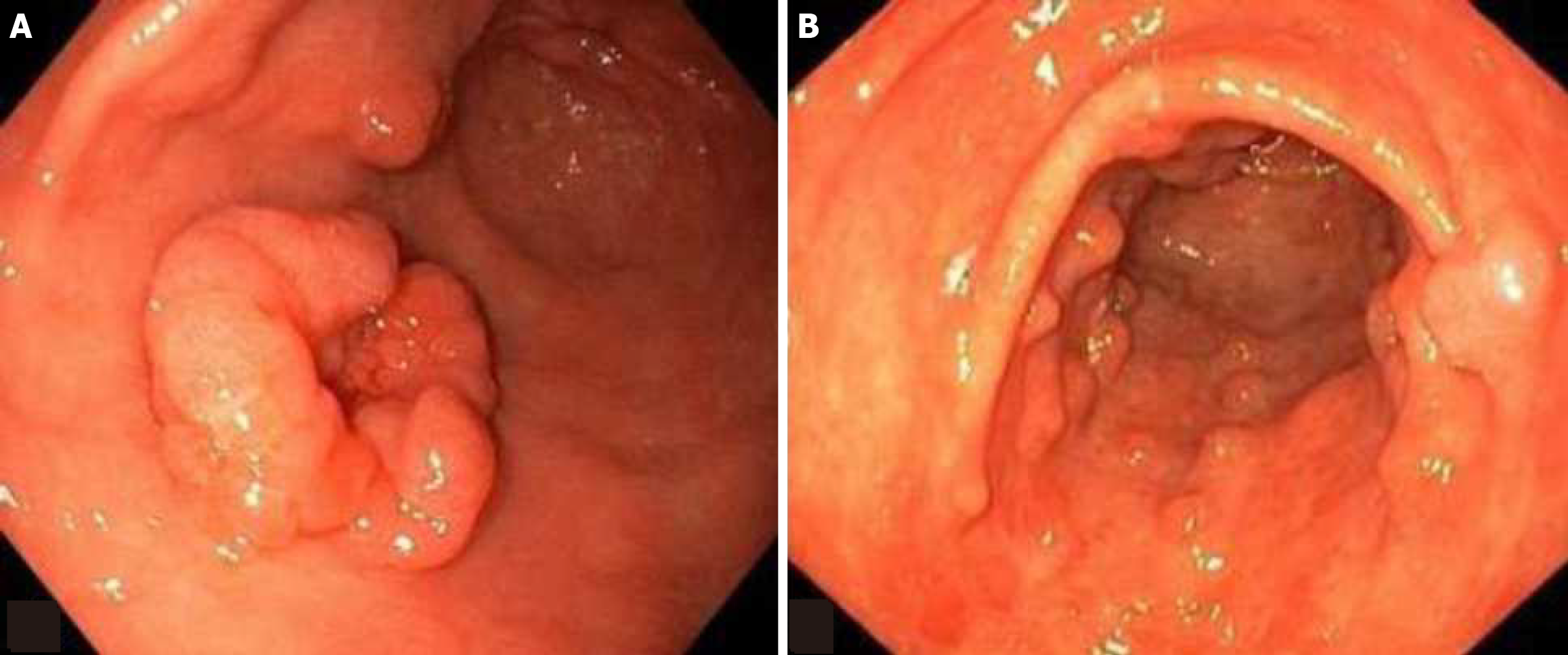
Figure 4 Portal hypertensive polyps.
A: Polypoid lesion in the gastric antrum up to 1 cm in diameter with an indentation in the middle, and of a soft-elastic consistency; B: Multiple polypoid lesions in the gastric antrum on a wide base from 3 mm to 7 mm in diameter.
- Citation: Olevskaya ER, Dolgushina AI, Garbuzenko DV, Khikhlova AO, Saenko AA, Khusainova GM, Kuznetsova AS. Role of endoscopy in the diagnosis and treatment of gastric mucosal lesions in liver cirrhosis patients. World J Gastrointest Endosc 2025; 17(9): 108787
- URL: https://www.wjgnet.com/1948-5190/full/v17/i9/108787.htm
- DOI: https://dx.doi.org/10.4253/wjge.v17.i9.108787


