©The Author(s) 2025.
World J Gastrointest Endosc. Nov 16, 2025; 17(11): 111770
Published online Nov 16, 2025. doi: 10.4253/wjge.v17.i11.111770
Published online Nov 16, 2025. doi: 10.4253/wjge.v17.i11.111770
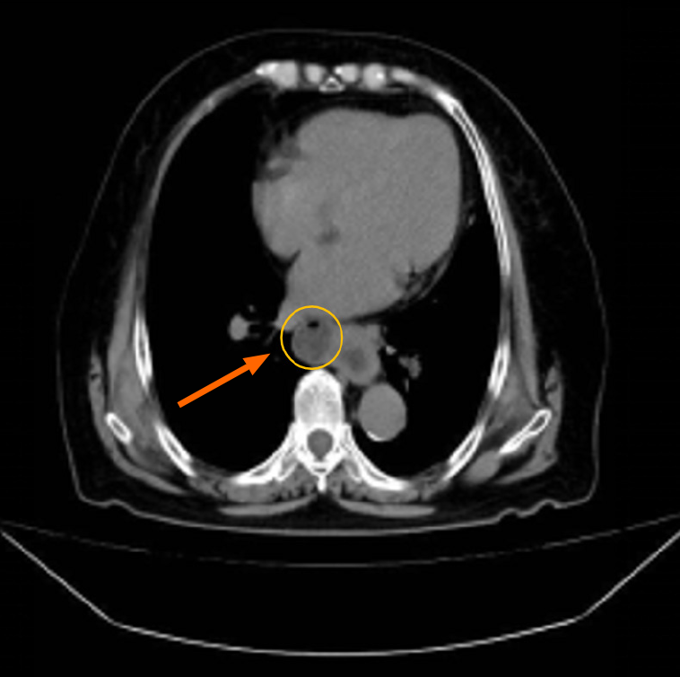
Figure 1 Computed tomography image of the whole abdomen.
The cystic bulge on the right side of the middle esophagus was considered an esophageal diverticulum, and local thickening of the lower esophagus was observed.
Figure 2 Upper gastrointestinal pan-glucosamine contrast.
A: A cystic shadow is visible on the right side of the middle esophagus, measuring approximately 3.0 cm × 3.0 cm. This was connected to the lumen of the esophagus, and barium could be seen entering and exiting the esophagus; B: A saccular pouching exophthalmos is visible on the right side of the esophagus, with rigidity of the esophageal wall and narrowing of the lumen in the lower part of the esophagus; C: A cystic shadow measuring approximately 2.1 cm × 2.6 cm was seen in the right side of the middle esophagus. String-like changes in the body of the esophagus were visible, accompanied by narrowing of the lumen in the lower and middle esophagus.
Figure 3 Gastroscopy.
A: A large diverticulum was seen in the esophagus; B: Slight tension in the lower and middle esophagus.
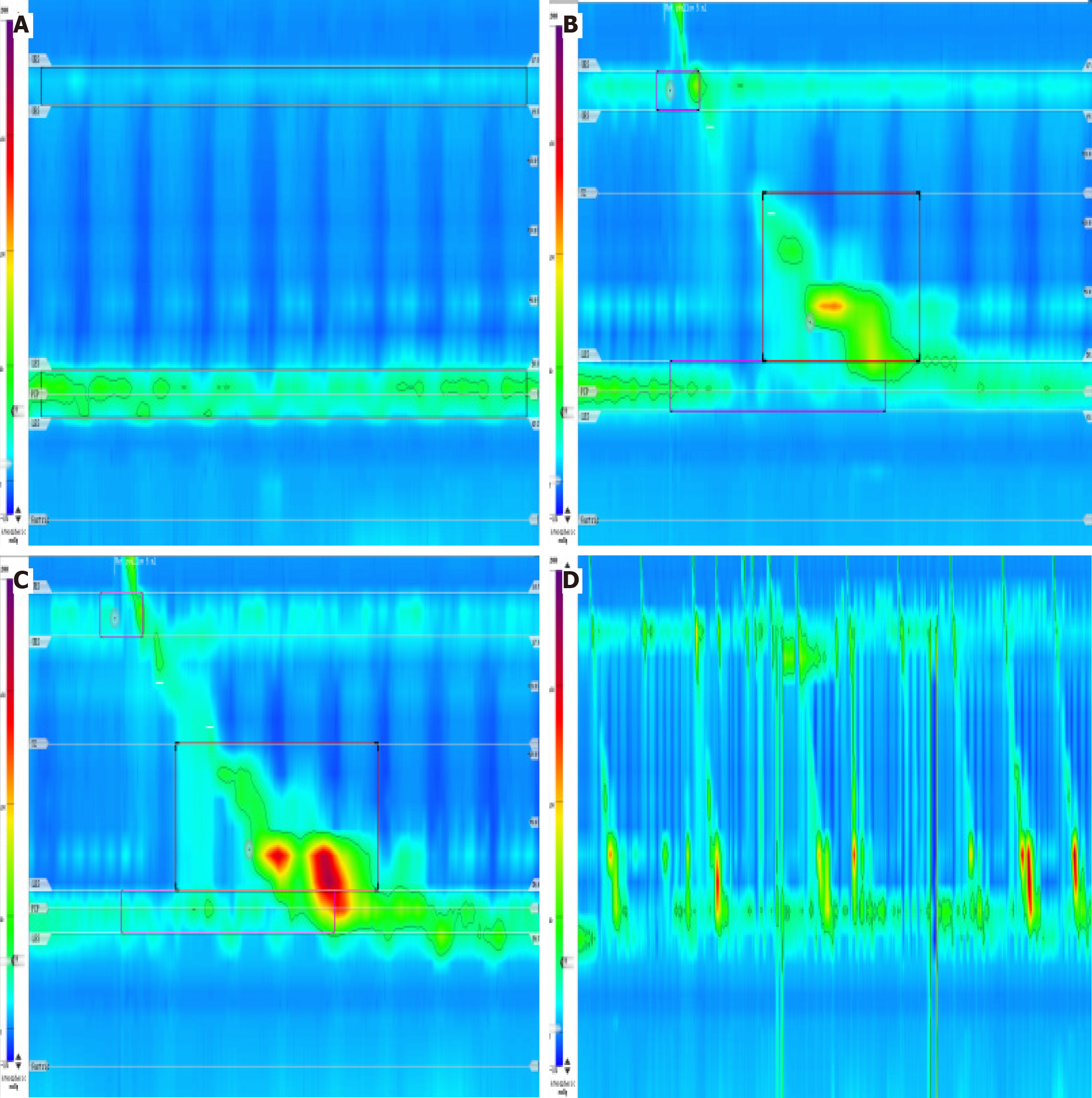
Figure 4 Esophageal manometry.
A: Static pressure: At rest, the upper esophageal sphincter pressure was low (8 mmHg in this patient, normal value 33-180 mmHg) and the low esophageal sphincter pressure was normal (27 mmHg in this patient, normal value 10-45 mmHg); B: 5 mL wet swallow: While normal 4s integrated relaxation pressure and normal distal contractile integral suggest normal peristalsis in the body of the esophagus, an interruption of the contraction wave on the 20 mmHg isobaric line with a defect length greater than 5 cm (6.5 cm) suggests segmental contraction; this swallow was ineffective; C: 5 mL wet pharynx: Normal 4s integrated relaxation pressure, normal distal contractile integral, distal latency, and maximal interruptions are in the normal range, suggesting normal esophageal motility; D: Comprehensive analysis: The patient performed 10 5-mL wet swallows, with 6 (60%) normal contractions (450 mmHg.s.cm < distal contractile integral < 8000 mmHg.s.cm) and 6 (60%) ineffective swallows (including 2 peristaltic failures, 2 weak contractions, and 2 fragmentary contractions). This suggests an indeterminate ineffective esophageal motility. Performed 3 multiple rapid swallows, with the distal contractile integral after performing multiple rapid swallows being less than the mean distal contractile integral of a single swallow, suggesting poor esophageal contractile reserve function.
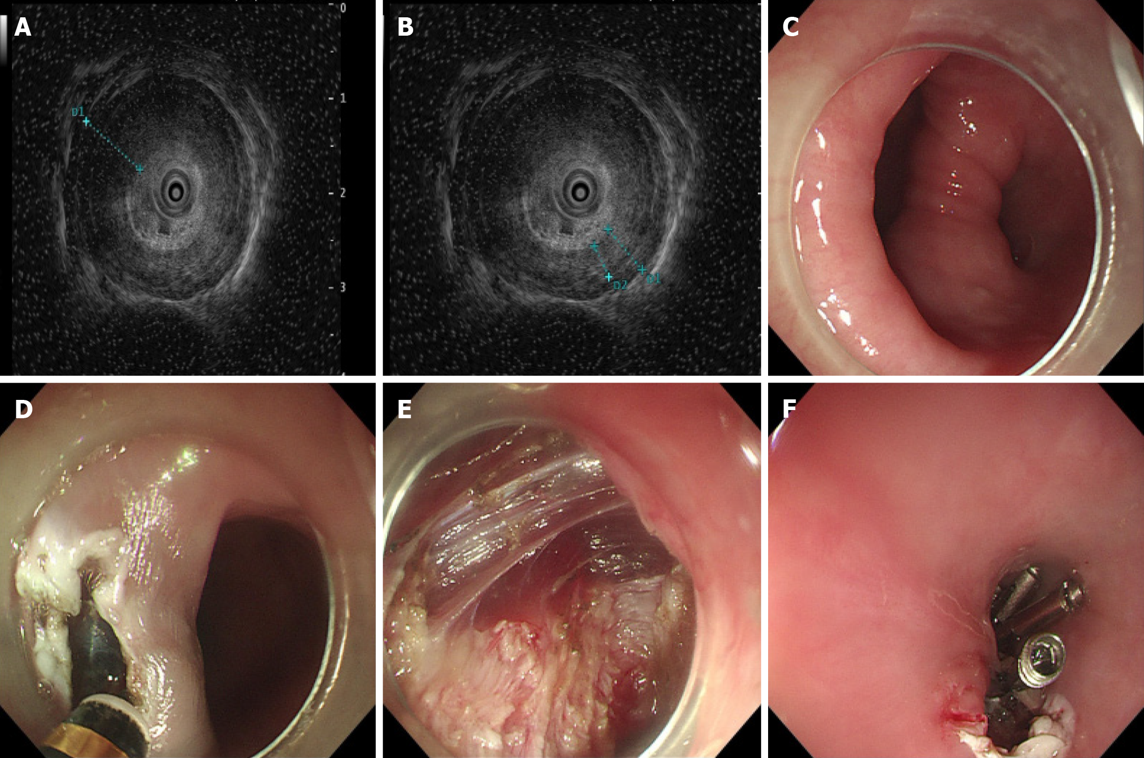
Figure 5 The submucosal tunneling endoscopic septum division surgical procedure.
A and B: Ultrasonic gastroscopy of the layers of the diverticulum; C: Giant esophageal diverticulum; D: Construction of the tunnel; E: Incision of the muscle; F: Closure of the wound with titanium clips.
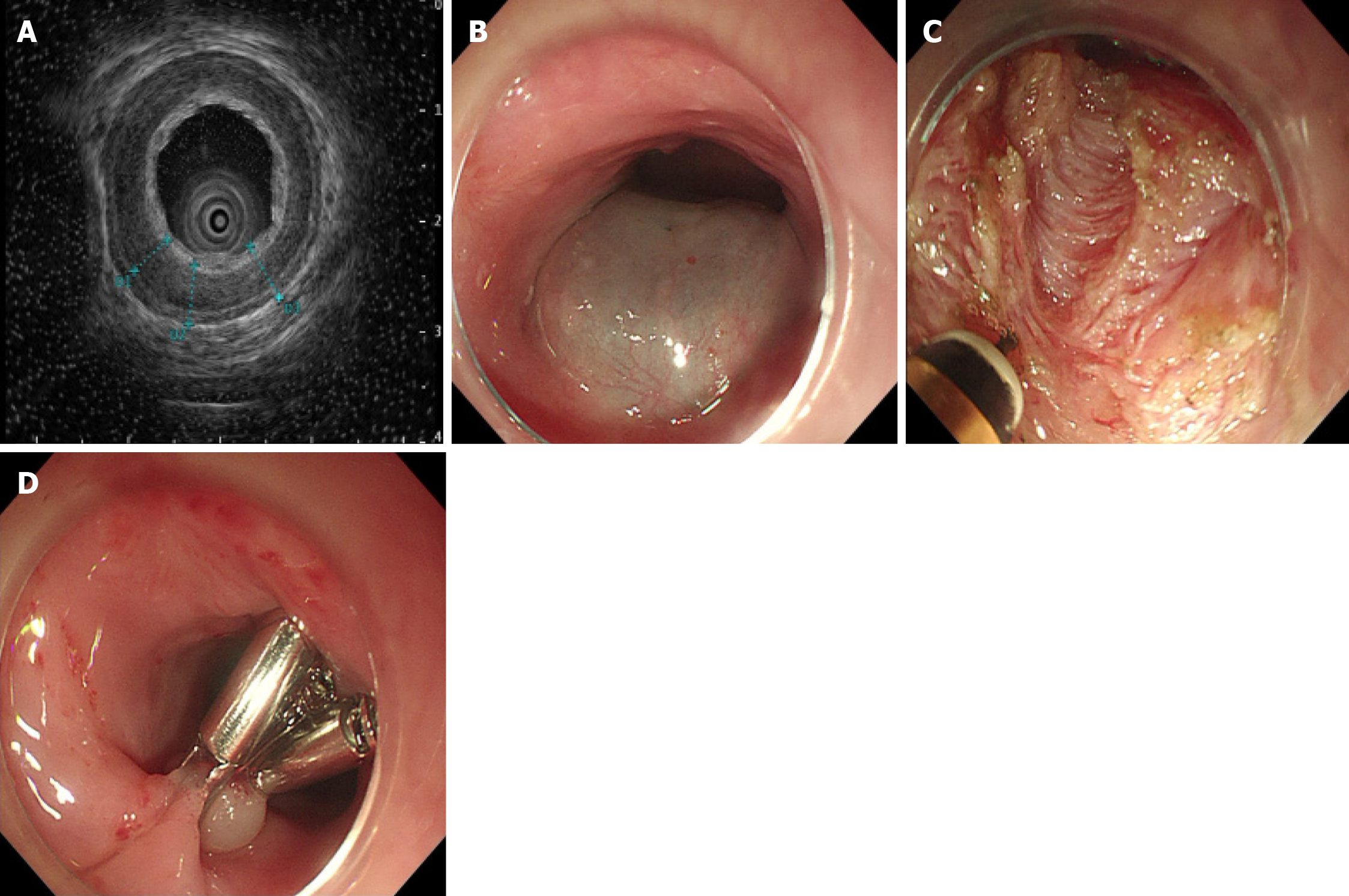
Figure 6 The per-oral endoscopic myotomy surgical procedure.
A: Ultrasonic gastroscopy of the layers of the esophagus; B: Construction of a tunnel; C: Incision of the muscle; D: Closure of the wound with titanium clips.
- Citation: Liu XR, Chen XZ, Fan MW, Zhang SH, Shi N, Liu CX, Chen Y, Wang XM. Endoscopic treatment for dysphagia caused by mid-esophageal diverticulum and diffuse esophageal spasm: A case report. World J Gastrointest Endosc 2025; 17(11): 111770
- URL: https://www.wjgnet.com/1948-5190/full/v17/i11/111770.htm
- DOI: https://dx.doi.org/10.4253/wjge.v17.i11.111770


