©The Author(s) 2025.
World J Gastrointest Endosc. Oct 16, 2025; 17(10): 111408
Published online Oct 16, 2025. doi: 10.4253/wjge.v17.i10.111408
Published online Oct 16, 2025. doi: 10.4253/wjge.v17.i10.111408
Figure 1 Abdominal computed tomography findings in a 58-year-old woman with elevated carbohydrate antigen 19-9.
A: The non-contrast-enhanced coronal view demonstrated a 7.1 cm × 4.8 cm hypodense cystic lesion (orange arrow) in the right upper quadrant; B and C: An arterial-phase contrast-enhanced coronal image reveals the lesion’s (orange arrow) intimate anatomical relationship with both the pancreatic head (P in B) and the greater curvature of the gastric antrum (G in C). The absence of a septum was found, and no significant arterial enhancement or suspicious nodularity of the cyst wall was found.
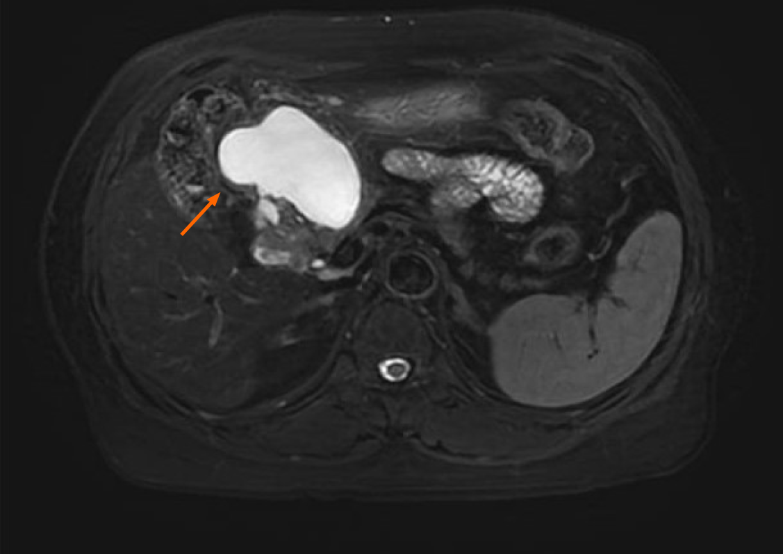
Figure 2 Contrast-enhanced magnetic resonance imaging findings in a 58-year-old woman with elevated carbohydrate antigen 19-9.
Magnetic resonance imaging revealed a well-circumscribed, oval cystic lesion (62 mm × 38 mm, SE3 IM9) in the right upper abdomen, with prolonged T2 signal intensity. The lesion was closely adjacent to the pancreatic head and the gastric antrum, with no fat plane separation. Peripheral wall enhancement was observed postcontrast.
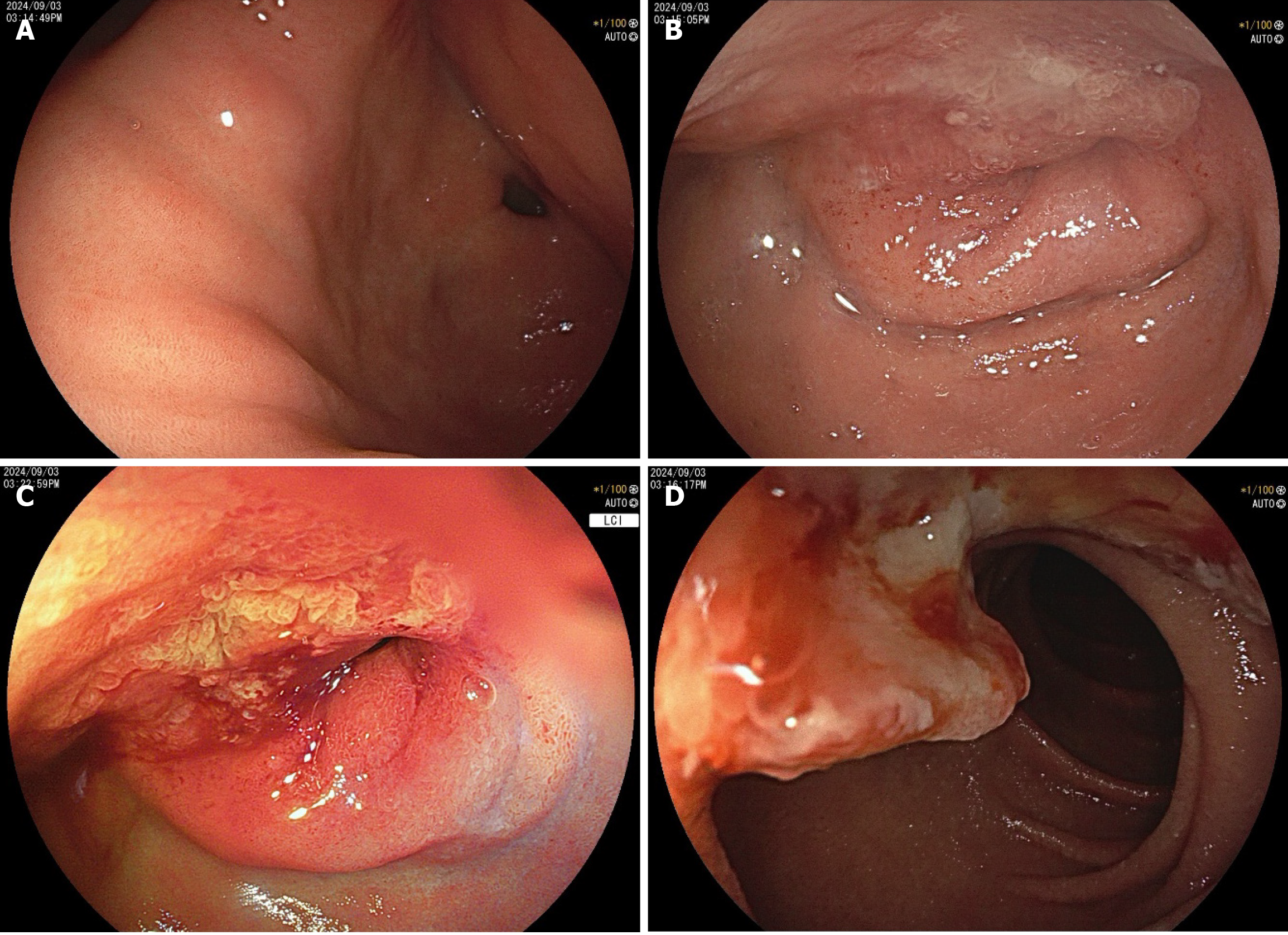
Figure 3 Endoscopic findings of ectopic pancreatic carcinoma in a 58-year-old woman.
A: A submucosal lesion with central umbilication was observed at the greater curvature of the gastric antrum, accompanied by extrinsic compression near the pylorus (soft on probing, with pyloric patency preserved); B and C: A giant ulcer with raised margins and a white fibrinoid base was found at the duodenal bulb-descending junction, causing significant luminal stenosis; D: The stenosis was nearly obstructive, the tumor in the descending duodenum exhibited mucosal breach and a coarse, irregular surface.
Figure 4 Endosonographic features of an abdominal solid cystic mass in a 58-year-old woman.
A: A multilobulated solid cystic mass (7.0 cm × 4.8 cm) was identified; B: The mass extended to the duodenal bulb-descending junction, causing luminal stenosis. The duodenal wall showed tumor infiltration (irregular hypoechoic thickening of up to 1 cm). The cyst wall exhibited hypoechoic unevenness, suggesting mural involvement; C: The lesion had ill-defined borders with the fourth layer of the gastric wall (as indicated by green arrow).
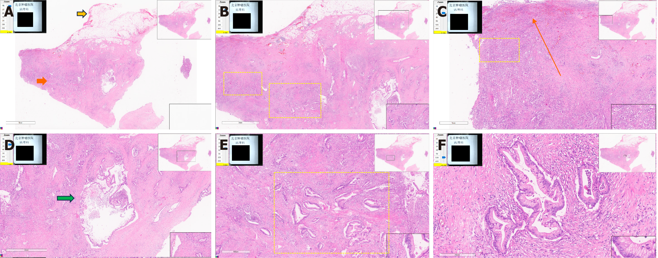
Figure 5 Histopathological analysis of a resected specimen from a 58-year-old woman with a gastric mass.
A-F: Histopathology confirmed a nonpancreatic primary adenocarcinoma with predominant gastric involvement. The lesion exhibited cystic degeneration and extended to the pyloroduodenal region, forming mucosal ulcers. Representative section demonstrating the lesion’s architecture, including the serosal layer (yellow arrow) and the muscularis propria (orange arrow) (A). Infiltrative irregular glands (yellow dashed box) exhibiting malignant cytological features (nuclear pleomorphism and a crowded growth pattern) were observed within the muscularis propria (B and E). Tumor invasion through the mucosal to subserosal layers (orange arrow) of the gastrointestinal wall was apparent, with multiple tumorous cells and glands in the muscularis propria (yellow dashed box) (C). Cystic dilation (green arrow) of the ectopic pancreatic ductal structures (D). A high-power view revealed glandular epithelial cells with abundant eosinophilic cytoplasm and elongated nuclei, consistent with pancreatobiliary adenocarcinoma (F).
- Citation: Wang YX, Wang J, Liang SX, Wu Q. Primary adenocarcinoma from a gastric heterotopic pancreas: A case report. World J Gastrointest Endosc 2025; 17(10): 111408
- URL: https://www.wjgnet.com/1948-5190/full/v17/i10/111408.htm
- DOI: https://dx.doi.org/10.4253/wjge.v17.i10.111408


