©The Author(s) 2025.
World J Gastrointest Endosc. Oct 16, 2025; 17(10): 108729
Published online Oct 16, 2025. doi: 10.4253/wjge.v17.i10.108729
Published online Oct 16, 2025. doi: 10.4253/wjge.v17.i10.108729
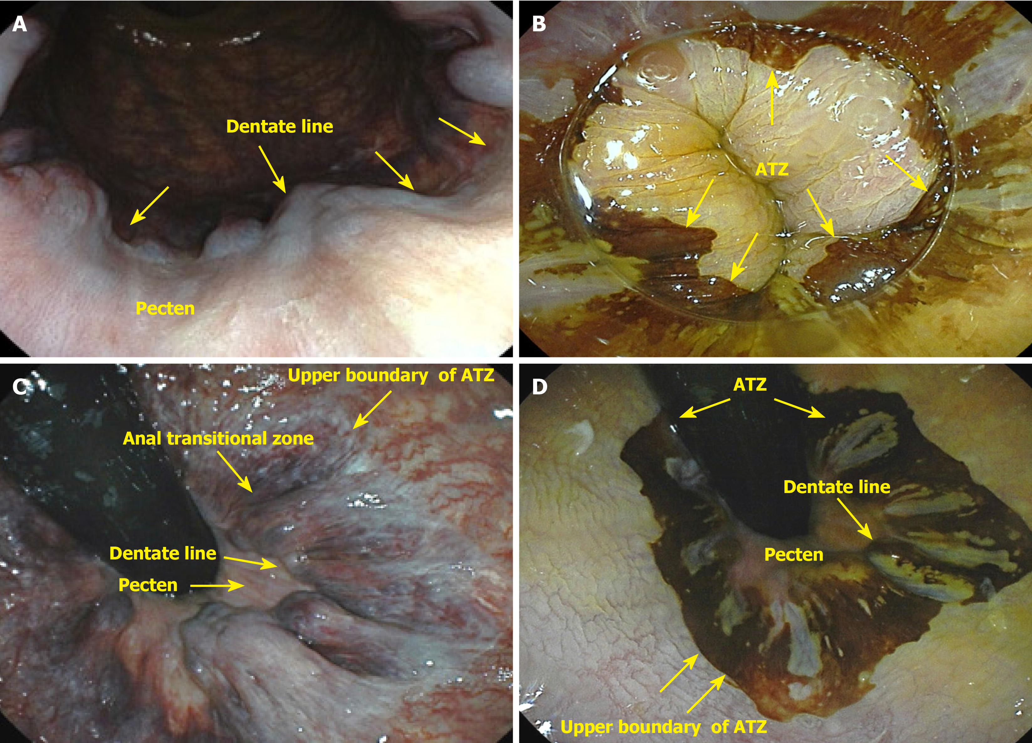
Figure 1 Normal appearance of the anal canal.
A: Frontal view of anal canal, pecten, and dentate line; B: Anoscopy, anal transitional zone, and dentate line (lugol); C: Rectal retroflexion of the anorectal junction, anal transitional zone, dentate line (yellow), and pecten; D: Rectal retroflexion of the anorectal junction, anal transitional zone, dentate line, and pecten (lugol). ATZ: Anal transitional zone.
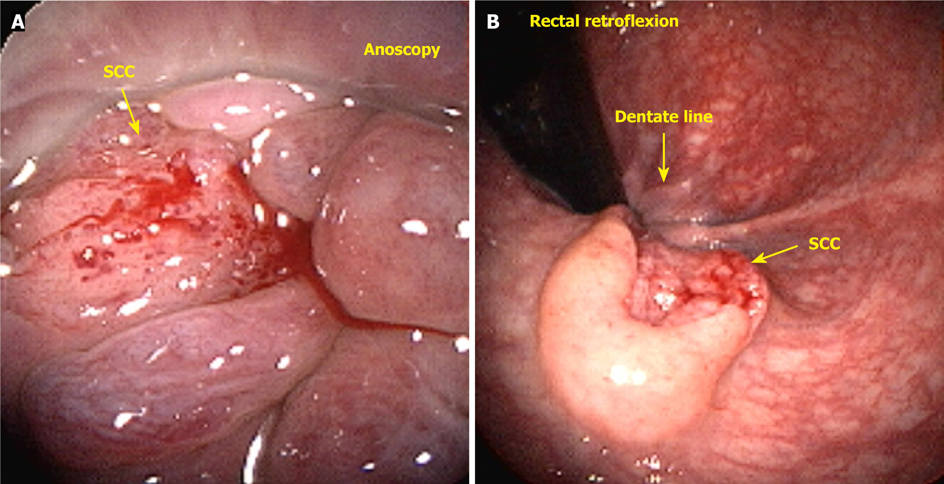
Figure 2 Squamous cell carcinoma of the anal canal.
A: Anoscopy; B: Rectal retroflexion. SCC: Squamous cell carcinoma.
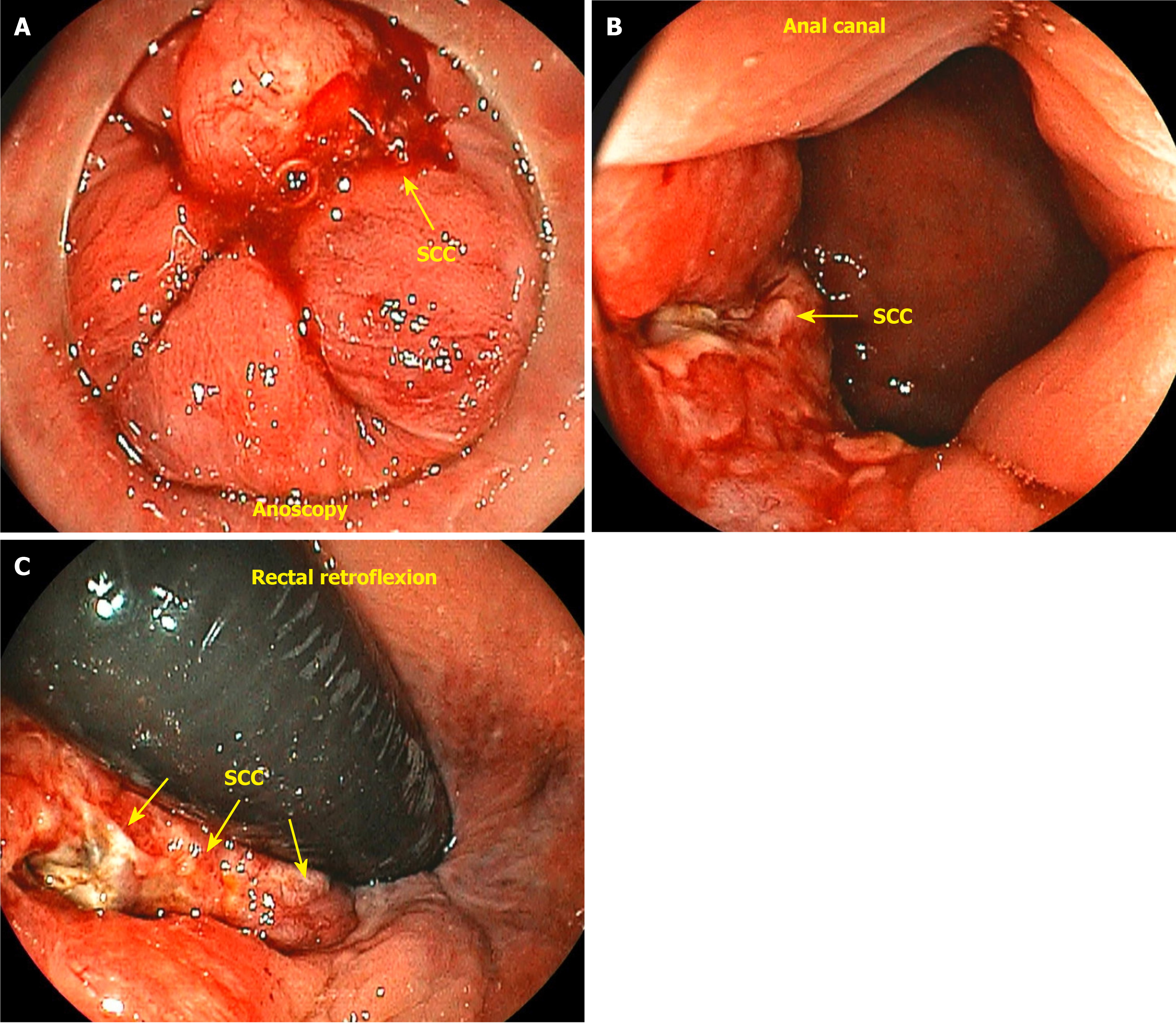
Figure 3 Cloacogenic carcinoma.
A: Anoscopy; B: Anal canal frontal view; C: Rectal retroflexion. SCC: Squamous cell carcinoma.
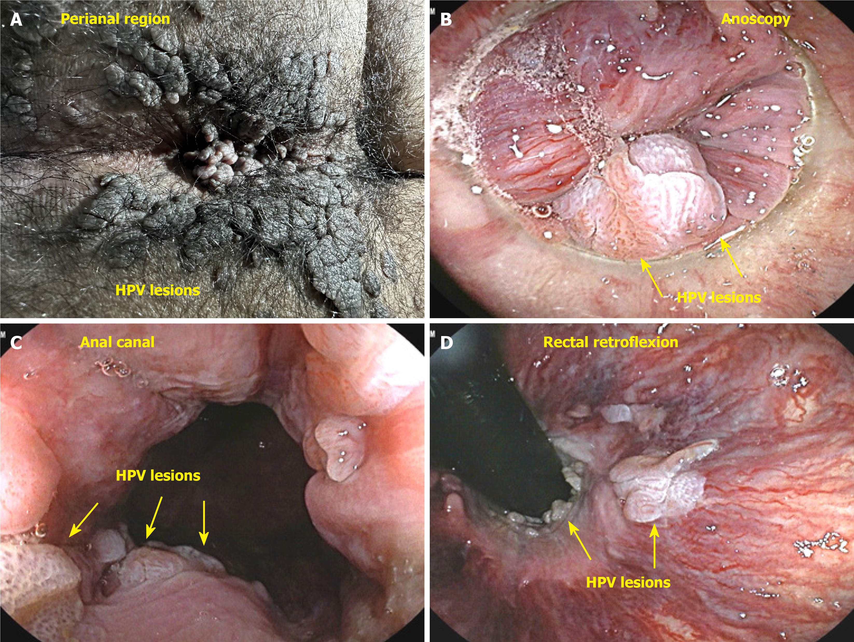
Figure 4 Giant condylomata acuminata of Bushke-Löwenstein and anal canal human papilloma virus lesions in patient human immunodeficiency virus.
A: Perianal lesions; B: Anoscopy; C: Frontal view; D: Rectal retroflexion.
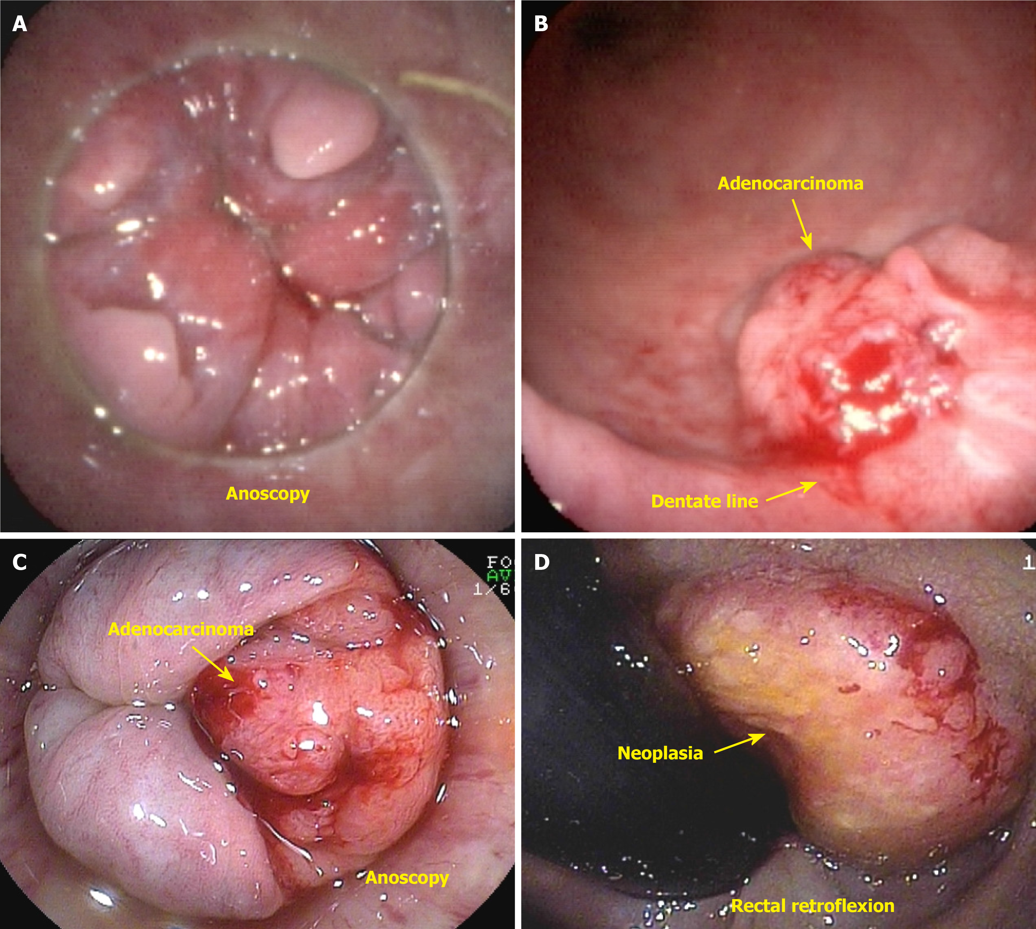
Figure 5 Adenocarcinoma arising in tubulovillous adenoma with high-grade dysplasia of the anal canal.
A: Anoscopy view showing focal adenocarcinoma within tubulovillous adenoma with high-grade dysplasia in the anal canal; B: Frontal view of the anal canal lesion with focal adenocarcinoma arising in tubulovillous adenoma; C: Anoscopy view of anal canal adenocarcinoma; D: Rectal retroflexion view demonstrating anal canal adenocarcinoma.
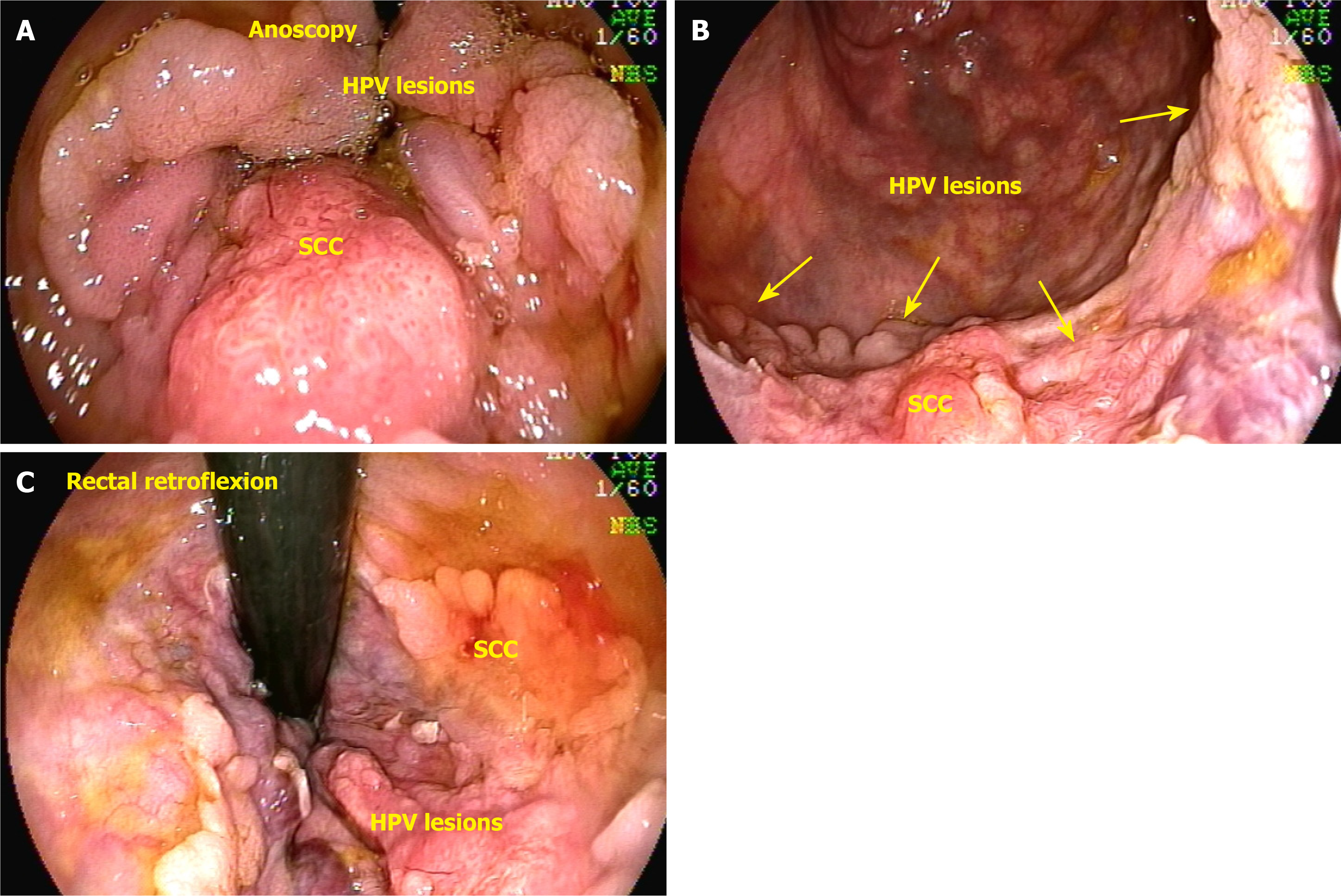
Figure 6 Anal canal lesions associated with human papilloma virus and squamous cell carcinoma.
A: Anoscopy; B: Frontal view; C: Retroflexed view. SCC: Squamous cell carcinoma; HPV: Human papilloma virus.
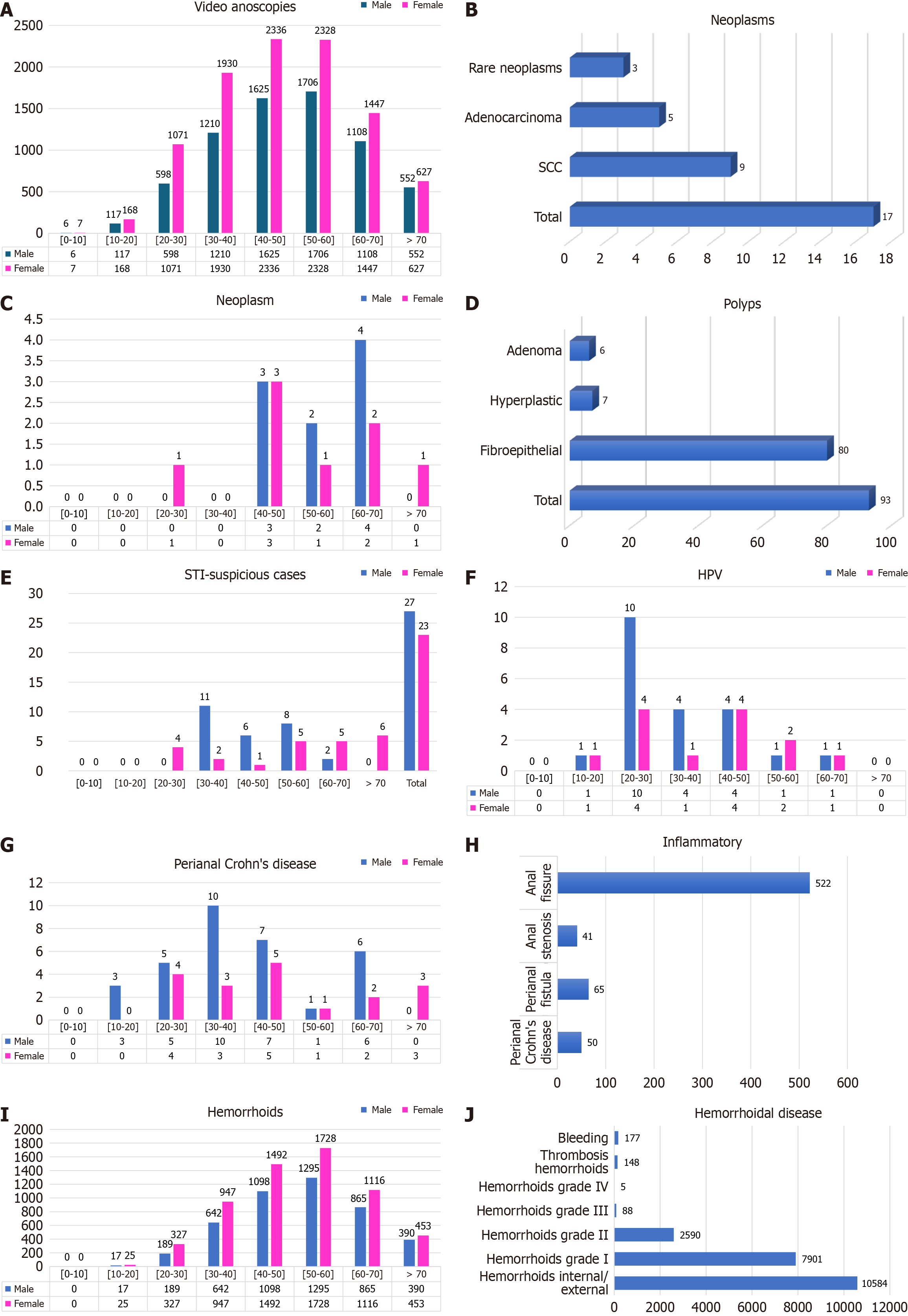
Figure 7 Bar chart.
A: Findings of the video anoscopies. The females (9914 cases, 58.89%) and male (6922 cases, 41.11%) groups were divided by sex and classified into 10-year age groups; B: Findings of 17 neoplasms. 3 cases of rare neoplasia (0.02%), 5 cases of adenocarcinoma (0.03%), and 9 cases of squamous cell carcinoma (0.05%); C: Findings of neoplasms in females and males classified by 10-year age groups; D: Findings of 93 polyps; E: Distribution of patients with suspected sexually transmitted infections according to sex and age; F: Distribution of patients with human papilloma virus lesions according to sex and age; G: Distribution of patients with perianal Crohn’s disease according to sex and age; H: Findings of inflammatory diseases. Perianal Crohn’s disease, perianal fistula, anal stenosis, and anal fissure; I: Distribution of 10585 patients with hemorrhoidal disease according to sex and age; J: Findings and number of cases of hemorrhoidal disease. SCC: Squamous cell carcinoma; STI: Sexually transmitted infection; HPV: Human papilloma virus.
Figure 8 Serrated adenoma.
A and B: Sessile polypoid lesion measuring approximately 12 mm and located close to the dentate line (anoscopy; rectal retroflexion); C-E: Lateral spreading tumors located in the anal canal (anoscopy; frontal view; rectal retroflexion).
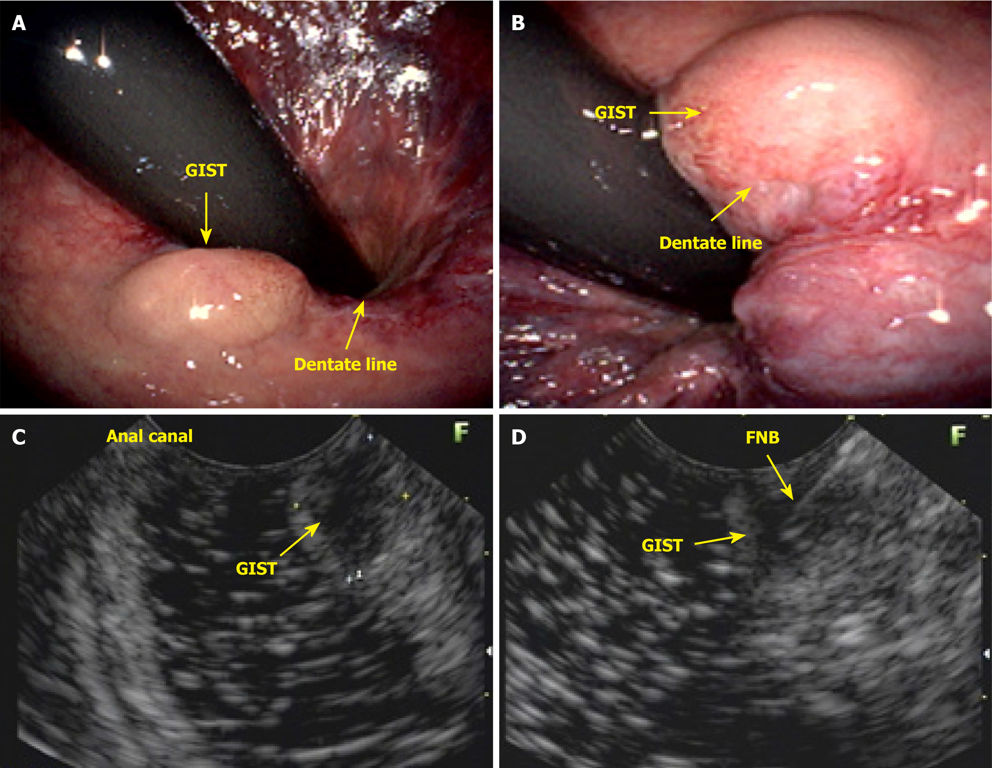
Figure 9 Gastrointestinal stromal tumor of the anal canal.
A and B: Rectal retroflexion; C: Endoscopic ultrasound image showing a 1.02 cm hypoechoic lesion in the anal canal; D: Endoscopic ultrasound and fine needle biopsy. GIST: Gastrointestinal stromal tumor; FNB: Fine-needle biopsy.
Figure 10 Endoscopic and morphological features of anorectal lesions.
A and B: Small ulcerated lesion (squamous cell carcinoma), measuring approximately 10 mm (anoscopy; rectal retroflexion); C and D: Anal canal polyp with verrucous epithelial hyperplasia with associated koilocytosis and high-grade dysplasia (anoscopy; frontal view); E: High-resolution anoscopy of high-grade intraepithelial neoplasia; F: Electrofulguration ablation; G and H: Verrucous epithelial hyperplasia with associated koilocytosis and high-grade dysplasia; 3% acetic acid was used to identify positive acetowhite lesions of the coarse speckled or mosaic type (anoscopy; rectal retroflexion). SCC: Squamous cell carcinoma; HSIL: High-grade squamous intraepithelial lesion.
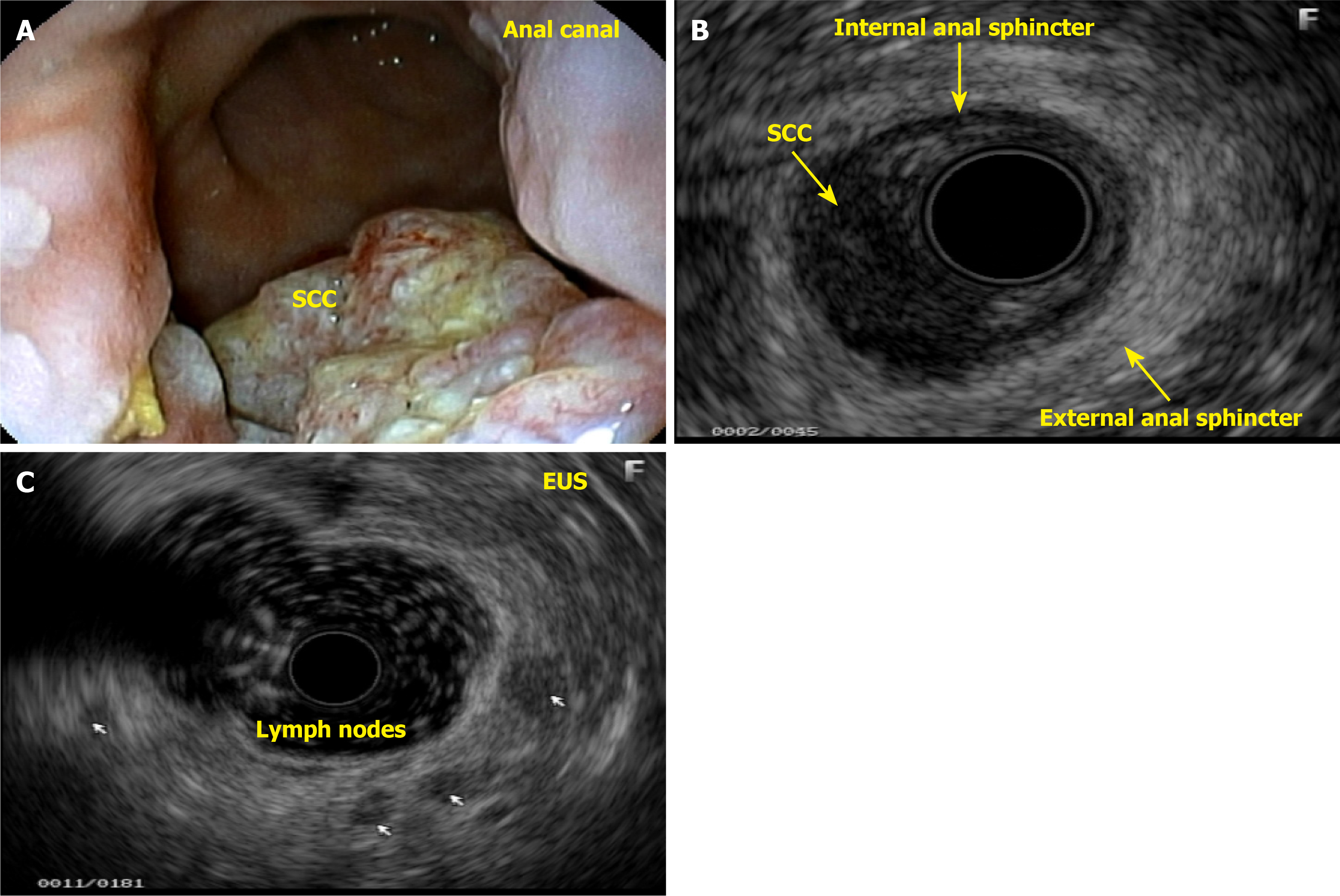
Figure 11 Squamous cell carcinoma in the anal canal.
A: Ulcerative-vegetative lesion measuring approximately 3 cm in length; B: Endoscopic ultrasound showing a hypoechoic, irregular, and heterogeneous lesion extending to the anterior wall of the lower rectum and infiltrating the internal and external anal sphincters, as well as the perirectal fat, affecting approximately 30% of the circumference of the wall, anterior face; C: Presence of 5 hypoechoic, regular, and homogeneous lymph nodes, some larger than 1 cm, bilateral, inguinal, and perirectal (-uT2N3Mx). SCC: Squamous cell carcinoma; EUS: Endoscopic ultrasound.
- Citation: Gomes A, Bara J, da Matta CHKT, Paiva PP, de Carvalho LM, Bononi BM, Pinto PCC, Rodrigues JMS, Borghesi RA. Anal neoplasm in colonoscopy: What endoscopists need to know. World J Gastrointest Endosc 2025; 17(10): 108729
- URL: https://www.wjgnet.com/1948-5190/full/v17/i10/108729.htm
- DOI: https://dx.doi.org/10.4253/wjge.v17.i10.108729


