Copyright
©2010 Baishideng Publishing Group Co.
World J Hepatol. Dec 27, 2010; 2(12): 428-433
Published online Dec 27, 2010. doi: 10.4254/wjh.v2.i12.428
Published online Dec 27, 2010. doi: 10.4254/wjh.v2.i12.428
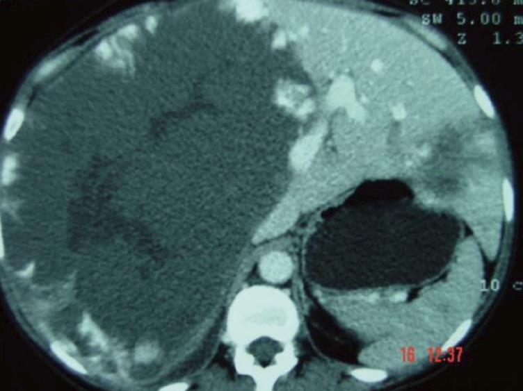
Figure 1 Computed tomography scan of a huge liver cavernous hemangioma compromising the right liver lobe.
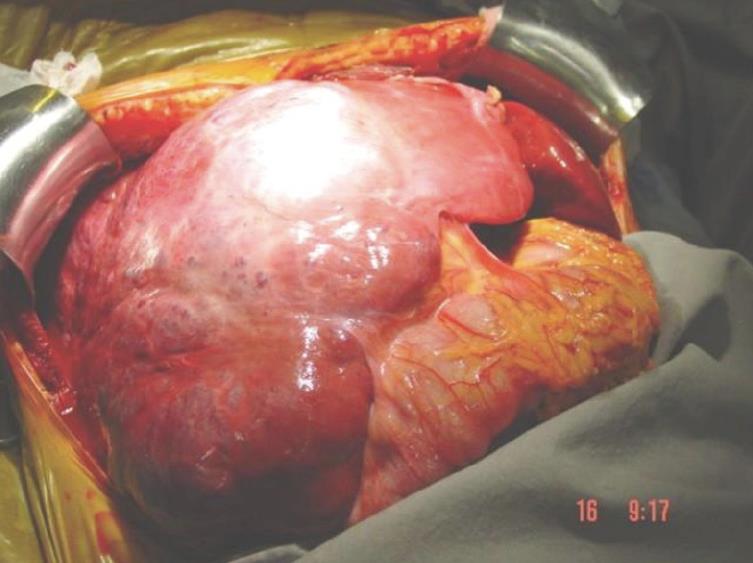
Figure 2 Intraoperative finding of a huge liver cavernous hemangioma compromising the right liver lobe.
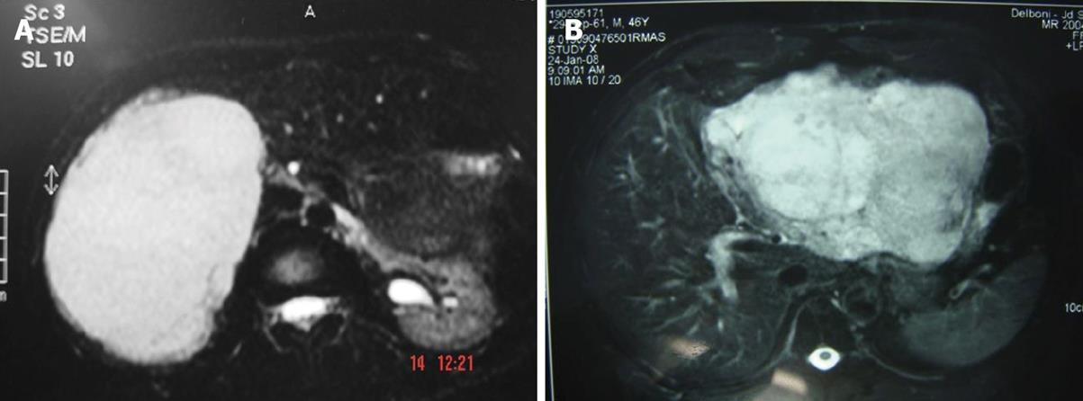
Figure 3 Magnetic resonance imaging of hemangioma.
A: Right lobe; B: Left lobe.
- Citation: Jr MAR, Papaiordanou F, Gonçalves JM, Chaib E. Spontaneous rupture of hepatic hemangiomas: A review of the literature. World J Hepatol 2010; 2(12): 428-433
- URL: https://www.wjgnet.com/1948-5182/full/v2/i12/428.htm
- DOI: https://dx.doi.org/10.4254/wjh.v2.i12.428


