
















Copyright: ©Author(s) 2026.
World J Hepatol. May 27, 2026; 18(5): 117157
Published online May 27, 2026. doi: 10.4254/wjh.v18.i5.117157
Published online May 27, 2026. doi: 10.4254/wjh.v18.i5.117157
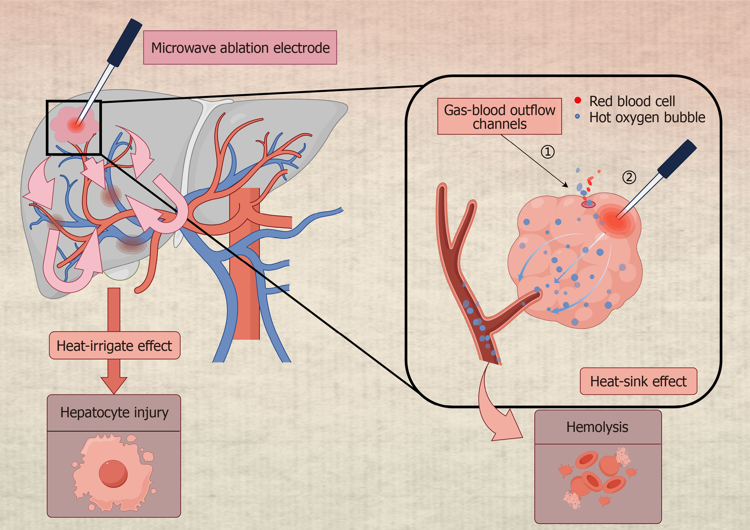
Figure 1 Schematic diagram illustrating microwave ablation of a hepatic hemangioma.
The image shows a microwave electrode inserted into the hemangioma and the generated heat distribution. It depicts heat transfer along the portal vein and heat accumulation within surrounding blood. During the ablation process, the heat from the vessel’s spreads to other liver segments, causing thermal damage to the liver and the accumulation of heat leading to intravascular hemolysis. A channel pre-drilled near the ablation point, known as the gas-blood outflow channel, is conducive to the outflow of hot oxygen bubbles and blood. The image created by FigDraw (Supplementary material).
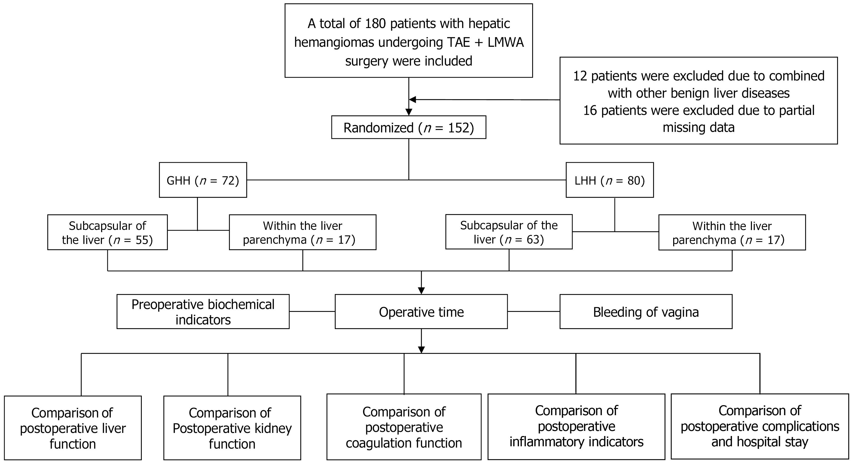
Figure 2 A total of 180 patients who underwent trans-arterial embolization + laparoscopic microwave ablation surgery for hepatic heman
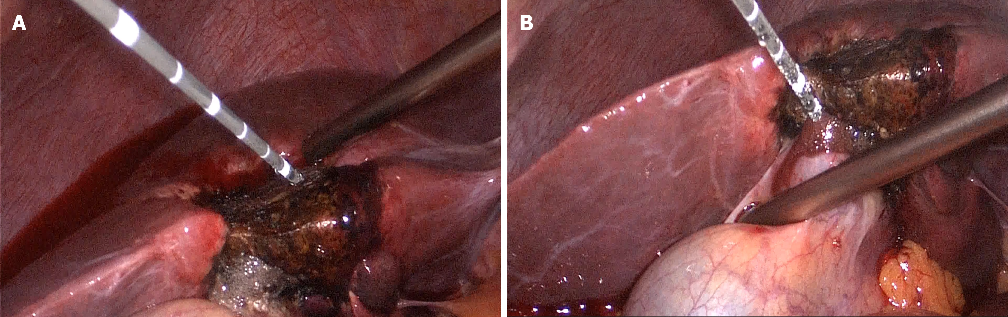
Figure 3 Establishment of the gas-blood outflow channel.
A: Prior to the initiation of each microwave ablation cycle, a gas-blood outflow channel is created using a standard microwave ablation needle. The specific depth of this channel is determined based on the maximum diameter of the hemangioma as measured from preoperative imaging, rather than a fixed length, to ensure tailored channel placement according to individual tumor characteristics (the depth is approximately half of the maximum diameter of the tumor); B: The blood flowing out of the channel, and the thermal oxygen bubbles caused by the heat conduction effect are observed.
- Citation: Wu CL, Chen XC, Zhao W, Hu B, Huan HB, Tu YL, Zhu ZM, Jiang K. Laparoscopic microwave ablation for hepatic hemangiomas with different locations: A comparative study. World J Hepatol 2026; 18(5): 117157
- URL: https://www.wjgnet.com/1948-5182/full/v18/i5/117157.htm
- DOI: https://dx.doi.org/10.4254/wjh.v18.i5.117157