
















Copyright: ©Author(s) 2026.
World J Hepatol. May 27, 2026; 18(5): 117141
Published online May 27, 2026. doi: 10.4254/wjh.v18.i5.117141
Published online May 27, 2026. doi: 10.4254/wjh.v18.i5.117141
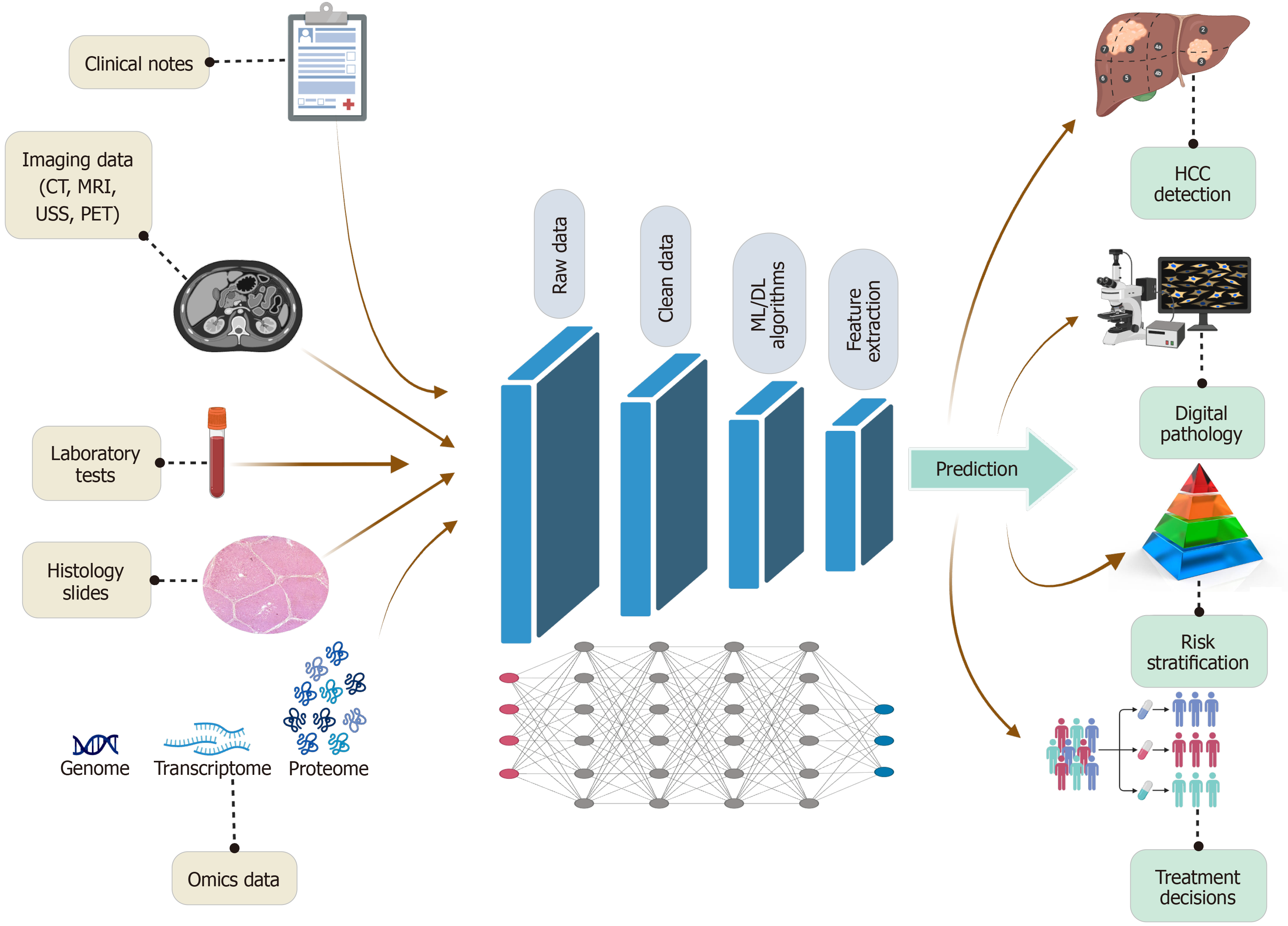
Figure 1 End-to-end pipeline of data processing and prediction in hepatology artificial intelligence models.
This figure illustrates the end-to-end pipeline by which artificial intelligence processes multimodal clinical data in hepatology. Diverse inputs, including clinical notes, laboratory tests, imaging (computed tomography, magnetic resonance imaging, ultrasound, positron emission tomography), histopathology, and omics, are aggregated and cleaned. Cleaned datasets are then analyzed using machine learning and deep learning algorithms to enable automated feature extraction. Feature extraction encompasses both handcrafted variables (e.g., liver stiffness, liver imaging reporting and data system category, radiomic texture metrics, fibrosis stage) and automatically learned features from deep networks. These computationally derived features feed predictive models that support downstream clinical applications such as hepatocellular carcinoma detection, digital pathology interpretation, risk stratification, grading of portal hypertension, prediction of variceal bleeding or ascites infection, and post-transplant graft or patient survival. The workflow highlights how artificial intelligence integrates complex datasets into actionable outputs that enhance precision hepatology. CT: Computed tomography; MRI: Magnetic resonance imaging; PET: Positron emission tomography; USS: Ultrasound; ML: Machine learning; DL: Deep learning; HCC: Hepatocellular carcinoma.
- Citation: Elsayed MO, Elshabrawi AY. Artificial intelligence and machine learning in hepatology: Revolutionizing diagnosis and treatment. World J Hepatol 2026; 18(5): 117141
- URL: https://www.wjgnet.com/1948-5182/full/v18/i5/117141.htm
- DOI: https://dx.doi.org/10.4254/wjh.v18.i5.117141