
















Copyright: ©Author(s) 2026.
World J Hepatol. May 27, 2026; 18(5): 116712
Published online May 27, 2026. doi: 10.4254/wjh.v18.i5.116712
Published online May 27, 2026. doi: 10.4254/wjh.v18.i5.116712
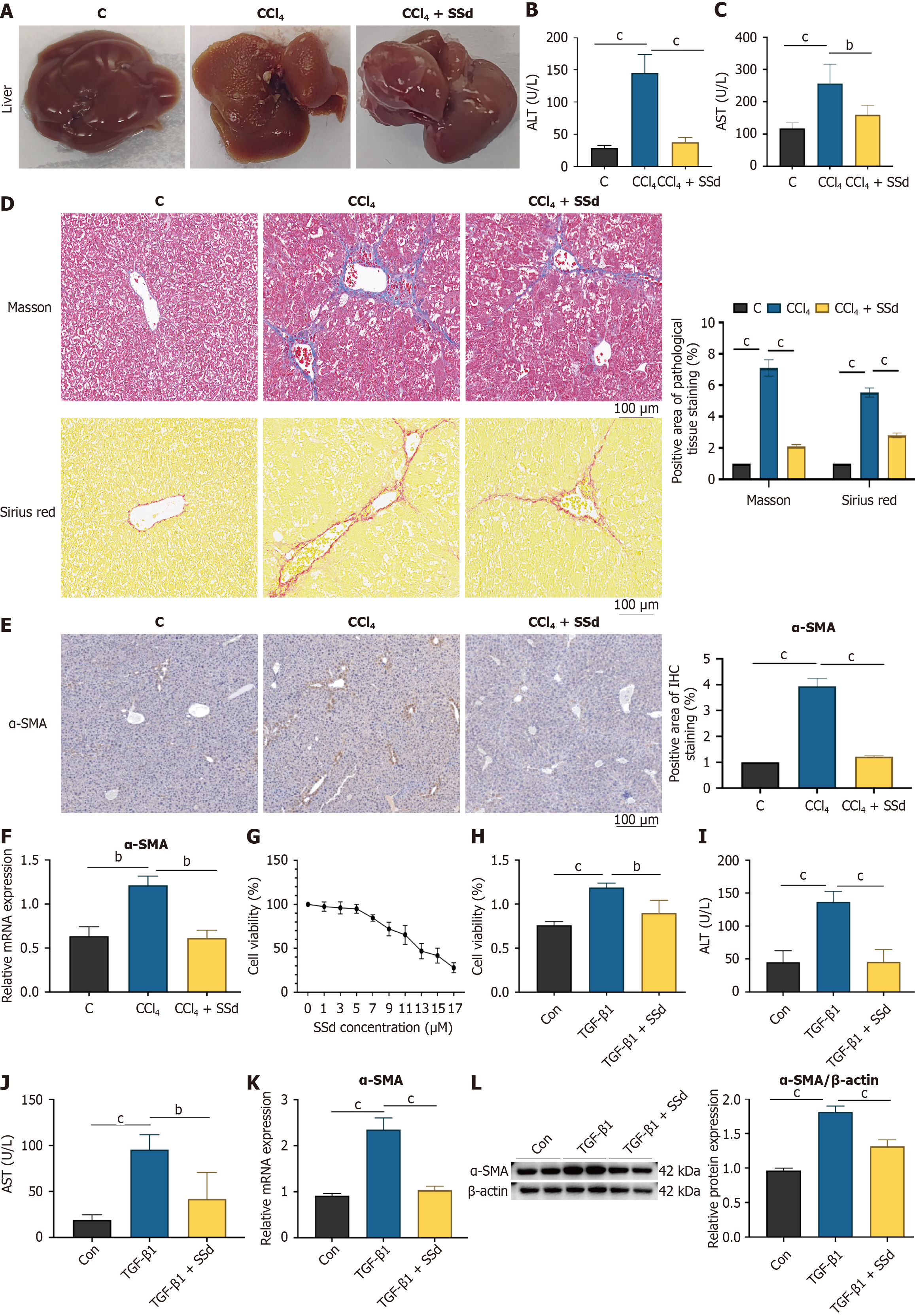
Figure 1 Saikosaponin-d attenuates the progression of hepatic fibrosis by inhibiting hepatic stellate cells activation.
A: Fresh liver images of mice in the control (C), carbon tetrachloride (CCl4), and CCl4 + saikosaponin-d (SSd) groups; B: Serum alanine aminotransferase levels in mice from the C, CCl4, and CCl4 + SSd groups, n = 6; C: Serum aspartate aminotransferase levels in mice from the C, CCl4, and CCl4 + SSd groups, n = 6; D: Masson’s trichrome and Sirius red staining of liver tissue from mice in the C, CCl4, and CCl4 + SSd groups, magnification × 200; E: Immunohistochemical staining for α-smooth muscle actin (α-SMA) in liver tissue from mice in the C, CCl4, and CCl4 + SSd groups, magnification × 100; F: MRNA expression levels of α-SMA in liver tissue from mice in the C, CCl4, and CCl4 + SSd groups, n = 6; G: The changes in cell viability of LX-2 cells after 24 hours of intervention with different concentrations of SSd, n = 6; H: Cell viability in the control (Con), transforming growth factor-β1 (TGF-β1), and TGF-β1 + SSd groups, n = 4; I: Aspartate aminotransferase levels in LX-2 cells from the Con, TGF-β1, and TGF-β1 + SSd groups, n = 4; J: Alanine aminotransferase levels in LX-2 cells from the Con, TGF-β1, and TGF-β1 + SSd groups, n = 4; K: MRNA expression levels of α-SMA in LX-2 cells from the Con, TGF-β1, and TGF-β1 + SSd groups, n = 4; L: Protein expression levels of α-SMA in LX-2 cells from the Con, TGF-β1, and TGF-β1 + SSd groups, n = 4. C: Control; CCl4: Carbon tetrachloride; SSd: Saikosaponin-d; ALT: Alanine aminotransferase; AST: Aspartate aminotransferase; α-SMA: Α-smooth muscle actin; TGF-β1: Transforming growth factor-β1.
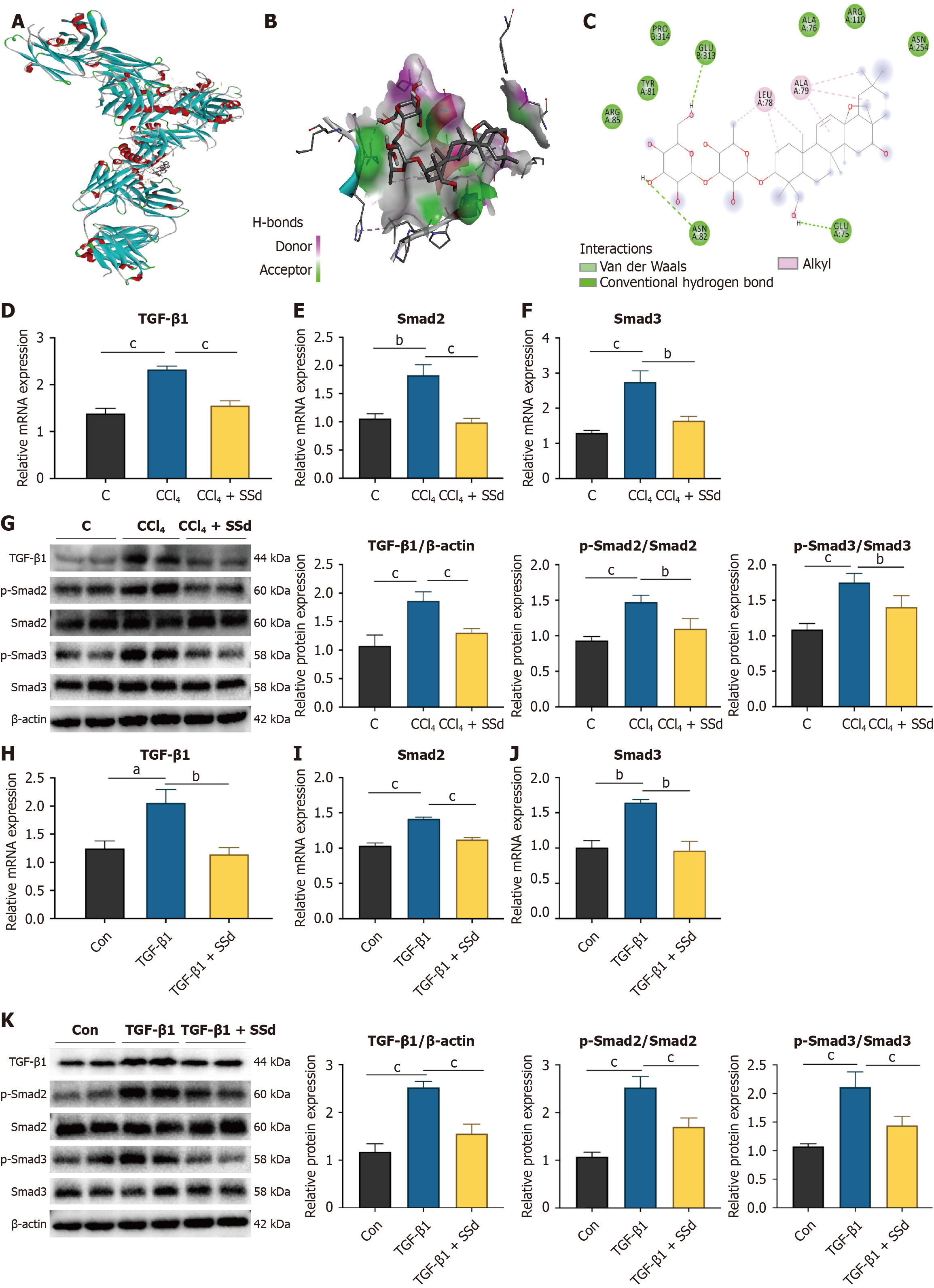
Figure 2 Saikosaponin-d attenuates hepatic fibrosis progression by inhibiting the transforming growth factor-β1/Smad signaling pathway.
A and B: The overall three-dimensional structure of the interaction between silver sulfadiazine molecules and transforming growth factor-β1 (TGF-β1) protein; C: The overall 2D structure of the interaction between silver sulfadiazine molecules and TGF-β1 protein; D: MRNA expression levels of TGF-β1 in mice from the control (C), carbon tetrachloride (CCl4), and CCl4 + saikosaponin-d (SSd) groups, n = 6; E: MRNA expression levels of Smad2 in mice from the C, CCl4, and CCl4 + SSd groups, n = 6; F: MRNA expression levels of Smad3 in mice from the C, CCl4, and CCl4 + SSd groups, n = 6; G: Protein expression levels of TGF-β1, p-Smad2, and p-Smad3 in mice from the C, CCl4, and CCl4 + SSd groups, n = 4; H: MRNA expression levels of TGF-β1 in LX-2 cells from the control (Con), TGF-β1, and TGF-β1 + SSd groups, n = 4; I: MRNA expression levels of Smad2 in LX-2 cells from the Con, TGF-β1, and TGF-β1 + SSd groups, n = 4; J: MRNA expression levels of Smad3 in LX-2 cells from the Con, TGF-β1, and TGF-β1 + SSd groups, n = 4; K: Protein expression levels of TGF-β1, p-Smad2, and p-Smad3 in LX-2 cells from the Con, TGF-β1, and TGF-β1 + SSd groups, n = 4. C: Control; CCl4: Carbon tetrachloride; SSd: Saikosaponin-d; TGF-β1: Transforming growth factor-β1.
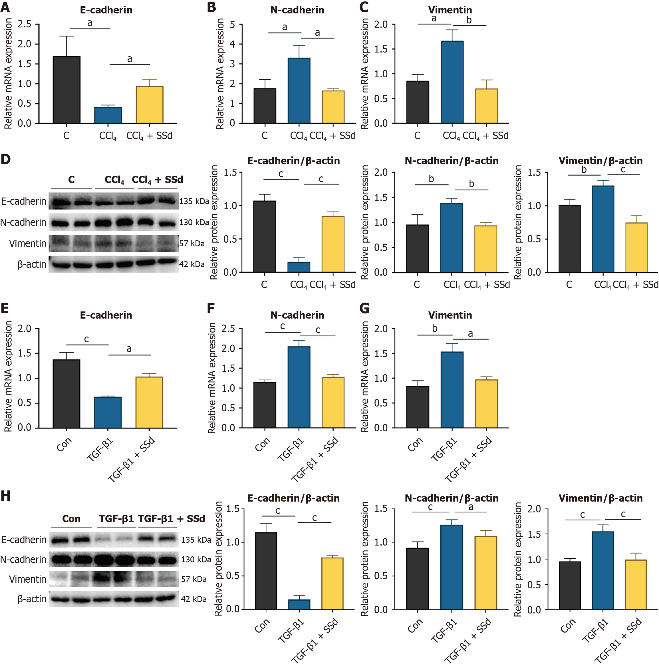
Figure 3 Saikosaponin-d inhibits epithelial-mesenchymal transition progression by suppressing the transforming growth factor-β1/Smad signaling pathway.
A: MRNA expression levels of E-cadherin in mice from the control (C), carbon tetrachloride (CCl4), and CCl4 + saikosaponin-d (SSd) groups, n = 6; B: MRNA expression levels of N-cadherin in mice from the C, CCl4, and CCl4 + SSd groups, n = 6; C: MRNA expression levels of vimentin in mice from the C, CCl4, and CCl4 + SSd groups, n = 6; D: Protein expression levels of E-cadherin, N-cadherin, and vimentin in mice from the C, CCl4, and CCl4 + SSd groups, n = 4; E: MRNA expression levels of E-cadherin in LX-2 cells from the control (Con), transforming growth factor-β1 (TGF-β1), and TGF-β1 + SSd groups, n = 4; F: MRNA expression levels of N-cadherin in LX-2 cells from the Con, TGF-β1, and TGF-β1 + SSd groups, n = 4; G: MRNA expression levels of vimentin in LX-2 cells from the Con, TGF-β1, and TGF-β1 + SSd groups, n = 4; H: Protein expression levels of E-cadherin, N-cadherin, and vimentin in LX-2 cells from the Con, TGF-β1, and TGF-β1 + SSd groups, n = 4. C: Control; CCl4: Carbon tetrachloride; SSd: Saikosaponin-d; TGF-β1: Transforming growth factor-β1.
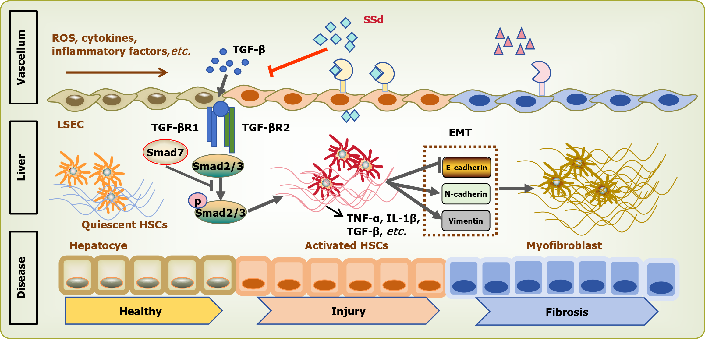
Figure 4 The mechanism diagram of saikosaponin-d suppressing epithelial-mesenchymal transition progression and alleviating liver fibrosis by inhibiting the transforming growth factor-β1/Smad signaling pathway.
Upon hepatic injury or stimulation by endotoxins and inflammatory cytokines, hepatic stellate cells (HSCs), Kupffer cells, hepatocytes, and liver sinusoidal endothelial cells secrete substantial amounts of transforming growth factor-β1 (TGF-β), contributing to hepatic immune regulation and inflammation. HSCs are key cellular players in the fibrogenic process. In response to liver injury, quiescent HSCs undergo activation and transition into activated HSCs, which secrete excessive extracellular matrix, driving fibrosis progression. Activated HSCs also secrete TGF-β, a major mediator of liver fibrosis. TGF-β signals through a heteromeric complex of two serine/threonine kinase receptors, type I and type II, which phosphorylate Smad2/Smad3 proteins. This leads to the activation of fibrogenic mediators, including collagen, α-smooth muscle actin, and fibronectin. Additionally, TGF-β1 is a potent inducer of the epithelial-mesenchymal transition, a process where epithelial cells lose their characteristic phenotype and acquire mesenchymal features, contributing to fibrosis. Saikosaponin-d attenuates HSC activation and mitigates liver fibrosis progression, partly by inhibiting the TGF-β1/Smad signaling pathway and the downstream epithelial-mesenchymal transition progression. ROS: Reactive oxygen species; TGF-β1: Transforming growth factor-beta 1; SSd: Saikosaponin-d; LSEC: Liver sinusoidal endothelial cell; TGF-βR: Transforming growth factor-beta receptor; EMT: Epithelial-mesenchymal transition; TNF-α: Tumor necrosis factor-alpha; IL-1β: Interleukin-1 beta; HSC: Hepatic stellate cell.
- Citation: Li J, Cao MX, Wang JM, Zheng YY, Que RY, Lin LB. Saikosaponin-d alleviates hepatic stellate cell activation and liver fibrosis by inhibiting the TGF-β1/Smads signaling pathway and blocking the EMT process. World J Hepatol 2026; 18(5): 116712
- URL: https://www.wjgnet.com/1948-5182/full/v18/i5/116712.htm
- DOI: https://dx.doi.org/10.4254/wjh.v18.i5.116712