Copyright
©The Author(s) 2020.
World J Hepatol. Dec 27, 2020; 12(12): 1349-1357
Published online Dec 27, 2020. doi: 10.4254/wjh.v12.i12.1349
Published online Dec 27, 2020. doi: 10.4254/wjh.v12.i12.1349
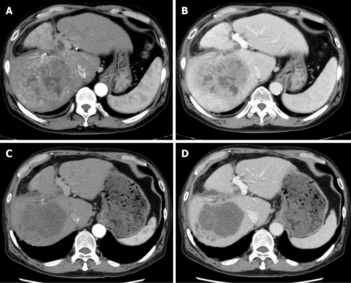
Figure 1 Preoperative computed tomography findings.
Before lenvatinib administration, tumor stain was observed in the arterial phase and washout was observed in the portal phase. After lenvatinib administration, the tumor stain disappeared in the arterial phase, and the tumor size was found to have decreased slightly in the portal phase. A and B: Images of the arterial phase (A) and portal phase (B) before administration of lenvatinib; C and D: Images of the arterial phase (C) and portal phase (D) after lenvatinib administration.
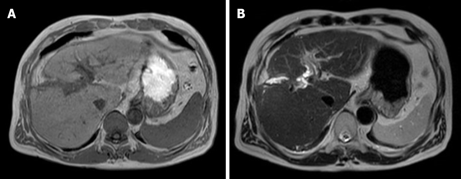
Figure 2 Magnetic resonance imaging findings at 5 mo before the second operation.
There was no evidence of the recurrent tumor on the magnetic resonance imagines. A: T1 image; B: T2 image.
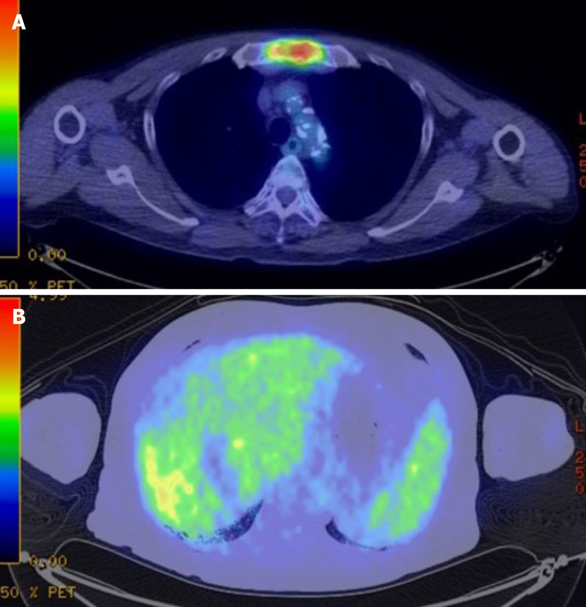
Figure 3 Preoperative fluorodeoxyglucose-positron emission tomography-computed tomography.
A: The positron emission tomography-computed tomography scan revealed accumulation of fluorodeoxyglucose in the sternum. The diagnosis was bone metastases of hepatocellular carcinoma; B: Slight uptake was observed in the liver.
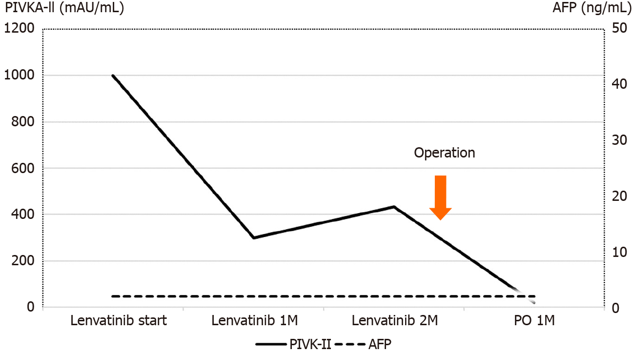
Figure 4 Perioperative tumor marker changes.
The protein-induced vitamin K absence or antagonist II level before lenvatinib administration was high at 998 mAU/mL. The protein-induced vitamin K absence or antagonist II level decreased to 434 mAU/mL at 2 mo after lenvatinib administration, and further decreased to 20 mAU/mL at 1 mo after the second surgery. AFP: α-Fetoprotein; M: Month; PO: Post-operation; PIVKA-II: Protein-induced vitamin K absence or antagonist II.
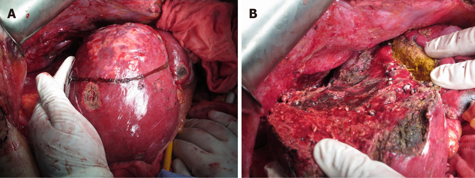
Figure 5 Surgical findings.
Findings before (A) and after (B) the second hepatectomy.
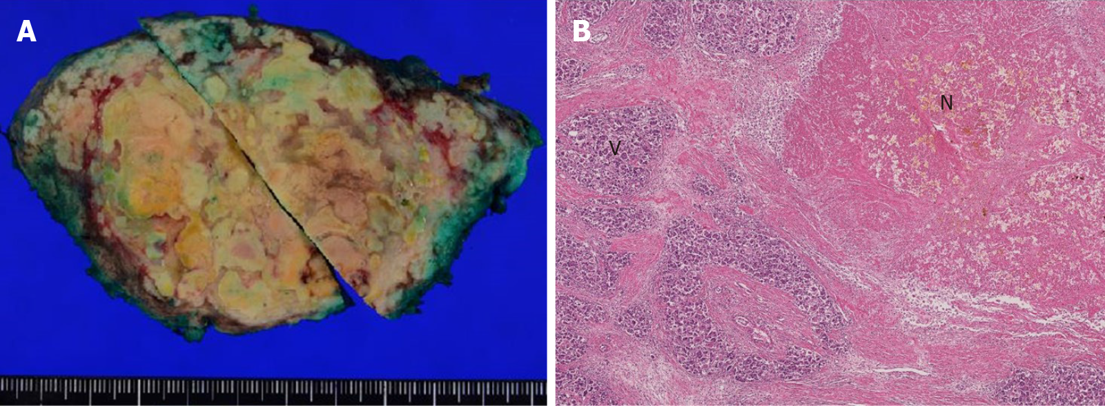
Figure 6 Macroscopic and pathological findings of the resected specimen.
The findings revealed that 80% of the tumor was necrotic and the resected margin of the cut liver surface showed no cancer component. A: Macroscopic findings; B: Pathological findings. N: Necrotic lesion; V: Viable lesion.
- Citation: Yokoo H, Takahashi H, Hagiwara M, Iwata H, Imai K, Saito Y, Matsuno N, Furukawa H. Successful hepatic resection for recurrent hepatocellular carcinoma after lenvatinib treatment: A case report. World J Hepatol 2020; 12(12): 1349-1357
- URL: https://www.wjgnet.com/1948-5182/full/v12/i12/1349.htm
- DOI: https://dx.doi.org/10.4254/wjh.v12.i12.1349


