©The Author(s) 2018.
World J Hepatol. Jul 27, 2018; 10(7): 517-522
Published online Jul 27, 2018. doi: 10.4254/wjh.v10.i7.517
Published online Jul 27, 2018. doi: 10.4254/wjh.v10.i7.517
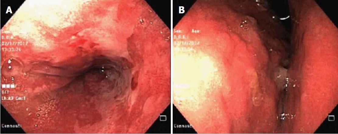
Figure 1 Upper digestive endoscopy showing intense erosive distal esophagitis and extrinsic compression of the stomach.
A: Intense erosive distal esophagitis; B: Extrinsic compression of the stomach.
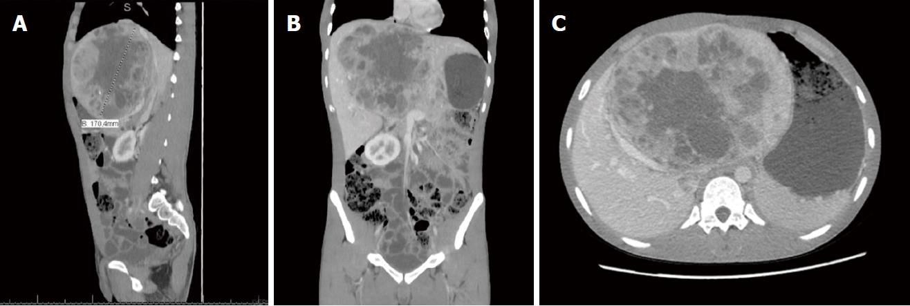
Figure 2 The computed tomography of hepatic lesion.
A and B: Computed tomography showing a voluminous heterogeneous hepatic lesion, with a neoplasic aspect and dimensions 19.5 cm × 12.5 cm × 17.0 cm; C: Compression of the right hepatic vein.
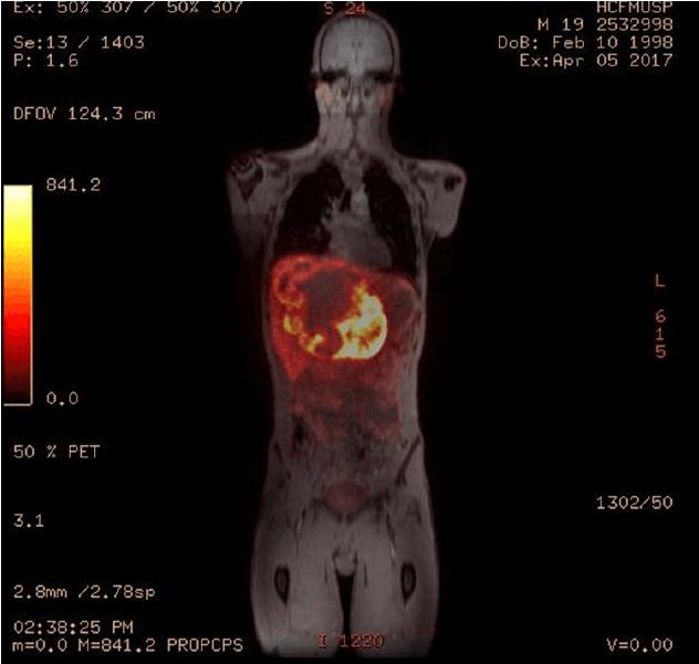
Figure 3 Positron emission tomography associated with magnetic resonance with somatostatin analogue confirmed that the liver was the primary site of the tumor.
Figure 4 Intraoperative appearances.
A: After hepatic posterior sector mass separation; B: Aspect of the remaining liver after surgical removal; C: Surgical piece.
- Citation: Pipek LZ, Jardim YJ, Mesquita GHA, Nii F, Medeiros KAA, Carvalho BJ, Martines DR, Iuamoto LR, Waisberg DR, D’Albuquerque LAC, Meyer A, Andraus W. Large primary hepatic gastrinoma in young patient treated with trisegmentectomy: A case report and review of the literature. World J Hepatol 2018; 10(7): 517-522
- URL: https://www.wjgnet.com/1948-5182/full/v10/i7/517.htm
- DOI: https://dx.doi.org/10.4254/wjh.v10.i7.517


