INTRODUCTION
The number of people with cancer is expected to increase from 12.7 million in 2008 (latest available figure) to 20.3 million in 2030[1]. Sixty percent of these patients receive radiation therapy with a chance of recovery of fifty percent. The number of radiotherapy centers in 2011 is about 7500, linear accelerators in clinical use are approximately 10000 (IAEA 2011). Up to 500000 patients per year undergo abdominal or pelvic radiotherapy worldwide. Eight out of ten will develop acute gastrointestinal symptoms and 5% to 10% will develop severe intestinal complications within 10 years after treatment. The efficacy of abdominal or pelvic radiotherapy requires an optimal compromise between normal tissue damage and tumor control that is the risk/benefit ratio. The lack of curative treatment at present and the potential severity for the disorder highlight the importance of novel and effective therapeutic strategies for gastrointestinal complications after radiation exposure[2]. Proton therapy is an attractive method to attenuate toxicity of radiotherapy because of the decrease of integral radiation dose to normal tissues, which should lead to fewer late side effects. Unlike other types of radiation therapy that use X-rays to destroy cancer cells, proton therapy uses a beam of special particles called protons, inducing less damage to the surrounding healthy tissue. There will be a lower risk of normal tissue toxicity associated with proton therapy because of a lower delivered dose outside of the target tissue[3].
POST-RADIATION DAMAGES
Radiolesions’ etiology focused on epithelial ulceration, microvascular destruction, mucosal and submucosal inflammation for the acute radiation enteropathy. The severity of acute radiation enteritis may be predictive for more severe chronic gastrointestinal symptoms. Acute or chronic side effects can also be aggravated after a radiotherapy accident such as an overdose. The risk factors for complications are age, irradiated volume, histories of abdominal surgery, androgenic hardship, diabetes, hemorrhoids and inflammatory intestinal diseases[4].
Treatments to manage post-radiotherapy pelvic damages
Treatments usually applied are only symptomatic. The systematic study of therapy complications shows: (1) coagulation with the plasma argon is insufficient in the long-term; (2) a moderated effect of the short chains fatty acids and of hyperbaric oxygen therapy; and (3) an insufficiency of proof for the efficiency of formaline, 5-amino-salicylic acid, sulfanazine, vitamin A and pentoxifylline. Corticoids remain scarcely effective, because the cause is more ischemic than inflammatory. Hence, more effective approaches are of primary clinical importance[5].
Targeted organ of pelvic radiation and chronic effects
The use of radiation therapy for the treatment of pelvic malignancy, including that of the prostate, cervix, uterus and ovaries, has increased in recent years. The frequency of complications is of 12% for uterus cancers and 7% for prostate cancers. The symptoms are proctopathies and radic cystitis. Intra-abdominal organs located close to cancerous tumors can be affected during radiotherapy. The most radiosensitive organ of the intra-abdominal area is the small intestine. Acute radiation responses affect patient quality of life, causing abdominal pain, diarrhea/constipation sequences, and malabsorption that may interrupt or delay the radiotherapy protocol. Radiotherapy is associated with a high incidence of undesirable acute and/or chronic gastrointestinal complications that can affect the patient’s quality of life and may even be life threatening. Exposure of the small intestinal tissue to ionizing radiation may lead to loss of its integrity by a dose-dependent stem cell depletion initiating gastrointestinal disorders. Radiation proctitis or proctopathy occur frequently and can be debilitating side effects of radiation therapy. There are 2 forms of radiation proctopathy, acute and chronic. The acute form occurs in nearly all patients. The incidence of the chronic form ranges from 2% to 20%. However, the true incidence may be underestimated. Chronic radiation proctopathy includes symptoms that occur as a continuation of early symptoms 3 mo after the completion of radiation therapy. The median onset is 8 to 12 mo, but the onset can occur as late as 30 years post therapy completion. Common symptoms include diarrhea, tenesmus, mucus/blood loss via the rectum, urgency, incontinence and pain. The most common symptom is rectal bleeding. Endoscopic finding include mucosal pallor, friability, spontaneous oozing, angiectasia and infrequently ulceration. These combinations of symptoms significantly affect patients’ quality of life[5].
Acute radiation tissue injury to the bladder is caused primarily by damage to the bladder mucosa, which contains cells that divide rapidly. This usually occurs during the treatment period. The underlying pathology of late adverse effects is different from that seen in acute reaction. Late responding tissues such as vascular and connective tissues, have a slow turnover rate, therefore, while they sustain radiation damages at the time of treatment, the effect are not expressed until repeated cell divisions are attempted. For this reason, late radiation tissue injury can take several months to many years to develop, and is largely function of the total radiation dose and fraction size. The pathological hallmark of late radiation tissue injury is obliterative endarteritis resulting in atrophy and fibrosis. Late radiation cystitis following radiation therapy for cancer in the pelvic region has an incidence of 5% to 10%. Late radiation cystitis can develop from 6 mo to as long as 20 years after radiation treatment, with a mean latent period of 35 mo. One chronic manifestation is recurrent haematuria or haemorrhagic cystitis, defined as diffuse vesical bleeding. Haemorrhagic cystitis present a significant clinical problem once established. A minority of patients will develop severe bleeding that may become life threatening; in such patients conservative treatment strategies are often inadequate and radical surgery may be the only curative option[6-8].
STEM CELL THERAPY
Multipotent (mesenchymal) stromal (stem) cell (MSC) therapy is currently among the most advanced cell therapy tools, with the availability of three Food and Drug Administration-approved products: Prochymal, provacel, and chondrogen. Stem cell-based approaches using MSCs are promising for the development of future therapy in therapeutics[9] to correct radiodermatitis[10], to improve haematopoiesis[11,12], and to prevent graft vs host disease (GVHD) post-haematopoietic stem cell transplantation[13]. Clinical studies have reported effects of MSCs on gastrointestinal healing such as the reversion of colon peritonitis in patients with GVHD or the treatment of rectovaginal and perianal fistulas in patients with Crohn’s disease[14].
Stem cell therapy of radiation damages
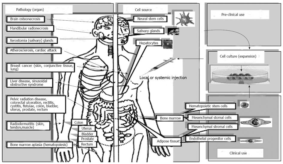
Stem cells can be used to treat toxic side effects induced by radiotherapy on healthy tissue of radio-sensitive patients. Figure 1 illustrates where stem cells could be used and where MSCs are already used. The organs impacted by irradiation side effects that are mostly studied are: brain salivary glands, mandibless skin, liver, heart, rectum/bladder and bone marrow. Stem cells isolated from bone marrow; adipose tissue are easily expanded in vitro. Expanded or un-expanded stem cells are injected locally or intravenously. Stem cells from bone marrow and adipose tissues are already in clinical use. Other sources of stem cells (neural stem cell, salivary gland stem cell, hepatocyte and embryonic stem cell) are only used in animal models (pre-clinical use).
Figure 1 Clinical trials to treat consequences of radiotherapy.
Clinical and pre-clinical use of stem cells to treat toxic side effects induced by radiotherapy on healthy tissue of radio-sensitive patients. Pathologies treated are (left side) osteonecrosis, xerostomia, atherosclerosis, cardiac attack, breast cancer, liver disease, pelvic radiation disease, radiodermatitis and bone marrow aplasia. Targeted organs (brain, salivary glands, mandibles, skin, liver, heart and rectum/bladder, bone marrow) are mentioned on the left side. On the right side, stem cells are isolated from several tissue sources, expanded in vitro, then injected locally (i loc) or intravenously (iv). Stem cells from bone marrow and adipose tissues are used in clinic. Other sources of stem cells (neural stem cell, salivary gland stem cell, hepatocyte and embryonic stem cell) are only used in animal models (pre-clinical use).
Preclinical treatment of pelvic radio-induced damages
As demonstrated in a preclinical model, MSCs may offer a novel strategy to treat pelvic radiation disease. After abdominal irradiation, MSCs have the capacity to engraft into the enteric mucosa[14-18]. MSCs are able to repair radiation-induced intestinal damage by inhibiting ulceration[19-21]. Mitigation of radiation-induced lethal intestinal injury can similarly be achieved by transplantation of bone marrow-derived adipose stromal cells (BMASC). BMASC increase blood levels of intestinal growth factors and induce regeneration of intestinal villi, thereby, accelerating functional recovery of the intestine[22]. Furthermore MSC injection improved muscle regeneration and increased contractile function of anal sphincters after injury[23]. The effects of MSCs are a consequence of their ability to improve the renewal capability of the small intestine epithelium. MSC treatment favors the re-establishment of cellular homeostasis by both increasing endogenous proliferation processes and inhibiting radiation-induce apoptosis of the small intestine epithelial cells[24]. MSCs release cytokines and growth factors such as, interleukin (IL)-11, human hepatocyte growth factor, fibroblast growth factor-2 and insulin-like growth factors. Each of these factor may be involved; they have been described earlier as facilitating intestinal mucosa repair, either through enhancement of cell proliferation or inhibition of epithelial cell apoptosis. Repeated infusion of MSC-derived bioactive components after abdominal irradiation increased the survival rate, decreased diarrhea occurrence, and improved the small intestine structural integrity of irradiated mice. By reducing the levels of pro-inflammatory cytokines, while inducing anti-inflammatory cytokines, MSCs may also dampen the systemic inflammatory response syndrome in radiation-induced gastrointestinal syndrome[25]. François et al[24] evidenced the potential of MSC to limit radiation effects on the small intestine in an IL-6 dependent manner. MSC actions involve cellular homeostasis stabilization[24]. The rescue of epithelial cell integrity by MSC after total body irradiation or abdominal radiotherapy might favor renewal of healthy intestinal tissue. Furthermore MSC treatment of a target organ may have an effect on distant tissues. MSC regenerated the small intestine epithelium, which in turn restored the enterohepatic recirculation pathway initially damaged by irradiation. Another mechanism that should be considered is the role of cytokines and growth factors produced by MSCs that are homing to other organs, such as the observed distant hepatic protection without engraftment of MSC in the liver[26]. This might reduce acute and/or chronic side effects arising from ionizing radiation and may be of therapeutic interest[24,27].
Clinical treatment
In 2008, three Epinal patients presenting serious intestinal radiation induced lesions, after over dosage of radiotherapy, compassionately received MSC treatment. For all three patients, the systematic administration of MSCs was well tolerated; efficient analgesic and anti-inflammatory effects as well as hemorrhage reduction were observed. A fourth patient was successfully treated in 2012[14]. Compassionate trial demonstrated the feasibility of cell therapy treatment for patients overdosed during radiation therapy of prostate cancer as in Epinal Medical Center. A new protocol will be under taken in 2013 for the treatment of late severe damages of abdominal radiotherapy.
Untoward effects
There is interest in using these cells in critical illness, however, the safety profile of these cells is not well known. The resistance to transformation of MSCs produced in 4 cell therapy facilities was investigated during clinical trials. This study demonstrated that MSCs with or without chromosomal alterations showed progressive growth arrest and entered senescence without evidence of transformation either in vitro or in vivo[28]. Authors conclude that genomic stability of cultured adult stem cells is robust and not a significant source of concern[29]. Another related concern is the capacity of MSCs to potentially contribute to tumor growth and metastasis especially for cell therapy after cancer radiotherapy treatment. The question currently remains; do somatic adult stem cells support or suppress tumor growth? A variety of tumor models in which MSCs are added exogenously have been used[30]. Many studies have reported that MSCs either promoted or suppressed tumor growth. Mechanisms implied chemokine signaling, modulation of apoptosis, vascular support, and immune modulation and interaction with cells in the tumor microenvironment.
While it is true that MSC therapy has shown utility in the reversal of tissue injury in nearly every model examined, there is more to consider with radiation damage that is not as important with other types of tissue injury. Radiations induce DNA damage and long-term inhibition of growth of exposed cells. This period of quiescence is mediated by P53 and other pathways. The biological effect of P53 activation is to stop cell growth long enough for DNA repair enzymes to attempt to repair the DNA damage. MSC therapy does nothing to improve DNA repair, so that MSC therapy will allow cells to continue to grow and repair the tissue over the short term, however the long-term consequences of this “repair” are not known. It might well be that allowing the cells to progress through cell cycle with a damaged DNA template will result in severe long-term consequences including cancer induction. However, we believe that the short-term effects far outweigh the possible long-term effects.
Understanding mechanisms by which adult somatic stem cells modulate tumor growth and long-term effects of MSCs after irradiation is essential to safely develop cell therapy as a therapeutic tool to treat radiation damages.
Lessons from clinical trials must be taken into account; since the first reported trial in 1995, cultured MSCs have been used in 125 registered clinical trials (registered at http://www.clinicaltrial.gov/) without any reported side effect of the cell therapy treatment. Clinical data support the long-term safety of MSCs. Furthermore the follow up of patients after cell therapy treatments post-radiotherapy for breast[31], bladder or prostate cancers[32] have never revealed side effects after a long period. A methodical review of clinical trials that examined the safety of MSCs was conducted using MEDLINE, EMBASE and the Cochrane Central Register of Controlled Trials (to June 2011). Systematic examination for adverse effects related to the use of MSCs did not identify any significant events other than transient fever. This systematic review provides some insurance to investigators and health regulators that this innovative therapy appears safe[33]. The safety of cell therapy to treat the consequence of radiation on healthy tissue must involve a network of laboratories from the production to the clinical units.
CONCLUSION
The number of individual with cancer is expected to increase from 12.7 million in 2008 (latest available figure) to 20.3 million in 2030. Sixty percent of these patients receive radiation therapy with 50% chance of recovery. Radiotherapy is associated with a high incidence of undesirable acute and/or chronic gastrointestinal complications that can affect the patient’s quality of life and may even be life threatening. Currently, five percents will develop pelvic radiation disease. Some argue that modern therapy techniques will improve outcomes. However, chemo-radiation enhances survival but also increases the risk of pelvic radiation disease. Results will be an increasing cost for the society (repeated hospitalization for palliative care) and an ethical problem to help these patients with irreversibly degraded quality of life. The lack of curative treatment at present and the potential severity for the disorder highlight the need for novel and effective therapeutic strategies for gastrointestinal complications after radiation exposure. Stem cells can be used to treat toxic side effects induced by radiotherapy on healthy tissue of radio-sensitive patients. The expected results for patients will be protential/improved healing of chronic refractory diseases and an increase in their quality of life, leading to lower health expenses by reducing patient treatment and hospitalization. The interest of cellular therapy by injection of MSC in the treatment of pelvic pathologies and chronic severe damages of radiotherapy has already been established. Six clinical trials for the treatment of pelvic complications are currently in progress, three of which are in phases III.
MSCs provide a long-term effect in inhibition of chronic inflammation and a fistulisation, arrest of hemorrhagic syndromes for the hemorrhagic cystitis. MSCs are successfully used to treat the late effects of radiotherapy for breast cancer and radiodermatitis. Their efficiency was also demonstrated on pain reduction. Concerning clinical trials to cure abdominal late severe damages of radiotherapy, one compassionate trial has demonstrated the feasibility of cell therapy treatment for patients overdosed. A new protocol will be under taken in 2013 for the treatment of late severe damages of abdominal radiotherapy.
BIOGRAPHY
Alain Chapel, PhD, scientific investigator at Institut de Radioprotection et de Sûreté Nucléaire, is team leader in the Laboratory of radioptathology and experimental therapies. Over 20 years, he developed gene and cell therapy for non-human primate, immune-tolerant mice (NOD/SCID) and rats to protect against side effects of radiation. He has developed representative experimental models to investigate the effect of radiation on the radiosensitive hematopoietic cells and their bone marrow microenvironment, the skin and gut. He collaborates with clinicians to develop new strategies for treatment of patients after radiation accidents or radiotherapy overexposures. In compassional trials, he has participated in the first establishment of a proof of concept for the therapeutic efficacy of MSCs for the treatment of hematopoietic deficit, radiodermatitis and the over dosages of radiotherapy. In collaboration with Saint-Antoine Hospital (Paris, France), he has contributed to the first reported correction of deficient hematopoiesis in patients (graft failure and aplastic anemia) thanks to the intravenous injection of MSC that restored the bone marrow microenvironment, mandatory to sustain hematopoiesis after total body irradiation. Currently, his work focuses on the development of radio-induced bone marrow aplasia using human hematopoietic stem cell derived from human induced pluripotent stem. He is a member of various national and international societies: European Bone Marrow Transplantation Group (EBMT), American Society for Hematology, International Society of Stem Cell Research, and Société Francaise de Greffe de moelle et de thérapie cellulaire. He is an associate editor of 5 international review journals: World Journal of Stem Cells, World Journal of Gastrointestinal Surgery, World Journal of Radiology, The Open Gene Therapy Journal, and the Journal of Clinical Rehabilitative Tissue Engineering Research. He participated in the scientific organization of the international conference EBMT Paris 2011.


