©2014 Baishideng Publishing Group Inc.
World J Stem Cells. Sep 26, 2014; 6(4): 505-510
Published online Sep 26, 2014. doi: 10.4252/wjsc.v6.i4.505
Published online Sep 26, 2014. doi: 10.4252/wjsc.v6.i4.505
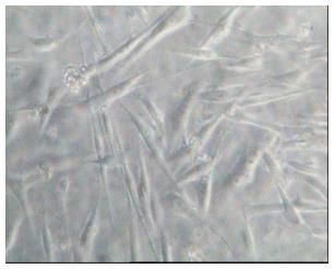
Figure 1 Colony derived from stem cells from human exfoliated deciduous teeth at the first passage.
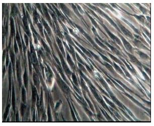
Figure 2 Stem cells from human exfoliated deciduous teeth at the third passage.
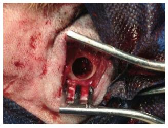
Figure 3 Through-and-through defects caused by trephine bur in the dog mandible.
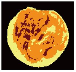
Figure 4 Percent of area 1 with brown color (connective tissue), area 2 with orange color (woven bone) and area 3 with yellow color (compact bone) in sample 3 from control group by image analysis software.
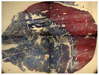
Figure 5 Histological view of regenerated bone (Masson’s trichrome stain, × 40).
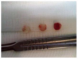
Figure 6 From left to right: buccal, middle and lingual sites of specimen in stem cells from human exfoliated deciduous teeth seeded groups.
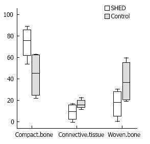
Figure 7 Mean of percentage in compact bone, connective tissue and woven bone in stem cells from human exfoliated deciduous teeth-seeded and control group.
- Citation: Behnia A, Haghighat A, Talebi A, Nourbakhsh N, Heidari F. Transplantation of stem cells from human exfoliated deciduous teeth for bone regeneration in the dog mandibular defect. World J Stem Cells 2014; 6(4): 505-510
- URL: https://www.wjgnet.com/1948-0210/full/v6/i4/505.htm
- DOI: https://dx.doi.org/10.4252/wjsc.v6.i4.505


