Copyright
©2013 Baishideng Publishing Group Co.
World J Stem Cells. Jul 26, 2013; 5(3): 86-97
Published online Jul 26, 2013. doi: 10.4252/wjsc.v5.i3.86
Published online Jul 26, 2013. doi: 10.4252/wjsc.v5.i3.86
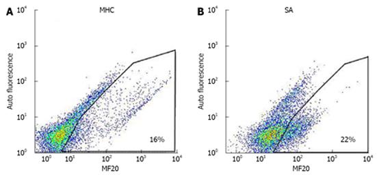
Figure 1 Dot plot of anti-myosin heavy chain and anti-sarcomeric α-actinin of HES-3 differentiated cardiomyocytes.
A: Myosin heavy chain (MHC); B: Sarcomeric α-actinin (SA).
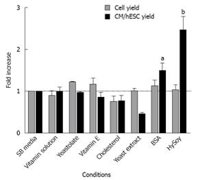
Figure 2 Effect of nutritional supplements added to SB medium on HES-3 differentiation to cardiomyocytes.
Differentiation efficiency is based on two FACS markers: myosin heavy chain (MHC) and sarcomeric α-actinin (SA). Cell and cardiomyocyte (CM) yields were measured at day 16 of culture. Results were normalized against the yields obtained in SB medium (control). (n = 3, aP < 0.05, bP < 0.01 vs cell yield). BSA: Bovine serum albumin; HySoy: Soy-hydrolysate.
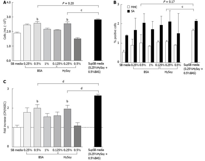
Figure 3 Additive and dose effect of Soy hydrolysate and bovine serum albumin supplementation on HES-3 differentiation to cardiomyocytes.
HES-3 cells differentiated in SB medium containing varying concentrations of bovine serum albumin (BSA) and Soy hydrolysate (HySoy), as well as a combination of both supplements (SupSB media), were harvested on day 16 and evaluated for total cell count (A) and cardiac specific markers, myosin heavy chain (MHC) and sarcomeric α-actinin (SA) (B); C: The results were summarized by calculating the normalized yield which takes into account the ratio of cardiomyocyte at day 16 compared to the initial human embryonic stem cell seeded. Normalized yields are based on both MHC and SA markers. Dot plots of both MHC and SA are shown in Figure 1. (n = 4. bP < 0.01 vs SB medium; cP < 0.05; dP < 0.01 vs SupSB media).
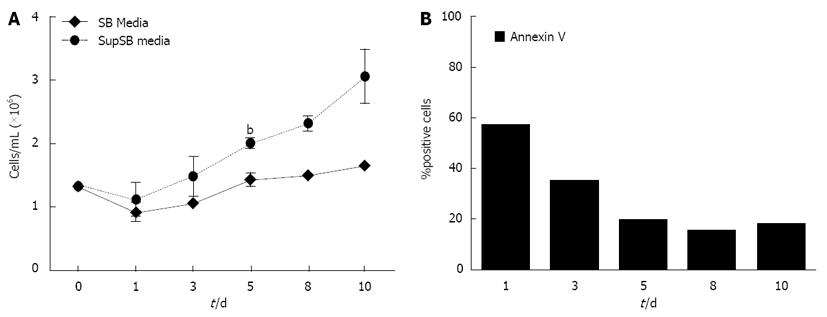
Figure 4 Cell growth and apoptosis in HES-3 cells during cardiomyocyte differentiation.
A: Cell growth kinetics of SB and SupSB media cultures; B: Kinetics of apoptosis in SupSB media cultures (measured by annexin V antibody) (n = 3, bP < 0.01 vs in ten days).
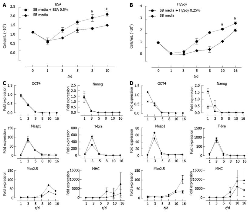
Figure 5 Cell growth and gene expression kinetics of HES-3 cells grown in cultures with and without supplements.
Cell densities at multiple times points over a period of 10 to 16 d were recorded with cultures with and without supplements bovine serum albumin (BSA) (A); Soy hydrolysate (Hysoy) (B). Gene expression profile recorded via quantitative real-time reverse transcription-polymerase chain reaction at multiple time points over 16 d during the course of cardiomyocytes (CM) differentiation. 6 genes: hESC pluripotency (OCT4, Nanog), Mesoderm (Mesp1, T-bra), cardiac progenitors (Mkx2.5), and cardiomyocytes (MHC) were monitored for BSA (C); Hysoy (D). n = 3, aP < 0.05, bP < 0.01 vs SB medium.
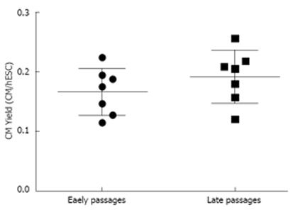
Figure 6 Robustness of SupSB medium over different cell ages (passage).
HES-3 cells from different passages (P12 to P39) were differentiated using the supplements, Soy hydrolysate (HySoy) and bovine serum albumin (BSA). The passages were divided into two groups: Early passage (P12 to P13) and Late Passage (P21 to P39). There is no significant difference in cardiomyocyte yield at different passages. Student t-test and one-way ANOVA test were used to calculate the statistical significance of the data.
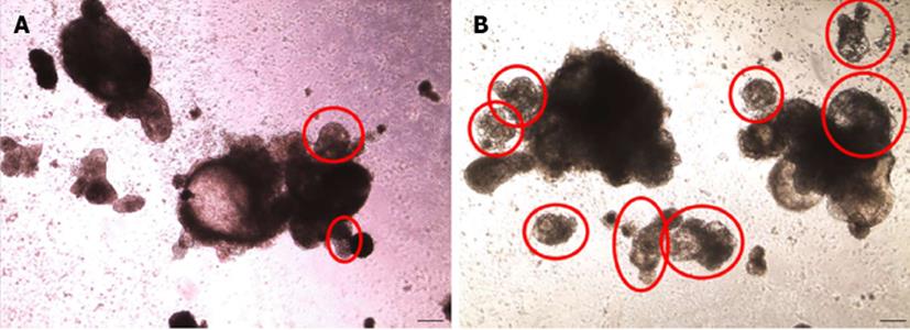
Figure 7 Phase contrast microscopy of cardiomyocytes obtained from differentiation of HES-3 cells.
Pictures of aggregates formed from human embryonic stem cells differentiated in SB media (A) and SupSB media (B) were taken after 16 d. Beating areas are indicated by red circles. Total number of beating aggregates was higher in cultures differentiated in SupSB medium(about 80% of total aggregates) compared to in SB medium (about 60% of total aggregates). Scale bar = 200 μm.
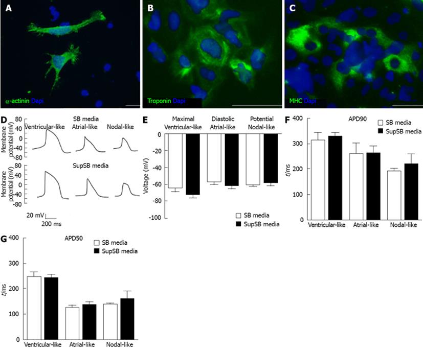
Figure 8 Characterization of cardiomyocytes: Immunocytochemistry and electrophysiology.
HES-3 differentiated cardiomyocyte aggregates at the end of differentiation (day 16) were mechanically dissociated and plated on 24-well plates and strained for markers. A: Sarcomeric α-actinin (Structural protein); B: Troponin-T (contractile function protein); C: Myosin heavy chain (MHC) (contractile function protein). Nuclei stained with DAPI (blue). Scale bar = 20 μm. MHC, myosin heavy chain; DAPI: 4’6-diamidino-2-phenylindole. D: Whole-cell patch-clamp recording was performed on beating cardiomyocyte aggregates at day 23 of differentiation. The recording shows the successful derivation of all three cardiac phenotypes as well as no difference between cells grown in SB media (control) and SupSB media (supplements); E: Maximal diastolic potential; F: Action potential duration at 90% repolarization (ADP90); G: Action potential duration at 50% repolarization (ADP50).
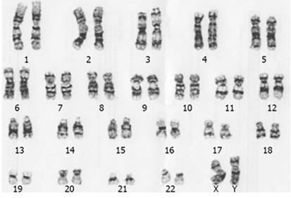
Figure 9 Karyotype of cardiomyocytes differentiated from HES-3 in SupSB media.
- Citation: Ting S, Lecina M, Chan YC, Tse HF, Reuveny S, Oh SK. Nutrient supplemented serum-free medium increases cardiomyogenesis efficiency of human pluripotent stem cells. World J Stem Cells 2013; 5(3): 86-97
- URL: https://www.wjgnet.com/1948-0210/full/v5/i3/86.htm
- DOI: https://dx.doi.org/10.4252/wjsc.v5.i3.86


