
















Copyright: ©Author(s) 2026.
World J Stem Cells. Oct 26, 2026; 18(10): 125673
Published online Oct 26, 2026. doi: 10.4252/wjsc.125673
Published online Oct 26, 2026. doi: 10.4252/wjsc.125673
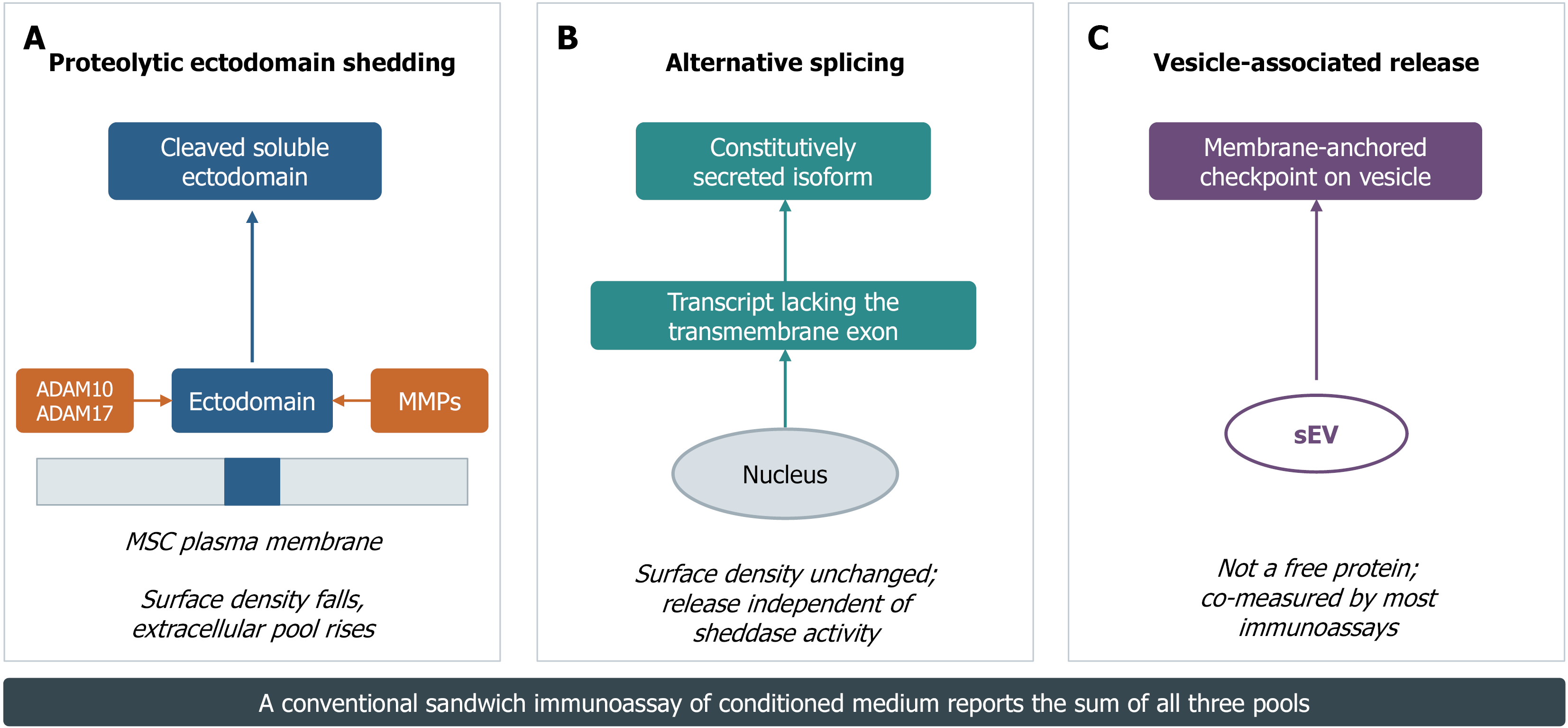
Figure 1 Three routes by which immune checkpoint proteins enter the extracellular space.
A: Proteolytic ectodomain shedding. Membrane-anchored checkpoints are cleaved within the juxtamembrane stalk by a disintegrin and metalloproteinase 10 (ADAM10), ADAM17 or matrix metalloproteinases, releasing an active ectodomain while reducing surface density; B: Alternative splicing. Transcripts lacking the transmembrane exon yield an isoform that is secreted constitutively, without any requirement for sheddase activity and without depleting the surface pool; C: Vesicle-associated release. The checkpoint remains membrane-anchored but is displayed on small extracellular vesicles. Because conventional sandwich immunoassays capture epitopes irrespective of the physical state of the analyte, a single reported concentration typically represents the sum of all three pools. ADAM10: A disintegrin and metalloproteinase 10; MMPs: Matrix metalloproteinases; MSC: Mesenchymal stromal cell; sEV: Small extracellular vesicle.
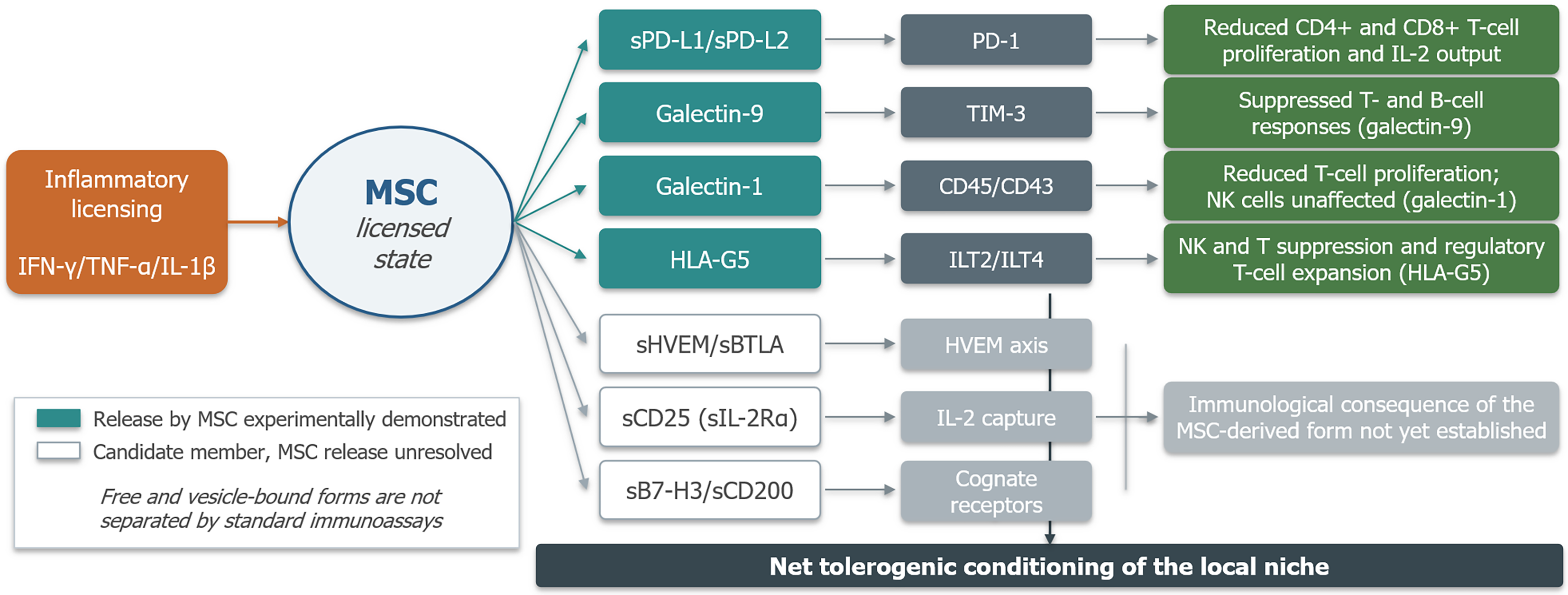
Figure 2 The mesenchymal stromal cell soluble checkpointome and its cellular targets.
Inflammatory licensing reconfigures the mesenchymal stromal cell (MSC) secretory programme. Established members (shaded) are proteins whose release by MSCs has been experimentally demonstrated and linked to a functional immunological outcome. Candidate members (unshaded) are checkpoint molecules of established immunological importance for which MSC-derived soluble forms have not yet been documented. Arrows denote engagement of the indicated receptor system and the downstream consequence. The net effect is tolerogenic conditioning of the local niche rather than the elimination of any single effector population. Each depicted outcome is bounded by the cited primary evidence: Galectin-1 knockdown restores T-cell proliferation but leaves non-alloreactive natural killer (NK) cells unaffected, so no NK or B-cell effect is attributed to galectin-1; galectin-9 is shown to suppress T and B cells as reported, and human leukocyte antigen-G5 to affect T and NK cells and regulatory T cells. Arrows denote reported functional associations, not proven direct molecular interactions. IFN-γ: Interferon gamma; TNF-α: Tumor necrosis factor alpha; IL: Interleukin; MSC: Mesenchymal stromal cell; PD-L1: Programmed death ligand 1; HLA-G5: Human leukocyte antigen-G5; HVEM: Herpesvirus entry mediator; BTLA: B- and T-lymphocyte attenuator; PD-1: Programmed death-1; TIM-3: T cell immunoglobulin and mucin domain-containing protein 3; ILT: Immunoglobulin-like transcript; NK: Natural killer.
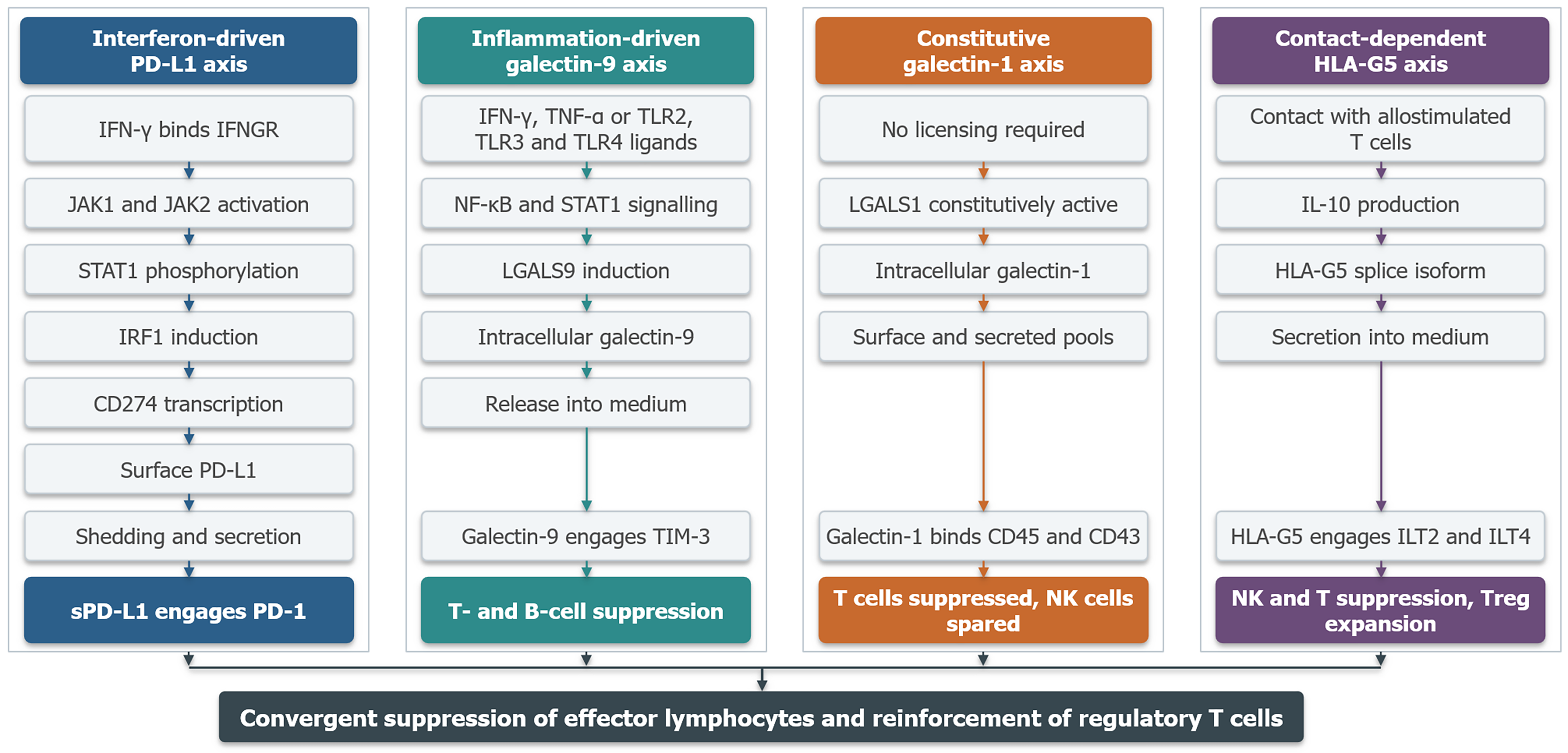
Figure 3 Parallel signaling routes through which the four established members of the soluble checkpointome are induced and act.
Each lane runs from the initiating signal at the top to the immunological outcome at the bottom, and the lanes converge only at the level of effector suppression. The interferon lane and the inflammation lane are licensed, the galectin-1 lane is constitutive, and the human leukocyte antigen-G5 lane requires cell contact. Terminal boxes denote the receptor engagement and its consequence. Pathway steps shown for each axis summarise the cited evidence; where a step is inferred from non-mesenchymal stromal cell (MSC) systems rather than demonstrated in MSCs it should be read as mechanistic context rather than established MSC biology, and the terminal outcomes do not exceed the effects reported in the cited studies. PD-L1: Programmed death ligand 1; IFN-γ: Interferon gamma; IFNGR: Interferon gamma receptor; JAK: Janus kinase; STAT1: Signal transducer and activator of transcription 1; IRF1: Interferon regulatory factor 1; PD-1: Programmed death-1; TNF-α: Tumor necrosis factor alpha; TLR: Toll-like receptor; TIM-3: T cell immunoglobulin and mucin domain-containing protein 3; NF-κB: Nuclear factor kappa B; NK: Natural killer; IL: Interleukin; HLA-G5: Human leukocyte antigen-G5; ILT: Immunoglobulin-like transcript; Treg: Regulatory T cell.
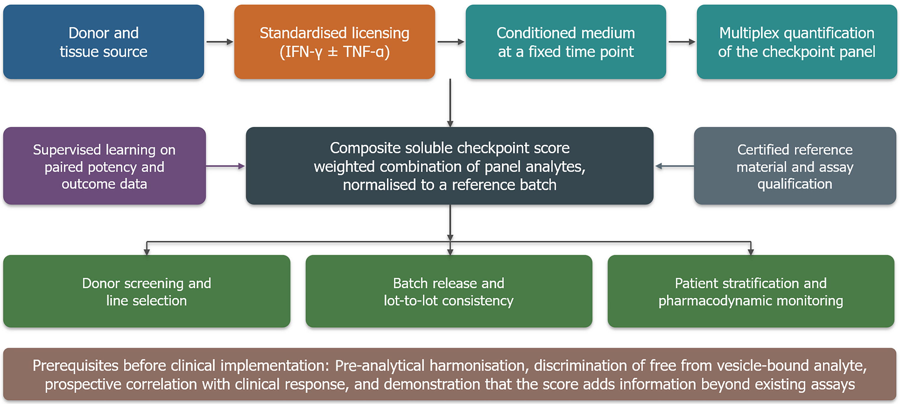
Figure 4 Proposed translational workflow for a checkpoint-based potency assay presents a hypothetical development framework rather than a validated clinical workflow.
It illustrates the experimental and analytical steps that would need to be evaluated before a checkpoint-based composite score could be considered for donor screening, batch qualification or patient stratification. No element of this workflow is validated; it defines future validation studies rather than a usable clinical procedure. IFN-γ: Interferon gamma; TNF-α: Tumor necrosis factor alpha.
- Citation: Al-Karawi AS, Al-Budairi AS. Soluble immunosuppressive checkpointome of mesenchymal stromal cells: Immunoregulatory mechanisms and a conceptual framework for potency assessment. World J Stem Cells 2026; 18(10): 125673
- URL: https://www.wjgnet.com/1948-0210/full/v18/i10/125673.htm
- DOI: https://dx.doi.org/10.4252/wjsc.125673