
















Copyright: ©Author(s) 2026.
World J Stem Cells. Oct 26, 2026; 18(10): 125423
Published online Oct 26, 2026. doi: 10.4252/wjsc.125423
Published online Oct 26, 2026. doi: 10.4252/wjsc.125423
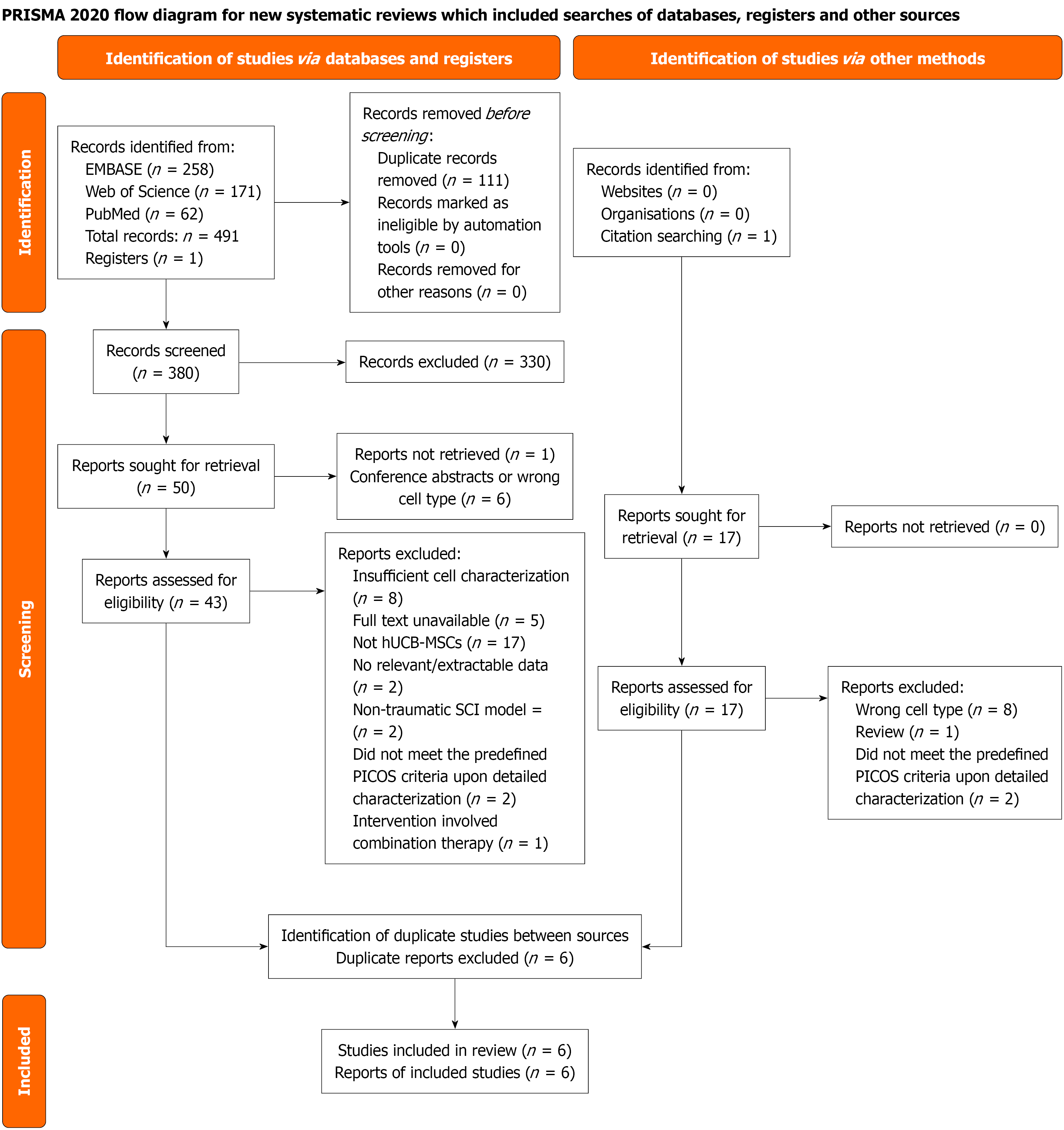
Figure 1 PRISMA flow diagram of the study selection process.
This diagram illustrates the identification, screening, and inclusion of studies evaluating human umbilical cord blood-derived mesenchymal stem cells for experimental spinal cord injury. Electronic searches of PubMed, EMBASE, and Web of Science (from inception to September 12, 2025) identified 491 records. After removal of 111 duplicates, 380 records were screened by title and abstract, resulting in 50 records categorized as “include” or “maybe”. Of these, 6 were excluded as conference abstracts or wrong cell type, leaving 44 reports for retrieval; one report could not be retrieved, leaving 43 full-text articles that were assessed for eligibility; 37 were excluded with reasons, and six unique studies met all eligibility criteria and were included in the qualitative synthesis. An updated search of all three databases performed on August 31, 2026 identified no additional eligible studies; the diagram depicts the primary search conducted on September 12, 2025. hUCB-MSCs: Human umbilical cord blood-derived mesenchymal stem cells; SCI: Spinal cord injury.
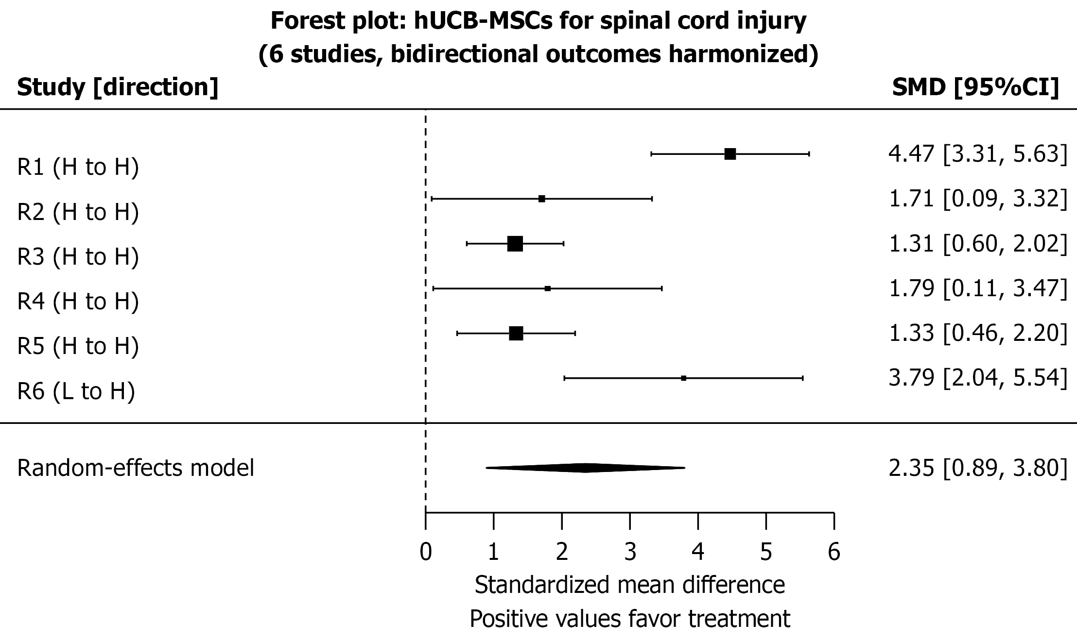
Figure 2 Forest plot of the primary meta-analysis evaluating the effect of human umbilical cord blood-derived mesenchymal stem cell transplantation on functional recovery in experimental spinal cord injury.
The plot displays the standardized mean difference (SMD) with 95% confidence intervals for each of the six included studies (R1-R6). All outcomes have been harmonized to the convention that higher values indicate better functional recovery. The size of each square represents the weight of the study in the random-effects model, with larger squares indicating greater weight. Horizontal lines represent 95% confidence intervals for individual studies. The diamond represents the pooled SMD (2.35, 95% confidence interval: 0.89-3.80). Substantial heterogeneity was observed (I2 = 81.8%, P < 0.001), indicating that while the average effect is positive, the true effect in a new study could range from possible harm to substantial benefit. The dashed vertical line at SMD = 0 indicates no effect. CI: Confidence interval; SMD: Standardized mean difference; hUCB-MSCs: Human umbilical cord blood-derived mesenchymal stem cells.
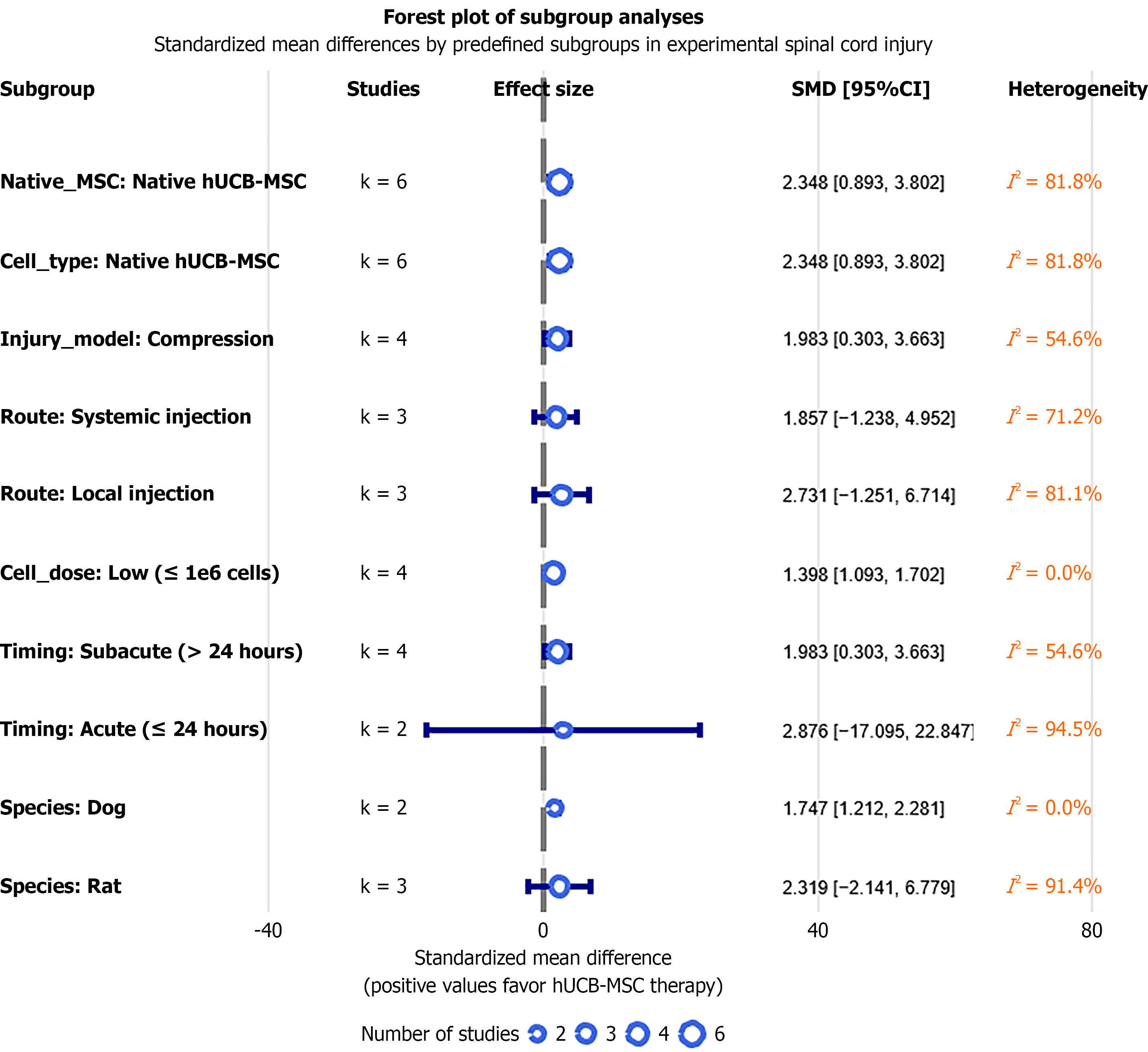
Figure 3 Forest plot of subgroup analyses for functional recovery.
The plot presents pooled standardized mean differences (SMDs) with 95% confidence intervals for predefined subgroups investigating potential sources of heterogeneity. Significant effects were observed in canine models (P = 0.015), subacute intervention timing (P = 0.033), low-dose therapy (P < 0.001), and compression injury models (P = 0.033). All outcomes have been harmonized to the convention that higher values indicate better recovery; positive SMD values favor human umbilical cord blood-derived mesenchymal stem cell therapy. The dashed vertical line at SMD = 0 represents the null effect. Note: No studies administered human umbilical cord blood-derived mesenchymal stem cells in the chronic phase (> 2 weeks post-injury); therefore, the “timing of intervention” subgroup includes only acute (≤ 24 hours) and subacute (> 24 hours to ≤ 2 weeks) categories. The low-dose subgroup comprised only four studies using absolute cell counts without interspecies dose normalization; the observed I2 = 0% should not be interpreted as evidence of true biological homogeneity (see the Limitations section for details). CI: Confidence interval; SMD: Standardized mean difference; hUCB-MSCs: Human umbilical cord blood-derived mesenchymal stem cells.
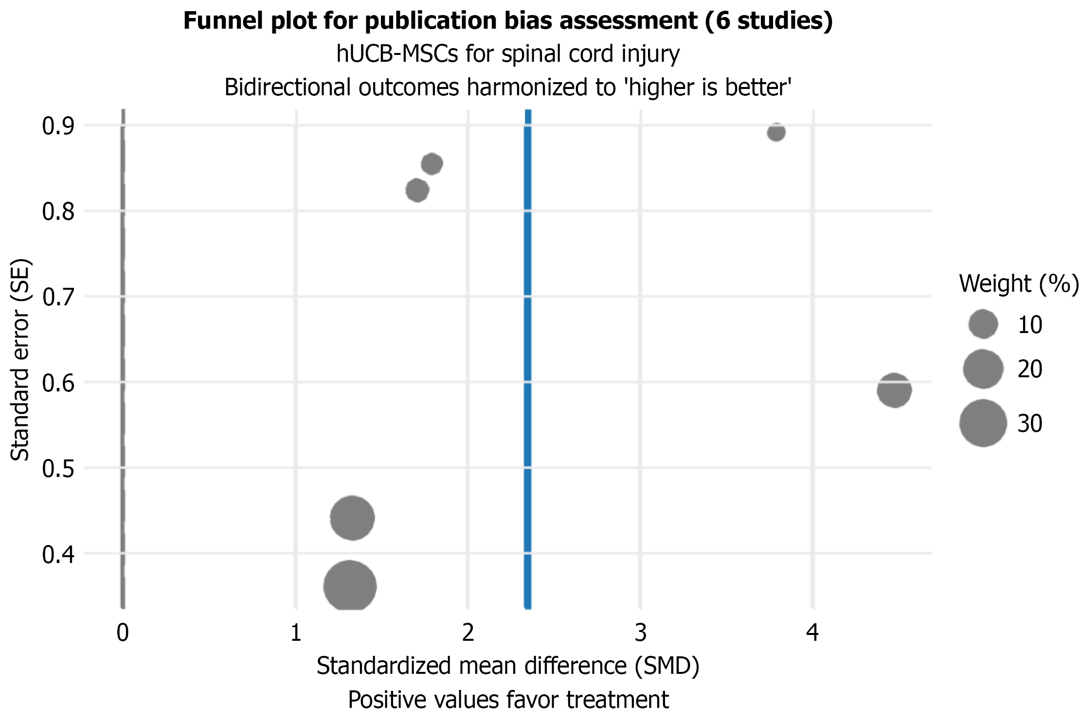
Figure 4 Funnel plot for the assessment of publication bias.
The funnel plot depicts the standard error of each study’s standardized mean difference against its effect size. In the absence of publication bias, studies are expected to distribute roughly symmetrically around the pooled effect estimate (represented by the vertical line at standardized mean difference = 2.35). The size of each dot is proportional to the weight (%) of the individual study in the random-effects model. The distribution appeared roughly symmetrical, and Egger’s test was nonsignificant (P = 0.311); however, with only six studies, neither visual inspection nor formal testing can reliably exclude small-study effects or publication bias. hUCB-MSCs: Human umbilical cord blood-derived mesenchymal stem cells.
- Citation: Liao LQ, Liang RX, Ren ZX, Wang YY, Wei W, Li YS. Human umbilical cord blood-derived mesenchymal stem cell therapy in experimental spinal cord injury: A systematic review and meta-analysis. World J Stem Cells 2026; 18(10): 125423
- URL: https://www.wjgnet.com/1948-0210/full/v18/i10/125423.htm
- DOI: https://dx.doi.org/10.4252/wjsc.125423