©The Author(s) 2025.
World J Stem Cells. Aug 26, 2025; 17(8): 109006
Published online Aug 26, 2025. doi: 10.4252/wjsc.v17.i8.109006
Published online Aug 26, 2025. doi: 10.4252/wjsc.v17.i8.109006
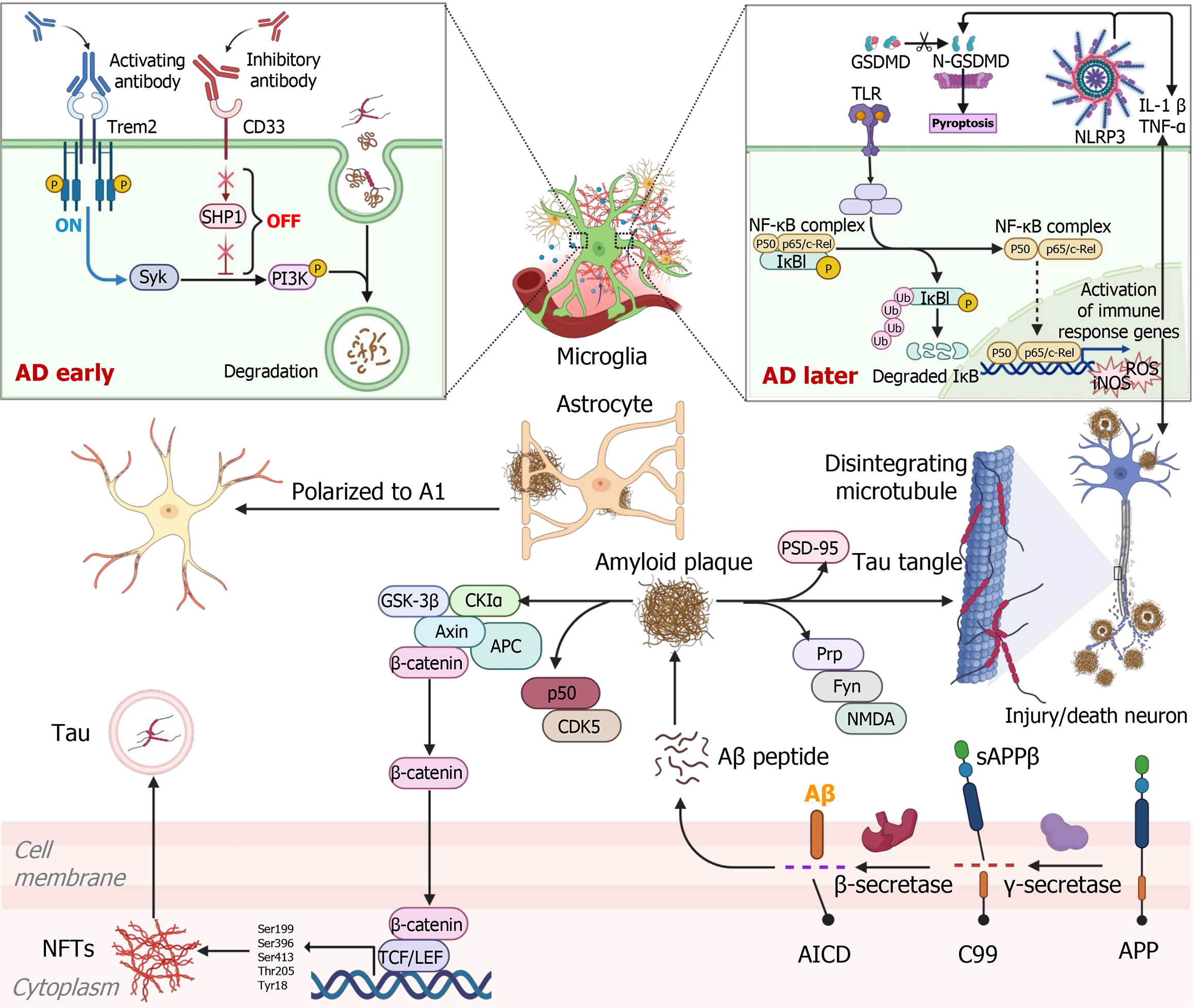
Figure 1 Alzheimer’s disease pathogenesis.
A combination of Aβ, tau proteins, neuroinflammation, and oxidative stress, the neural network in Alzheimer’s disease suffers severe dysfunction, leading to widespread brain atrophy and cognitive decline. Figure was created with BioRender (Supplementary material). Trem2: Triggering receptor expressed on myeloid cells 2; Syk: Spleen tyrosine kinase; SHP1: Src homology region 2 domain containing phosphatase-1; PI3K: Phosphatidylinositol-3-kinase; GSDMD: Gasdermin D; N-GSDMD: N-terminal gasdermin D; TLR: Toll-like receptor; NLRP3: Nod-like receptor protein 3; IL-1β: Interleukin-1β; TNF-α: Tumor necrosis factor α; NF-κB: Nuclear factor-kappa B; IκB: Inhibitor of nuclear factor kappa B; ROS: Reactive oxygen species; iNOS: Inducible nitric oxide synthase; PSD-95: Postsynaptic density protein-95; GSK-3β: Glycogen synthase kinase 3 beta; CKIα: Casein kinase I alpha; APC: Antigen-presenting cell; Prp: Prion protein; NMDA: N-methyl-D-aspartate; Aβ: Β-amyloid; sAPPβ: Soluble amyloid precursor proteins; AICD: Amyloid precursor protein intracellular domain; APP: Amyloid precursor protein; NFTs: Neurofibrillary tangles; TCF: T-cell factor; LEF: Lymphoid enhancer-binding factor; AD: Alzheimer’s disease.
- Citation: Shan XQ, He MH, Gao WL, Li YJ, Liu SZ, Liu Y, Wang CL, Zhao L, Xu SX. Mesenchymal stem cell-derived exosomes: Shaping the next era of Alzheimer’s disease treatment. World J Stem Cells 2025; 17(8): 109006
- URL: https://www.wjgnet.com/1948-0210/full/v17/i8/109006.htm
- DOI: https://dx.doi.org/10.4252/wjsc.v17.i8.109006


