©The Author(s) 2025.
World J Stem Cells. Jan 26, 2025; 17(1): 98777
Published online Jan 26, 2025. doi: 10.4252/wjsc.v17.i1.98777
Published online Jan 26, 2025. doi: 10.4252/wjsc.v17.i1.98777
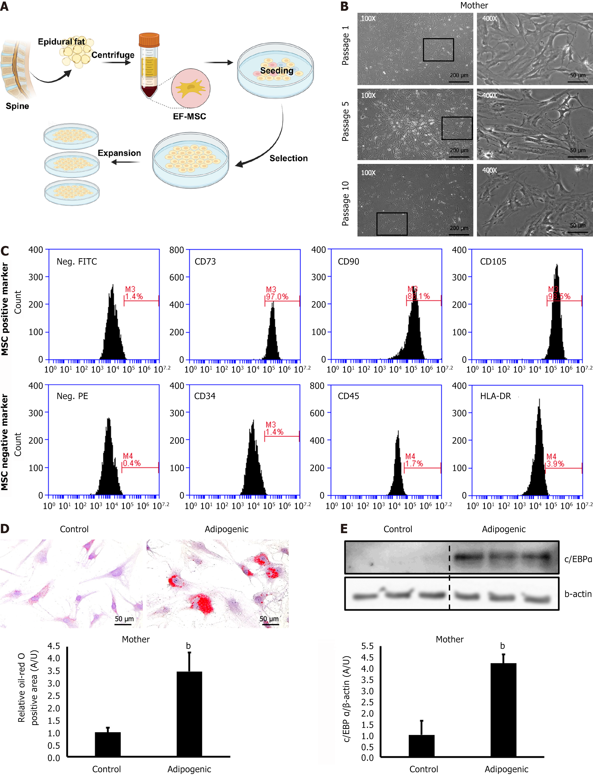
Figure 1 Isolation and characterization of epidural fat-derived mesenchymal stem cells.
Mesenchymal stem cells were successfully isolated from human epidural fat. A: Graphical representation of the mesenchymal stem cell (MSC) isolation protocol; B: Representative images of epidural fat-derived MSCs (EF-MSCs), scale bars = 200 μm and 50 μm; C: Fluorescence-activated single cell sorting analysis results showing the expression of SC surface markers; D: Representative images and quantification of Oil Red O staining of EF-MSCs, scale bar = 50 μm; E: Expression levels of adipogenesis marker protein CCAAT/enhancer-binding protein alpha. Data are presented as the mean ± standard deviation for each group. bP < 0.01.
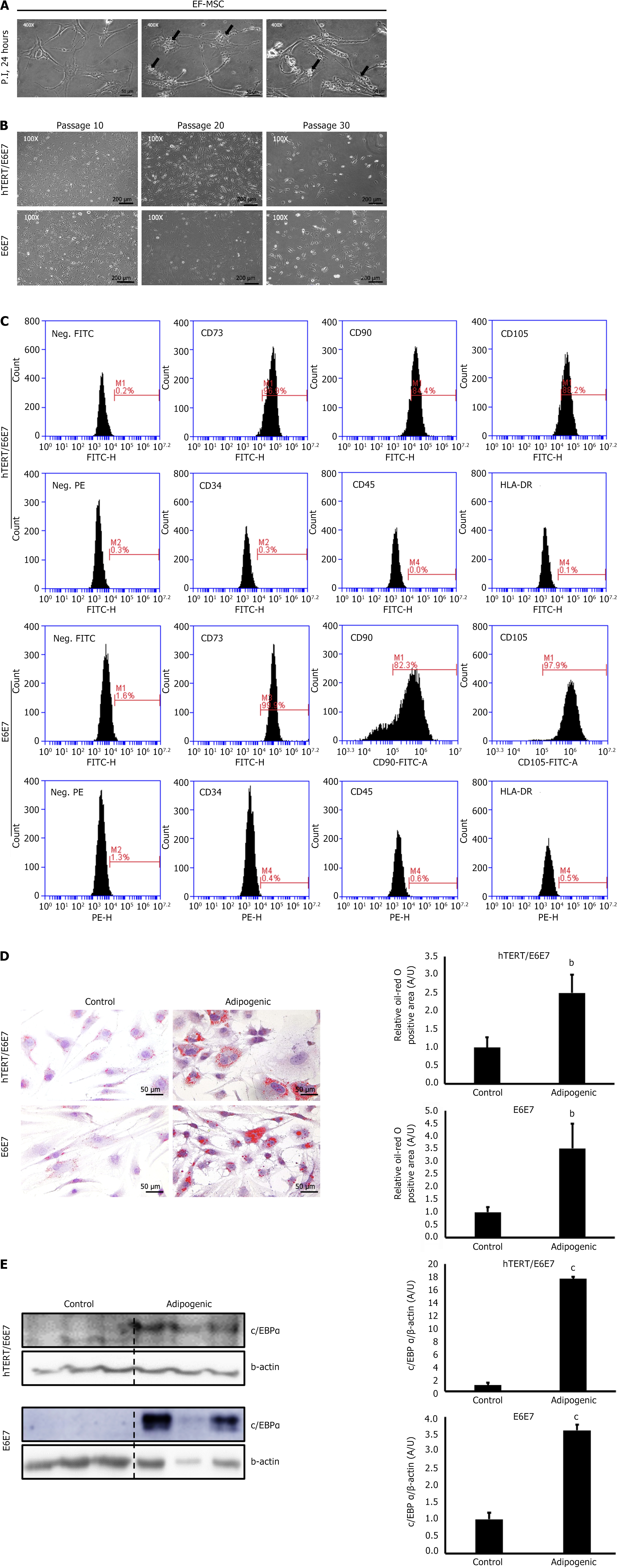
Figure 2 Preserved stem cell characteristics in immortalized epidural fat-derived mesenchymal stem cells.
Immortalized epidural fat-derived mesenchymal stem cells (EF-MSCs) maintained their SC surface markers and adipogenic potential. A: Representative images of immortalized EF-MSCs, showing vacuolar degeneration in the transfected MSCs (black arrow), scale bars = 50 μm; B: Representative images demonstrating the prolonged lifespan of EF-MSCs, scale bar = 200 μm; C: Fluorescence-activated single cell sorting analysis results showing the expression of stem cell surface markers in immortalized EF-MSCs (passage 20); D: Representative images and quantification of Oil Red O staining in immortalized EF-MSCs, scale bar = 50 μm; E: Expression levels of adipogenesis marker protein CCAAT/enhancer-binding protein alpha. Data are presented as the mean ± standard deviation for each group. bP < 0.01, cP < 0.001. hTERT: Human telomerase reverse transcriptase.
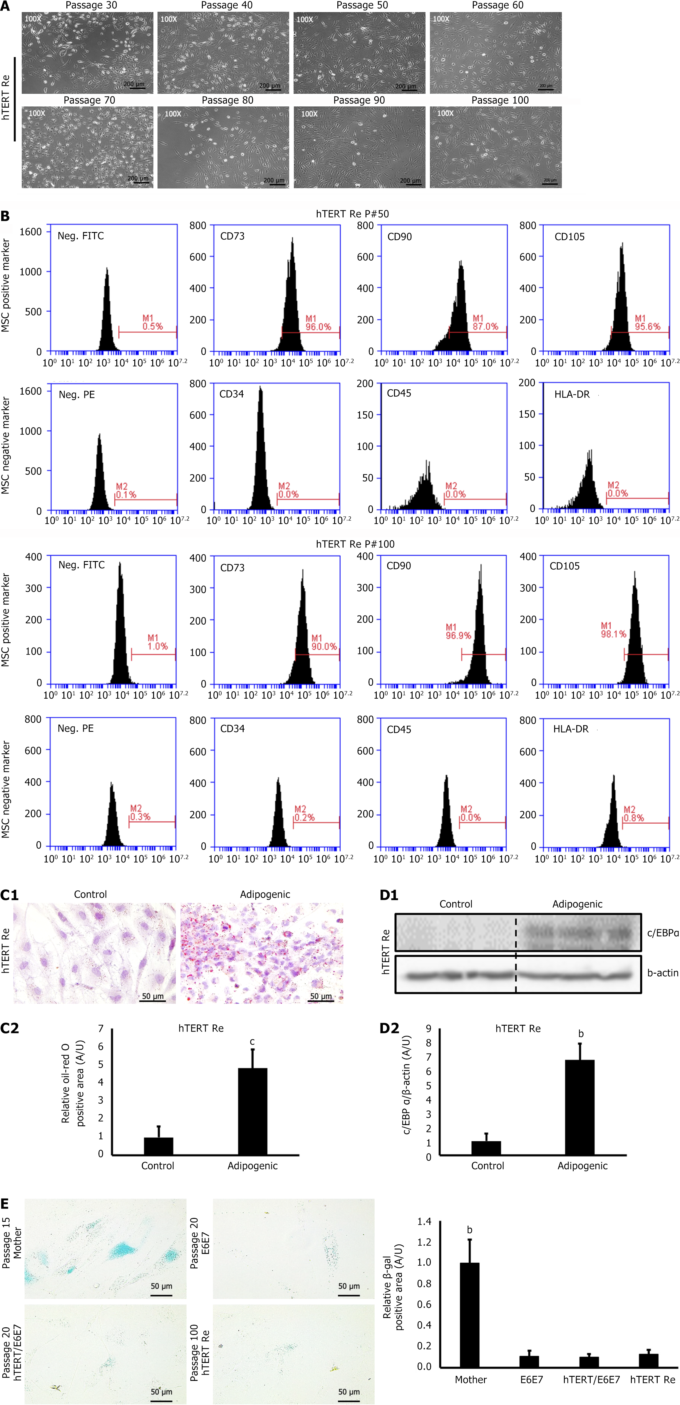
Figure 3 Retransfection of human telomerase reverse transcriptase restores growth arrest due to cell senescence.
Human telomerase reverse transcriptase (hTERT)-Re epidural fat-derived mesenchymal stem cells (EF-MSCs) exhibited a significantly enhanced proliferative capacity and extended lifespan, while maintaining their SC characteristics. A: Representative images of hTERT-Re EF-MSCs, demonstrating preserved proliferation beyond passage 100, scale bar = 200 μm; B: Fluorescence-activated single cell sorting analysis showing the expression of SC surface markers in hTERT-Re EF-MSCs; C: Representative images and quantification of Oil Red O staining in hTERT-Re EF-MSCs (passage 100), scale bar = 50 μm; D: Expression levels of the adipogenesis marker protein CCAAT/enhancer-binding protein alpha in hTERT-Re EF-MSCs (passage 100); E: Representative images and quantification of β-galactosidase staining in EF-MSCs, scale bar = 50 μm. Data are presented as the mean ± standard deviation for each group. bP < 0.01, cP < 0.001.
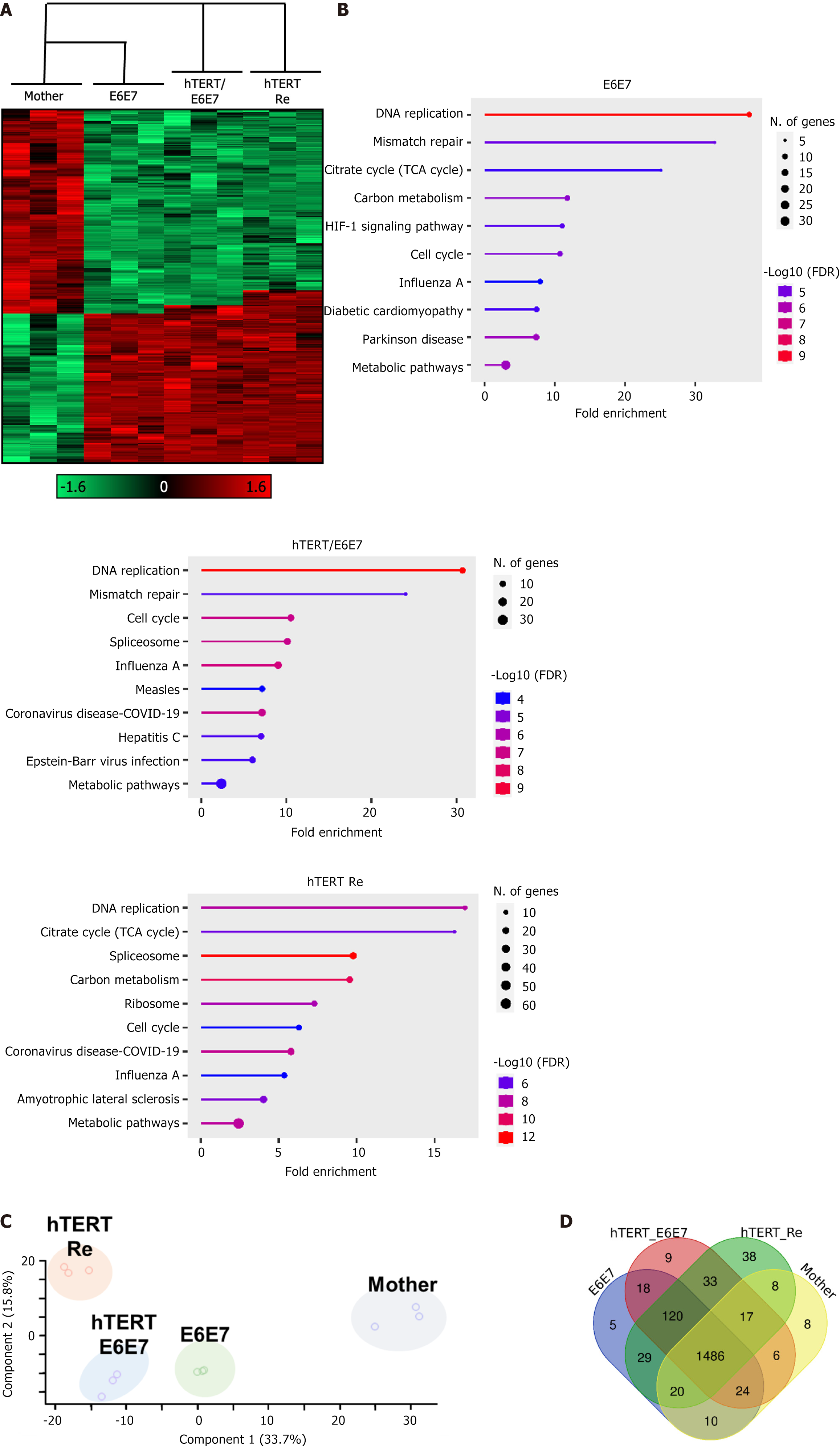
Figure 4 Results of proteomics analysis of isolated epidural fat-derived mesenchymal stem cells.
Immortalized cells exhibited significantly altered protein expression patterns and an increased expression of components involved in cell proliferation pathways. A: Heatmap results of the isolated epidural fat-derived mesenchymal stem cells (EF-MSCs); B: Kyoto Encyclopedia of Genes and Genomes pathway enrichment graph for isolated EF-MSCs, indicating increased DNA replication in immortalized cells; C and D: Protein expression patterns visualized through principal component analysis and Venn diagram plots. hTERT: Human telomerase reverse transcriptase.
- Citation: Lee SW, Lim YJ, Kim HY, Kim W, Park WT, Ma MJ, Lee J, Seo MS, Kim YI, Park S, Choi SK, Lee GW. Immortalization of epidural fat-derived mesenchymal stem cells: In vitro characterization and adipocyte differentiation potential. World J Stem Cells 2025; 17(1): 98777
- URL: https://www.wjgnet.com/1948-0210/full/v17/i1/98777.htm
- DOI: https://dx.doi.org/10.4252/wjsc.v17.i1.98777


