©The Author(s) 2023.
World J Stem Cells. Sep 26, 2023; 15(9): 876-896
Published online Sep 26, 2023. doi: 10.4252/wjsc.v15.i9.876
Published online Sep 26, 2023. doi: 10.4252/wjsc.v15.i9.876
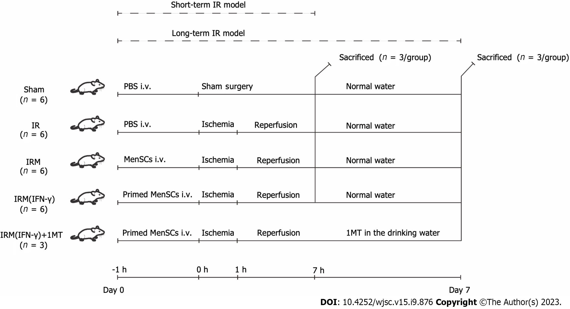
Figure 1 Animal experiment grouping and timeline.
Detailed descriptions are provided in the Materials and Methods. In brief, we established short-term and long-term liver ischemia-reperfusion mouse models to explore the therapeutic potential of menstrual blood-derived stromal cells (MenSCs) and primed MenSCs. MenSCs: Menstrual blood-derived stromal cells; IFN-γ: Interferon-γ; 1MT: 1-methyl-D-tryptophan; IR: Ischemia-reperfusion; IRM: Combination treated with ischemia-reperfusion and menstrual blood-derived stromal cells; IRM (IFN-γ): Combination treated with ischemia-reperfusion and interferon-γ-primed menstrual blood-derived stromal cells; PBS: Phosphate buffered saline; i.v.: Intravenous injection.
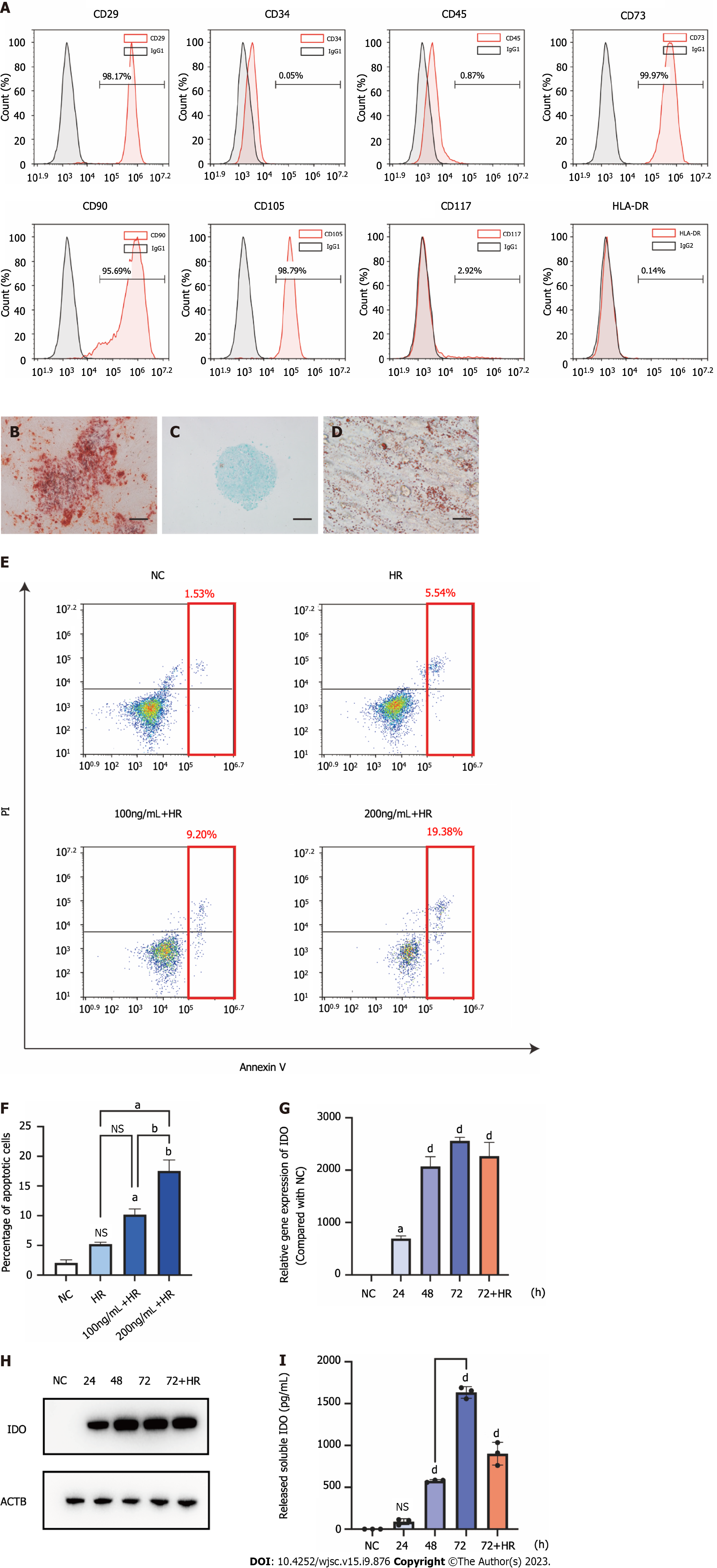
Figure 2 Characterization of menstrual blood-derived stromal cells and menstrual blood-derived stromal cells primed with interferon-γ.
A: The surface marker expression of menstrual blood-derived stromal cells (MenSCs) was assessed by flow cytometry analysis; B: The osteogenic differentiation potential of MenSCs. Scale bar 200 μm; C: The chondrogenic differentiation potential of MenSCs. Scale bar 200 μm; D: The adipogenic differentiation potential of MenSCs. Scale bar 200 μm; E: MenSC apoptosis was detected by flow cytometry. The red box indicates the sum of late and early apoptotic cells, which represent the overall level of apoptosis; F: The overall level of apoptosis was analyzed; G: After MenSC priming with 100 ng/mL interferon-γ for 24 h, 48 h, 72 h, and 72 h + hypoxia/reoxygenation, the mRNA levels of indoleamine 2,3-dioxygenase (IDO) in MenSCs were determined by quantitative real-time reverse transcription polymerase chain reaction analysis; H: The protein level of IDO in MenSCs was determined by western blotting; I: The level of IDO secreted by MenSCs into the cell culture supernatant was measured by ELISA. The data are shown as the means ± SEMs, n = 3. aP < 0.05, bP < 0.01, cP < 0.001, dP < 0.0001. NS represents not statistically significant. All P values were obtained by one-way ANOVA. HLA-DR: Human leukocyte antigen-DR; IgG: Immunoglobulin G; NC: Negative control; HR: Hypoxia/reoxygenation; PI: Propidium iodide; IDO: Indoleamine 2,3-dioxygenase.
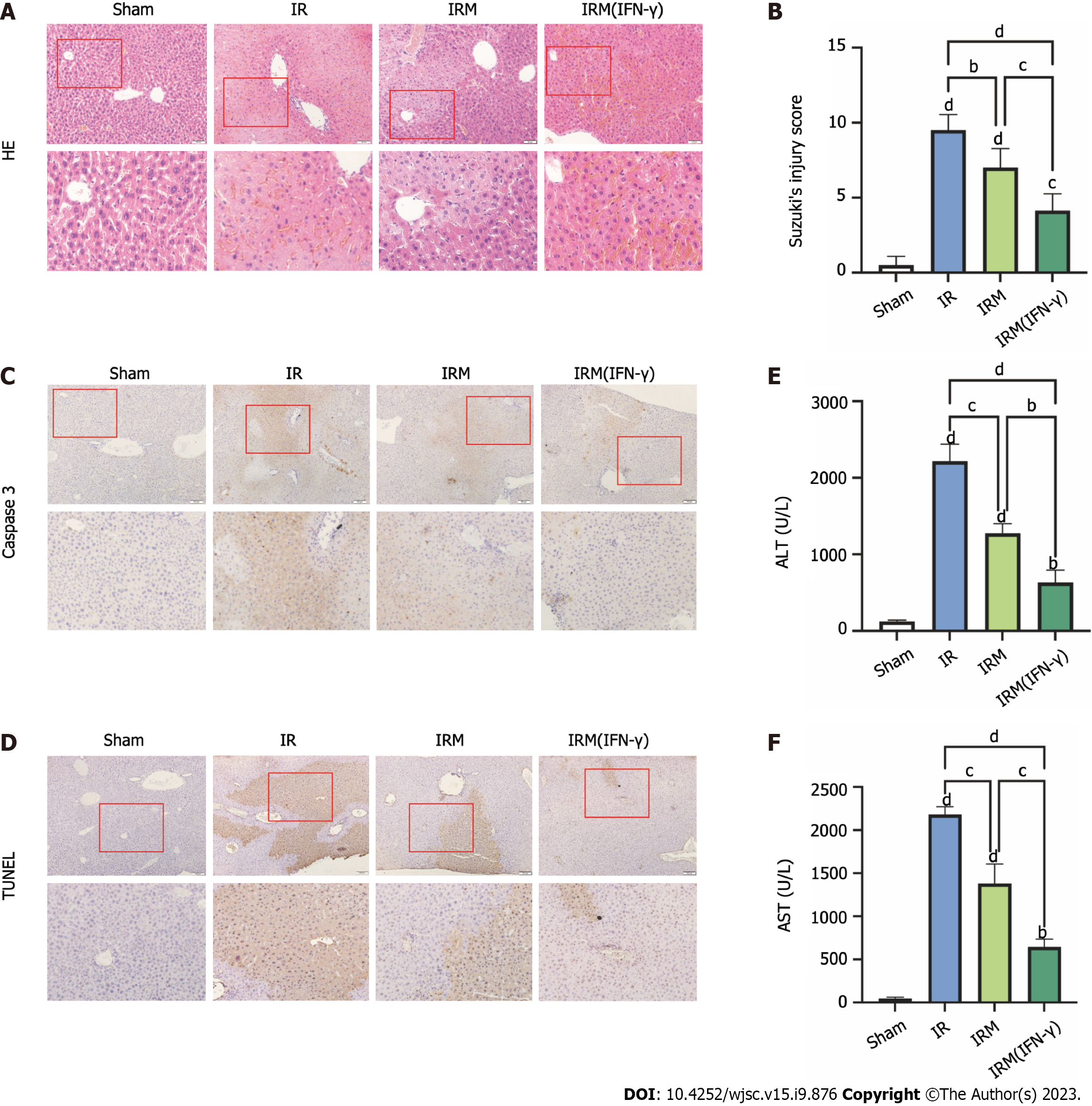
Figure 3 Interferon-γ-primed menstrual blood-derived stromal cells attenuated liver ischemia-reperfusion injury in the mouse short-term ischemia-reperfusion model.
A: Representative images of histological liver sections stained with hematoxylin and eosin (hematoxylin and eosin staining, magnification 200 ×, images in the second row are partial magnifications of regions indicated by the red box); B: The degree of liver injury in each group was determined according to Suzuki’s injury score. The data are expressed as the means ± SEMs (n = 10/group); C: Representative images of liver sections from each group stained for caspase-3 to evaluate the apoptosis level (magnification 200 ×, images in the second row are partial magnifications of regions indicated by the red box); D: Representative liver sections from each group stained with TUNEL were obtained to evaluate the apoptosis level (magnification 200 ×, images in the second row are partial magnifications of regions indicated by the red box); E: The serum alanine aminotransferase levels were measured to indicate liver function levels, the data are expressed as the means ± SEMs (n = 3/group); F: The serum aspartate aminotransferase levels were measured to indicate liver function levels, and the data are expressed as the means ± SEMs (n = 3/group). aP < 0.05, bP < 0.01, cP < 0.001, dP < 0.0001. NS represents not statistically significant. All P values were obtained by one-way ANOVA. MenSCs: Menstrual blood-derived stromal cells; IFN-γ: Interferon-γ; IR: Ischemia-reperfusion; IRM: Combination treated with ischemia-reperfusion and menstrual blood-derived stromal cells; IRM (IFN-γ): Combination treated with ischemia-reperfusion and interferon-γ-primed menstrual blood-derived stromal cells; HE: Hematoxylin and eosin; TUNEL: Terminal deoxynucleotidyl transferase-mediated dUTP-biotin Nick End Labeling; ALT: Alanine aminotransferase; AST: Aspartate aminotransferase.
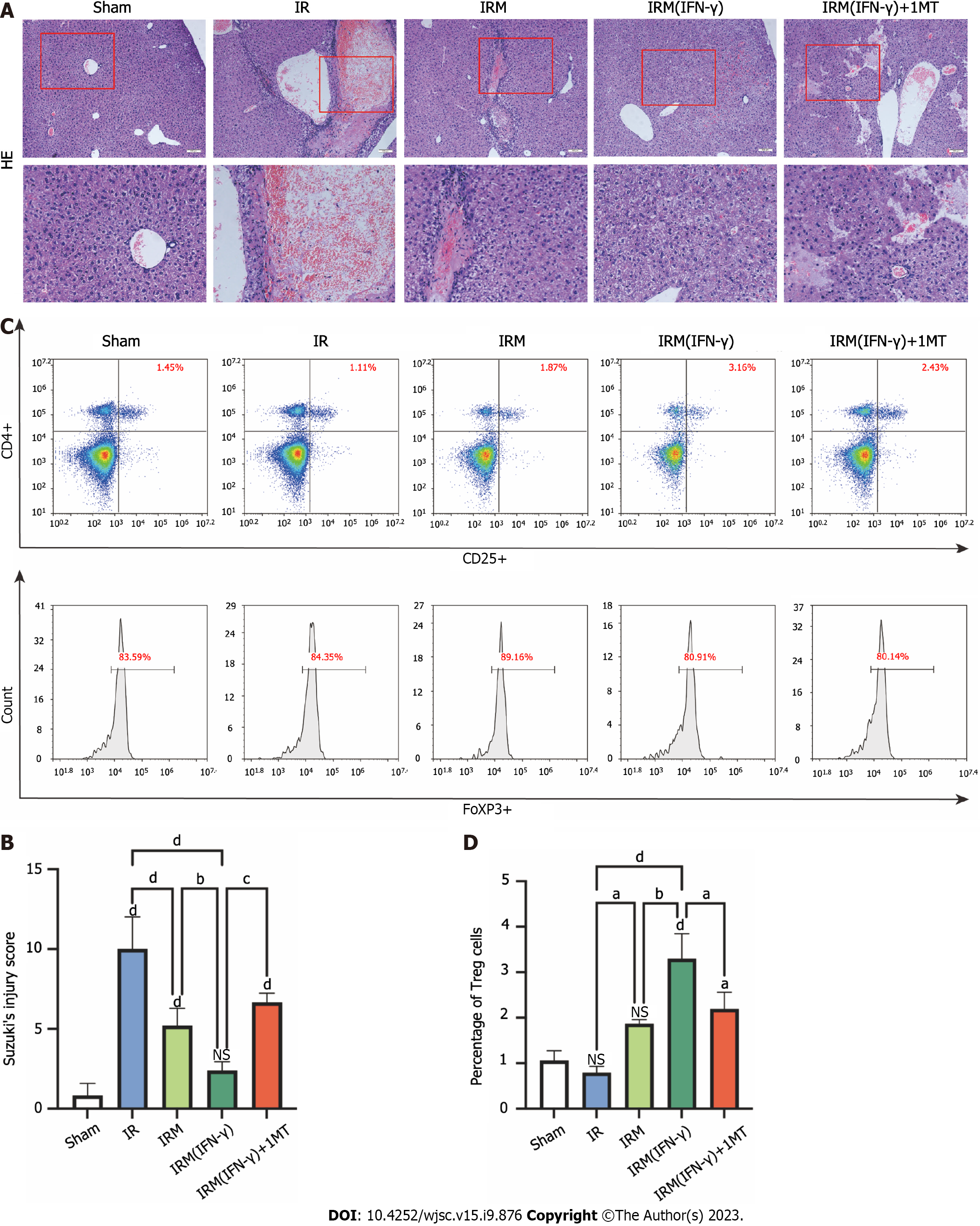
Figure 4 Interferon-γ-primed menstrual blood-derived stromal cells reduced hepatocellular damage and increased the Treg numbers in the mouse long-term ischemia-reperfusion model.
A: Representative images of histological liver sections stained with hematoxylin and eosin were obtained (hematoxylin and eosin staining, magnification 200×, images in the second row are partial magnifications of regions indicated by the red box); B: The degree of liver injury in each group was determined according to Suzuki’s injury score. The data are expressed as the means ± SEMs (n = 3-5/group); C: The numbers of CD4, CD25, and FoxP3 positive cells (regulatory T cells) in the spleen were determined by flow cytometry to estimate the ability of primed menstrual blood-derived stromal cells to induce immune tolerance; D: The percentages of Tregs are expressed as the means ± SEMs (n = 3/group). aP < 0.05, bP < 0.01, cP < 0.001, dP < 0.0001. NS represents not statistically significant. All P values were obtained by one-way ANOVA. MenSCs: Menstrual blood-derived stromal cells; IFN-γ: Interferon-γ; 1MT: 1-methyl-D-tryptophan; IR: Ischemia-reperfusion; IRM: Combination treated with ischemia-reperfusion and menstrual blood-derived stromal cells; IRM (IFN-γ): Combination treated with ischemia-reperfusion and interferon-γ-primed menstrual blood-derived stromal cells; HE: Hematoxylin and eosin; Treg: Regulatory T cell.
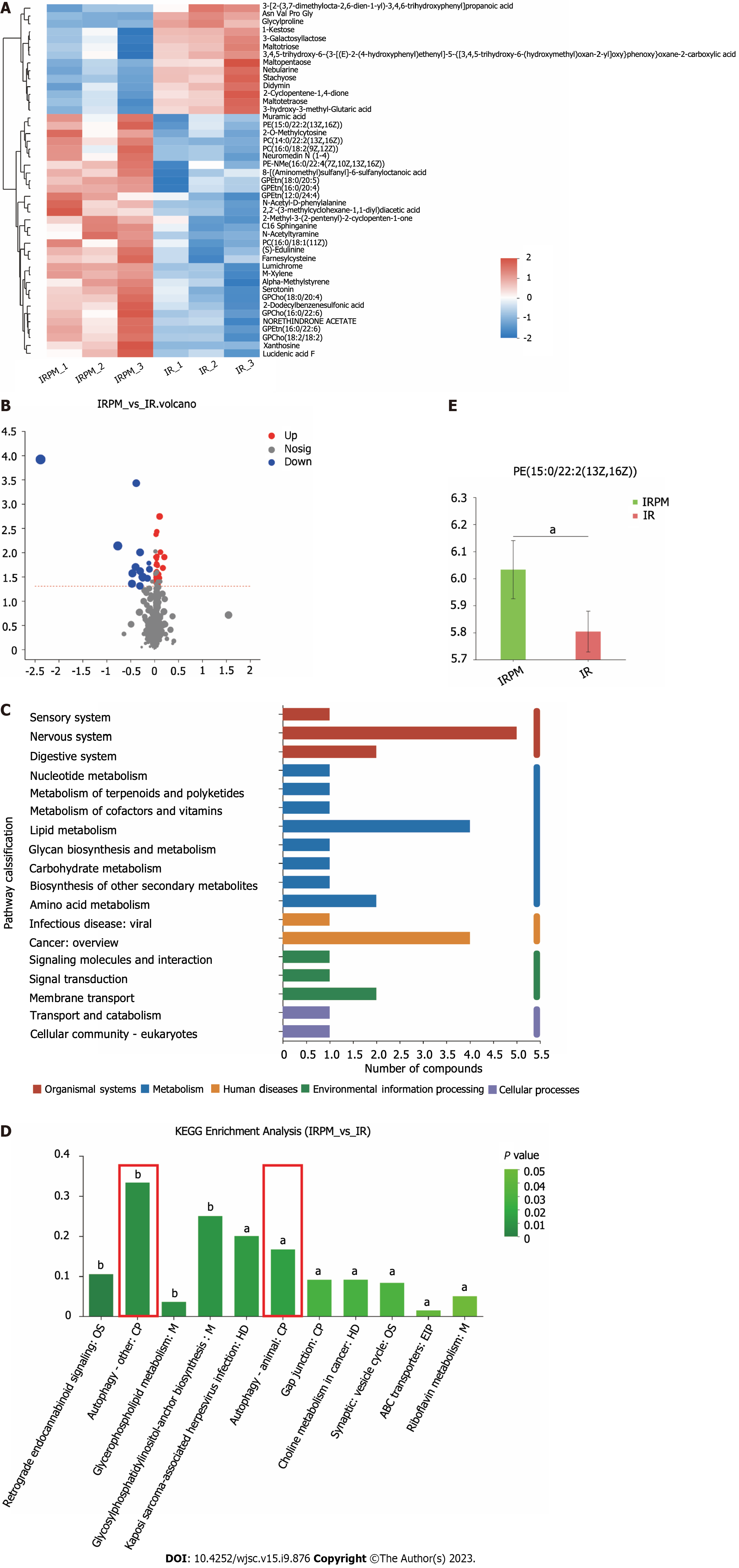
Figure 5 Metabolomic data analysis.
A: Heatmap of 45 differential metabolites between the ischemia-reperfusion (IR) group and IRM (interferon-γ) group; B: Volcano plot of 45 differential metabolites; C: The relevant pathways in which the differential metabolites are involved were analyzed by comparing the information in the Kyoto Encyclopedia of Genes and Genomes database; D: The main biological functions of differential metabolites were elucidated by pathway enrichment analysis; E: Relative quantitation of the differential metabolite phosphatidyl ethanolamine was performed to measure the effect of primed menstrual blood-derived stromal cells on the level of autophagy in liver tissues. MenSCs: Menstrual blood-derived stromal cells; IFN-γ: Interferon-γ; IR: Ischemia-reperfusion; IRM(IFN-γ) or IRPM: Combination treated with ischemia-reperfusion and interferon-γ-primed menstrual blood-derived stromal cells; PE: Phosphatidyl ethanolamine; OS: Organismal systems; M: Metabolism; HD: Human diseases; CP: Cellular processes; EIP: Environmental information processing; KEEG: Kyoto Encyclopedia of Genes and Genomes.
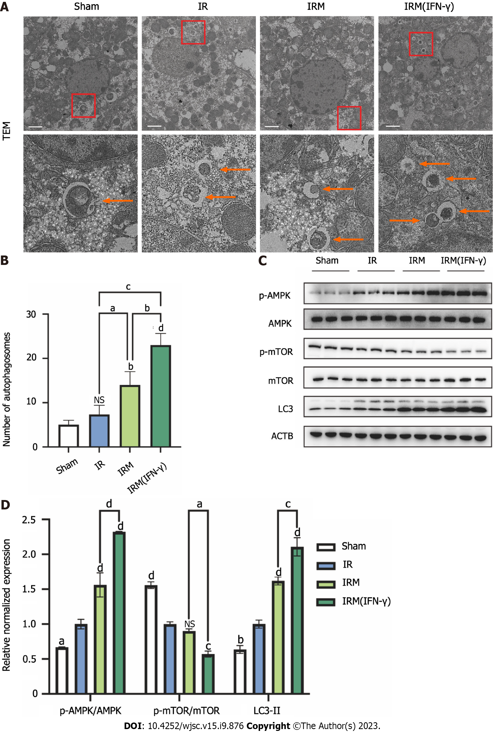
Figure 6 Interferon-γ-primed menstrual blood-derived stromal cells enhanced autophagy in mouse livers.
A: Representative TEM images were used to determine the number of autophagic vacuoles in liver tissues from each experimental group. Scale bar 2 μm. The images in the second row are partial magnifications of the regions indicated by the red box; B: The number of autophagosomes was counted by TEM. The data are expressed as the means ± SEMs (n = 3/group); C: The expression of p-AMPK, AMPK, p-mammalian target of rapamycin (p-mTOR), mTOR, LC3, and ACTB in liver tissues was determined by western blotting; D: The relative normalized expression was quantified based on the intensities of the western blot bands (n = 3/group). The data are presented as the means ± SEMs. aP < 0.05, bP < 0.01, cP < 0.001, dP < 0.0001. NS represents not statistically significant. All P values were obtained by one-way ANOVA. MenSCs: Menstrual blood-derived stromal cells; IFN-γ: Interferon-γ; IR: Ischemia-reperfusion; IRM: Combination treated with ischemia-reperfusion and menstrual blood-derived stromal cells; IRM (IFN-γ): Combination treated with ischemia-reperfusion and interferon-γ-primed menstrual blood-derived stromal cells; TEM: Transmission electron microscopy; AMPK: The AMP-activated protein kinase signaling pathway; mTOR: Mammalian target of rapamycin; Tregs: Regulatory T cells.
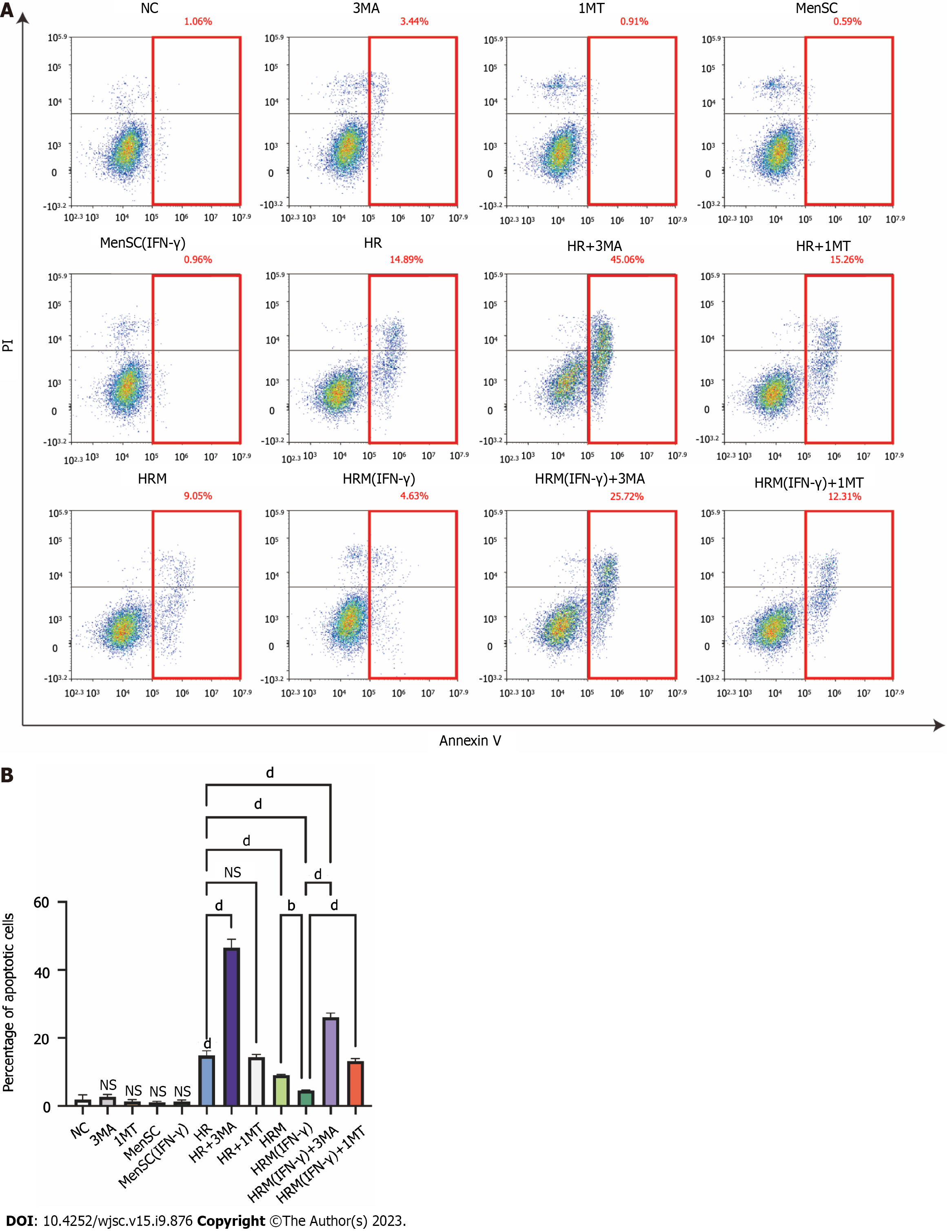
Figure 7 interferon-γ-primed menstrual blood-derived stromal cells decreased hypoxia/reoxygenation-induced cell apoptosis via autophagy and indoleamine 2,3-dioxygenase.
A: Apoptosis of L02 cells after different treatments was analyzed; B: The sum of the percentage of early apoptotic cells and late apoptotic cells was analyzed. The total percentage of apoptotic cells (sum of the percentage of early apoptotic cells and late apoptotic cells) was analyzed. The data are shown as the mean ± SEM, n = 3; bP < 0.01, dP < 0.0001. NS represents not statistically significant. All P values were obtained by one-way ANOVA. PI: Propidium iodide; NC: Negative control; 3MA: 3-methyladenine; 1MT: 1-methyl-D-tryptophan; MenSCs: Menstrual blood-derived stromal cells; IFN-γ: Interferon-γ; HR: Hypoxia/reoxygenation; HRM: Combination treated with hypoxia/reoxygenation and menstrual blood-derived stromal cells; HRM (IFN-γ): Combination treated with hypoxia/reoxygenation and I interferon-γ-primed menstrual blood-derived stromal cells.
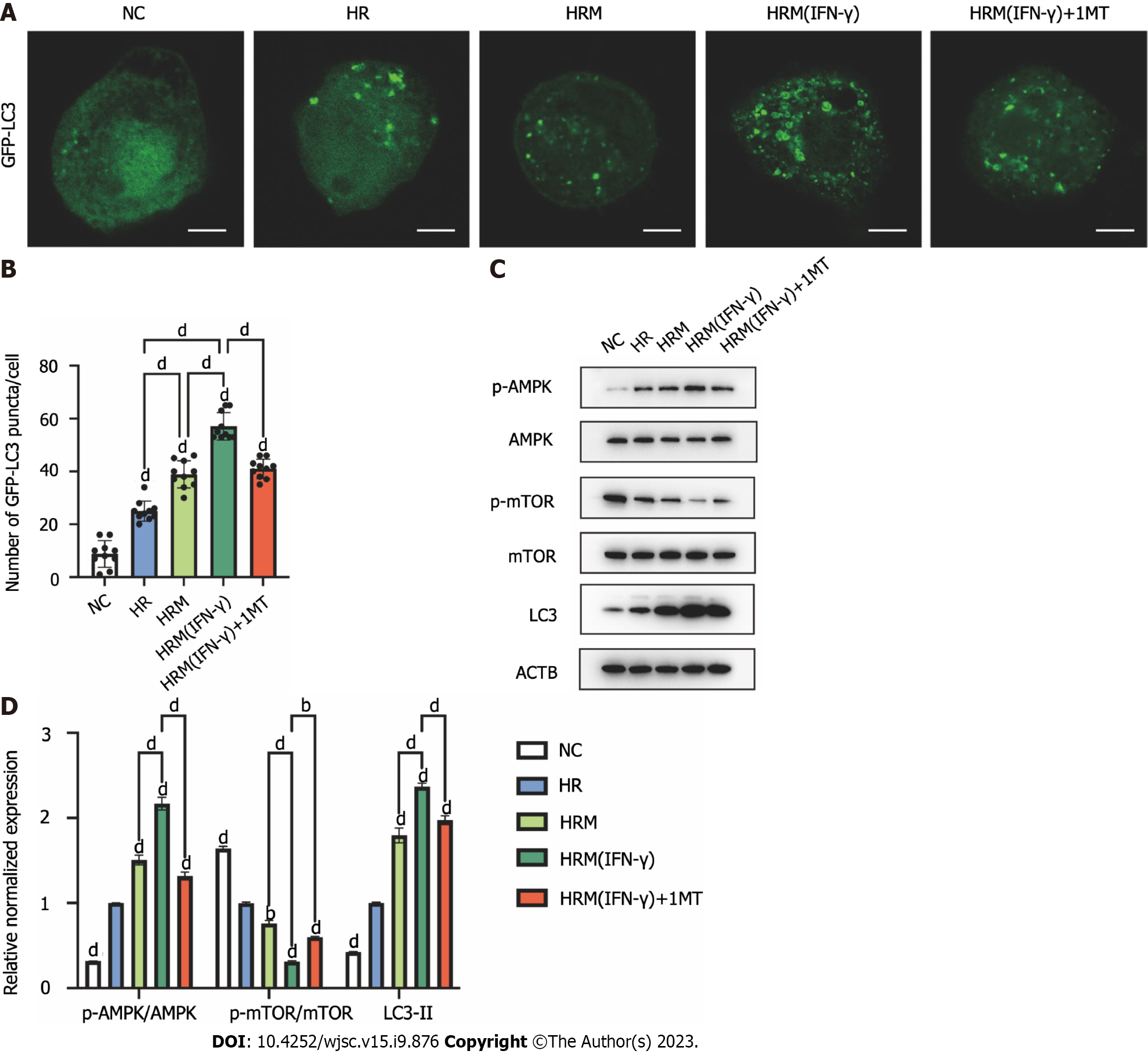
Figure 8 Indoleamine 2,3-dioxygenase secreted by interferon-γ-primed menstrual blood-derived stromal cells enhanced autophagy by inhibiting the mammalian target of rapamycin pathway and activating the AMPK pathway.
A: Immunofluorescence images of GFP-LC3. Scale bar 2 μm; B: The number of intense punctate GFP-LC3 aggregates in each cell was counted to determine the level of autophagy in each group. The data are expressed as the means ± SEMs (n = 10/group); C: The expression of p-AMPK, AMPK, p-mammalian target of rapamycin (p-mTOR), mTOR, LC3, and ACTB in L02 cells was determined by western blotting; D: The relative normalized protein expression was quantified based on the intensities of the western blot bands (n = 3/group). The data are presented as the means ± SEMs. aP < 0.05, bP < 0.01, cP < 0.001, dP < 0.0001. NS represents not statistically significant. All P values were obtained by one-way ANOVA. NC: Negative control; 3MA: 3-methyladenine; 1MT: 1-methyl-D-tryptophan; MenSCs: Menstrual blood-derived stromal cells; IFN-γ: Interferon-γ; HR: Hypoxia/reoxygenation; HRM: Combination treated with hypoxia/reoxygenation and menstrual blood-derived stromal cells; HRM (IFN-γ): Combination treated with hypoxia/reoxygenation and I interferon-γ-primed menstrual blood-derived stromal cells; AMPK: AMP-activated protein kinase; mTOR: the Mammalian target of rapamycin signaling pathway.
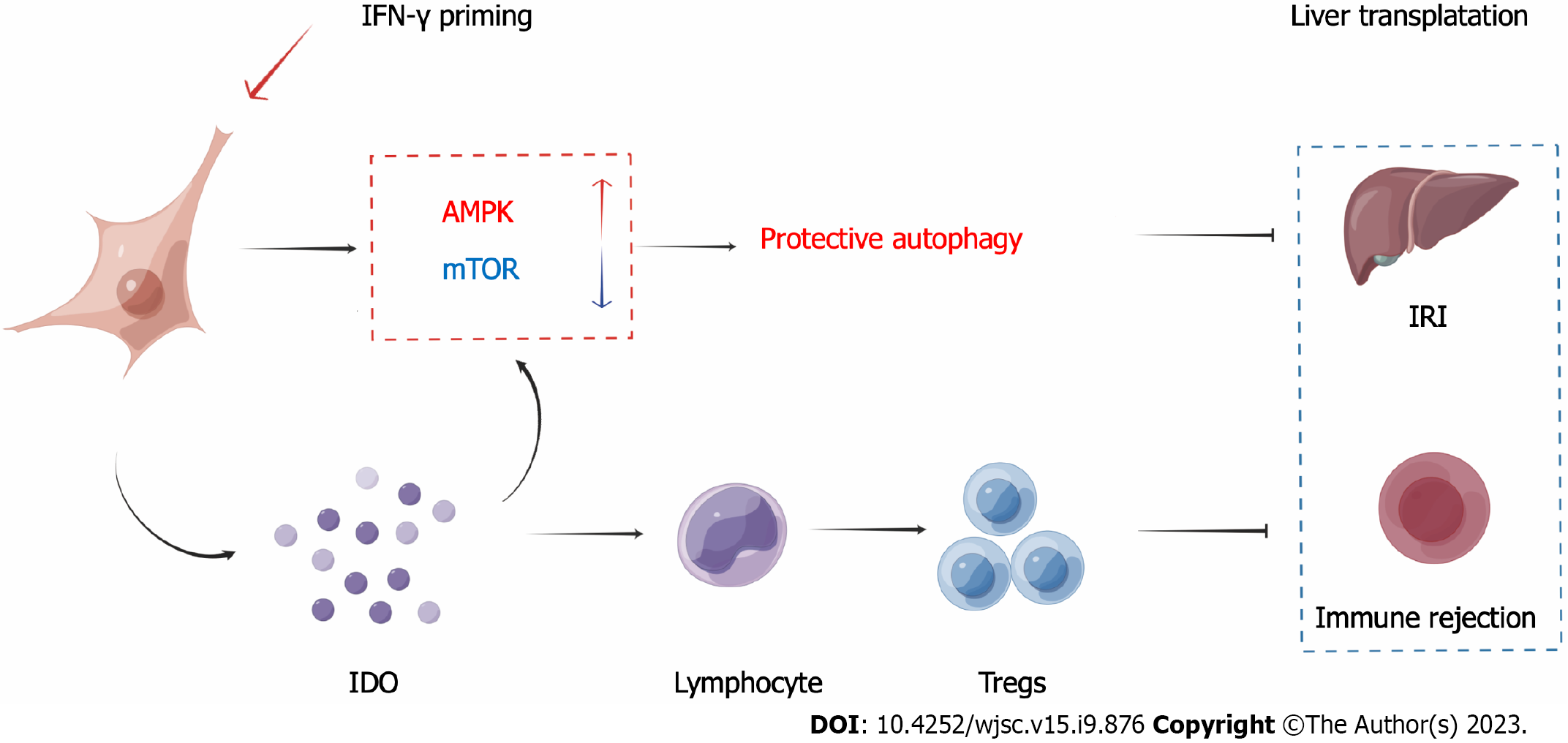
Figure 9 Schematic depiction of the mechanism.
Ischemia-reperfusion injury (IRI) and immune rejection are two important factors that affect the prognosis of liver transplantation. After priming menstrual blood-derived stromal cells (MenSCs) with interferon-γ, the MenSCs secreted high levels of indoleamine 2,3-dioxygenase (IDO). On the one hand, MenSCs and IDO enhanced the AMPK-mammalian target of rapamycin-autophagy axis to reduce IRI; on the other hand, IDO promoted the differentiation of lymphocytes into Tregs to reduce immune rejection. IRI: Ischemia-reperfusion injury; MenSCs: Menstrual blood-derived stromal cells; IFN-γ: Interferon-γ; IDO: Indoleamine 2,3-dioxygenase; AMPK: AMP-activated protein kinase signaling pathway; mTOR: Mammalian target of rapamycin signaling pathway; Tregs: Regulatory T cells.
- Citation: Zhang Q, Zhou SN, Fu JM, Chen LJ, Fang YX, Xu ZY, Xu HK, Yuan Y, Huang YQ, Zhang N, Li YF, Xiang C. Interferon-γ priming enhances the therapeutic effects of menstrual blood-derived stromal cells in a mouse liver ischemia-reperfusion model. World J Stem Cells 2023; 15(9): 876-896
- URL: https://www.wjgnet.com/1948-0210/full/v15/i9/876.htm
- DOI: https://dx.doi.org/10.4252/wjsc.v15.i9.876


