©The Author(s) 2023.
World J Stem Cells. Jul 26, 2023; 15(7): 768-780
Published online Jul 26, 2023. doi: 10.4252/wjsc.v15.i7.768
Published online Jul 26, 2023. doi: 10.4252/wjsc.v15.i7.768
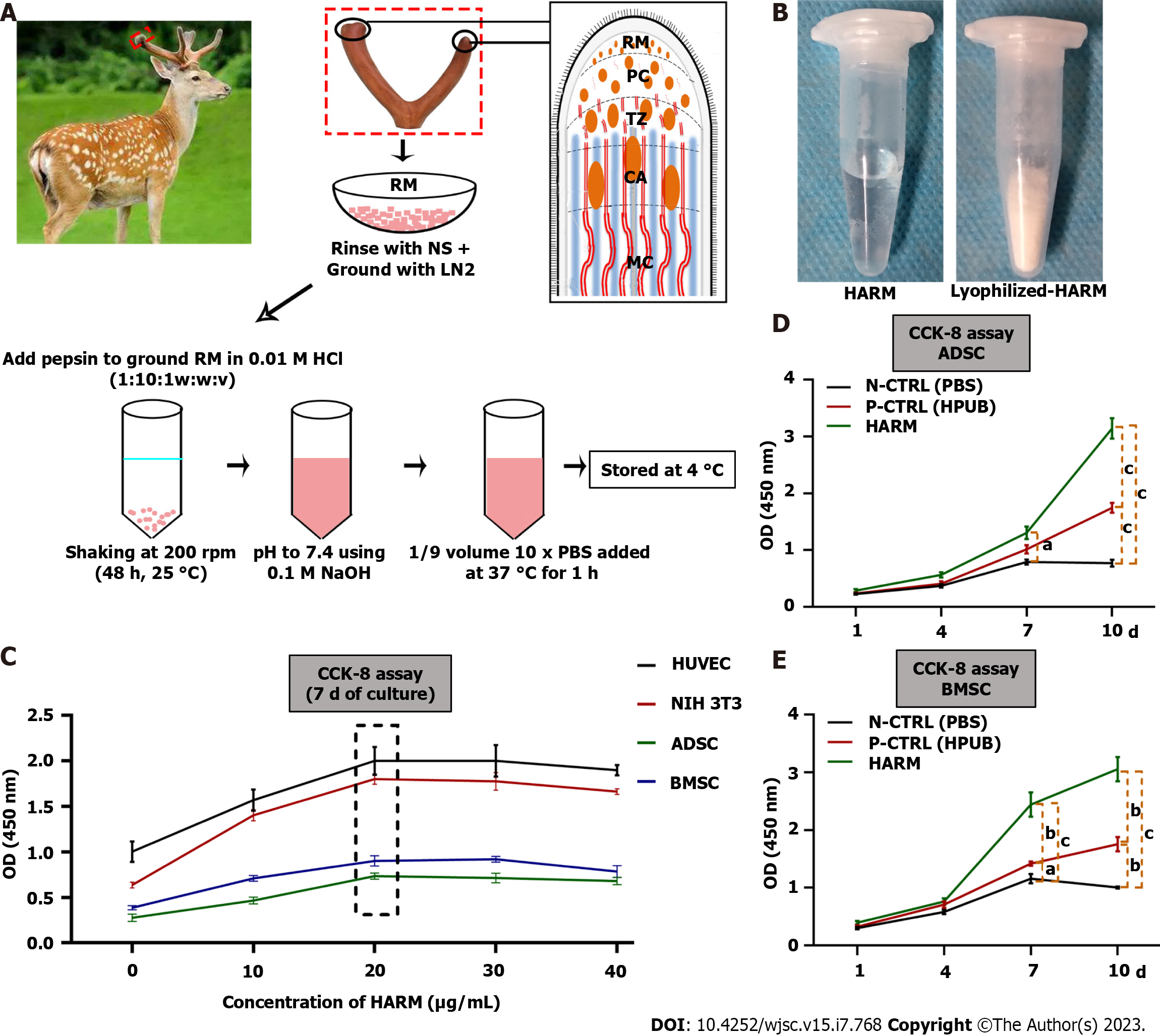
Figure 1 Preparation of hydrogels from the antler reserve mesenchyme matrix and its effects on cell proliferation invitro.
A: Location of reserve mesenchyme (RM) and fabrication of hydrogels from the antler reserve mesenchyme matrix (HARM); B: HARM (top) and lyophilized-HARM (bottom); C: Screening of the optimal concentration of HARM on cell [HUVEC, NIH 3T3, adipose mesenchymal stem cells (ADSC), and bone marrow mesenchymal stem cells (BMSC)] proliferation rate; D and E: Comparison of HARM with hydrogels from porcine urinary bladder matrix (HPUB) on cell (ADSC and BMSC) proliferation rate. Note that the concentration of 20 μg/mL HARM had the strongest stimulation over the others on proliferation rate of HUVEC, NIH 3T3, ADSC, and BMSC; moreover, proliferation rates of ADSC and BMSC in HARM were stronger than in N-CTRL (PBS) or in P-CTRL (HPUB). RM: Reserve mesenchyme; PC: Pre-cartilage; TZ: Transitional zone; CA: Cartilage; NS: Normal saline; LN2: Liquid nitrogen; HARM: Hydrogels derived from antler reserve mesenchyme; HPUB: Hydrogels from porcine urinary bladder matrix; HUVEC: Human umbilical vein endothelial cells; NIH 3T3: Mouse embryonic fibroblasts; ADSC: Adipose mesenchymal stem cells; BMSC: Bone marrow mesenchymal stem cells; N-CTRL: Negative control; P-CTRL: Positive control. n = 3; mean ± SEM; statistically significance set at aP < 0.05, bP < 0.01, and cP < 0.001.
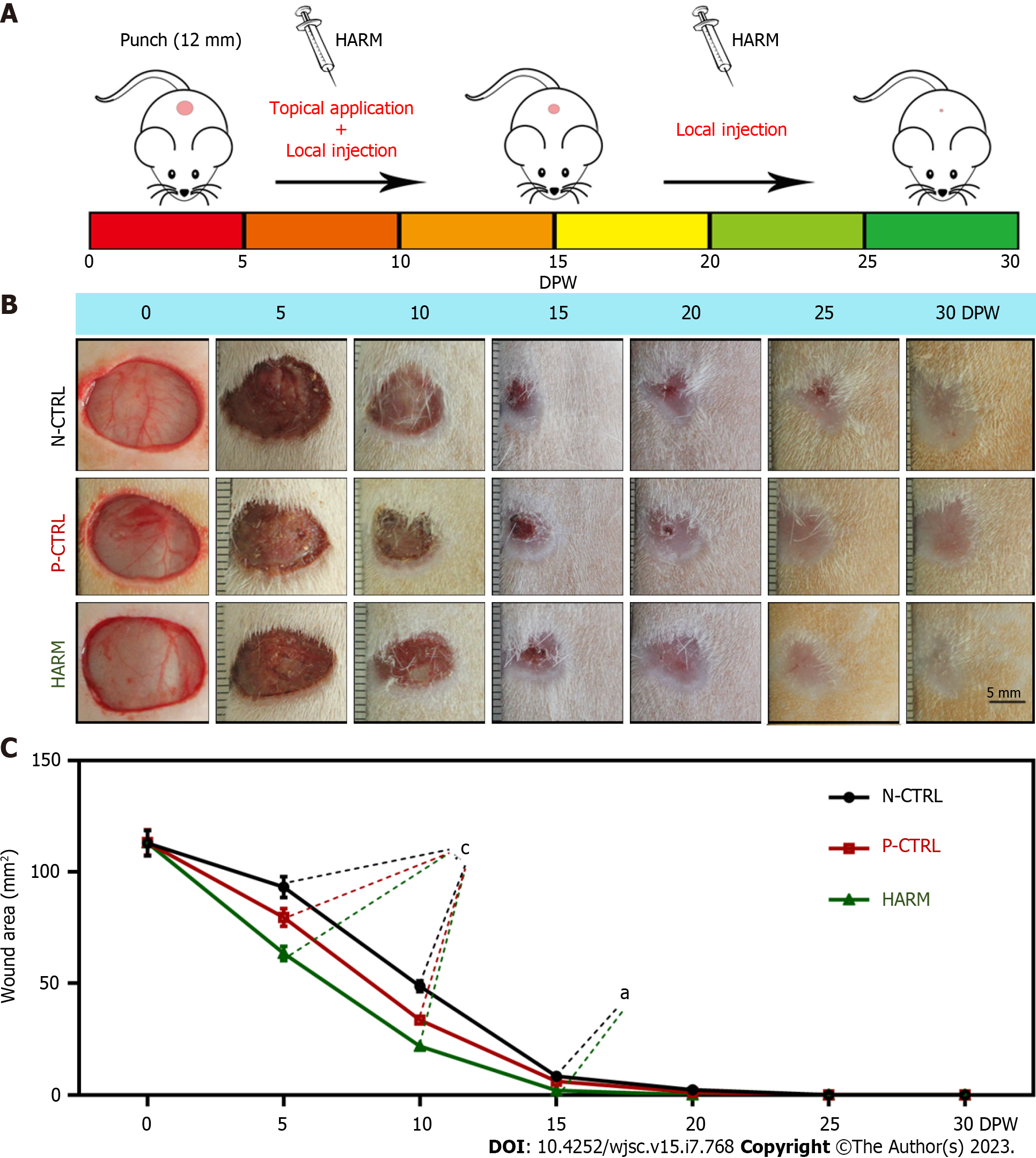
Figure 2 Effects of hydrogels from the antler reserve mesenchyme matrix on wound healing rate in full-thickness skin defects in rats.
A: Experimental procedure; B: Macroscopically observation of the wound healing status; C: Quantification of the wound area using the Image J software according to macroscopical images. Note that the fastest wound healing rate and the smallest scar area were observed in the hydrogels from the antler reserve mesenchyme group. DPW: Days post-wounding; HARM: Hydrogels derived from antler reserve mesenchyme; N-CTRL: Negative control; P-CTRL: Positive control. Mean ± SEM; statistical significance set at aP < 0.05, cP < 0.001.
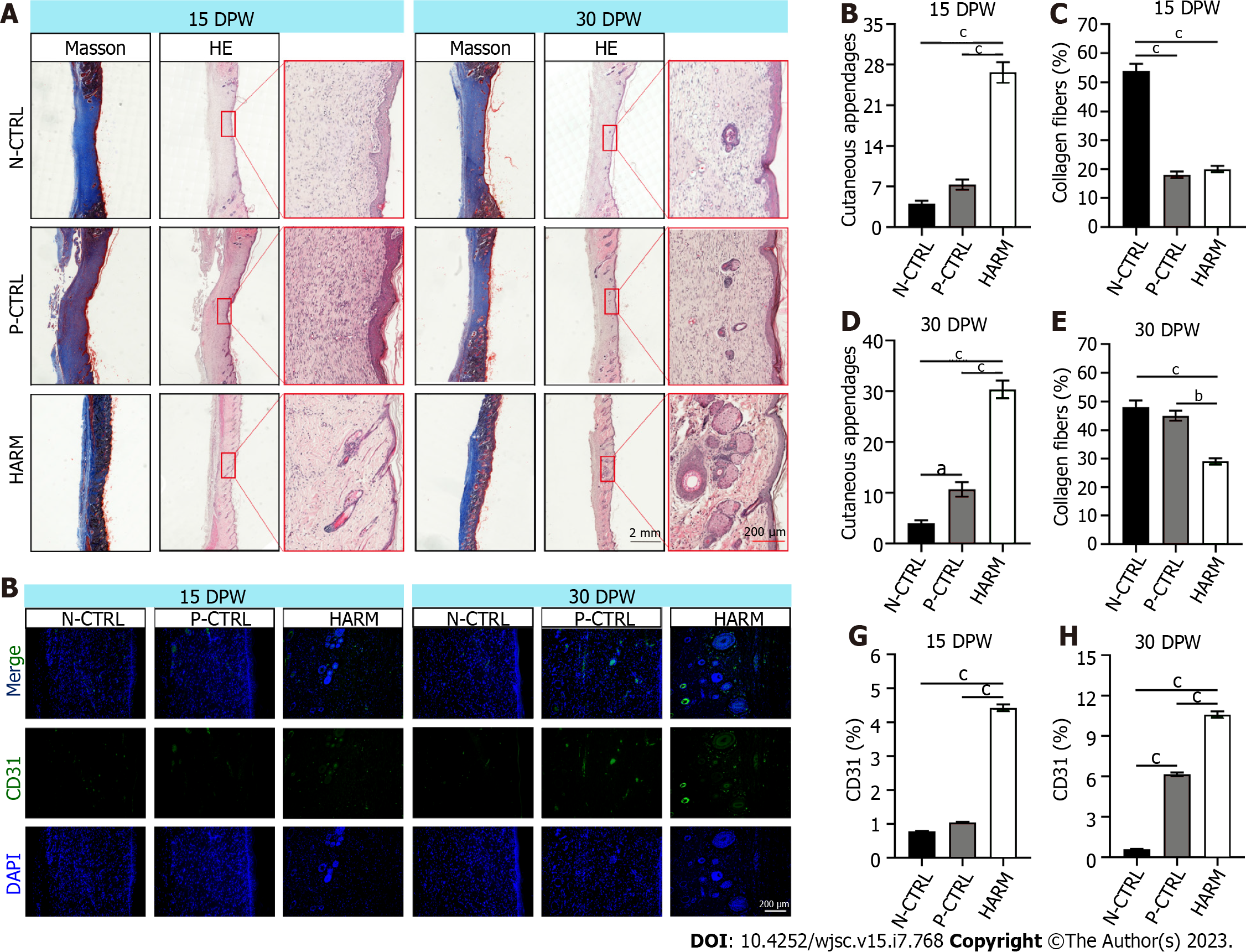
Figure 3 Effects of hydrogels from the antler reserve mesenchyme on the quality of wound healing.
A: Histological sections of the healed skin stained with HE and Masson; B-E: Quantification of cutaneous appendages and the collagen fiber accounts (blue area in Masson staining); F: CD31 immunofluorescence staining; G and H: Expression percentage of CD31. Note that the highest number of cutaneous appendages, least account of collagen fibers, and highest percent were found in the HARM group in the healed tissues compared to the N-CTRL and P-CTRL groups. DPW: Days post-wounding; HARM: Hydrogels derived from antler reserve mesenchyme; N-CTRL: Negative control; P-CTRL: Positive control. n = 3; mean ± SEM; statistical significance set at aP < 0.05, bP < 0.01, cP < 0.001.
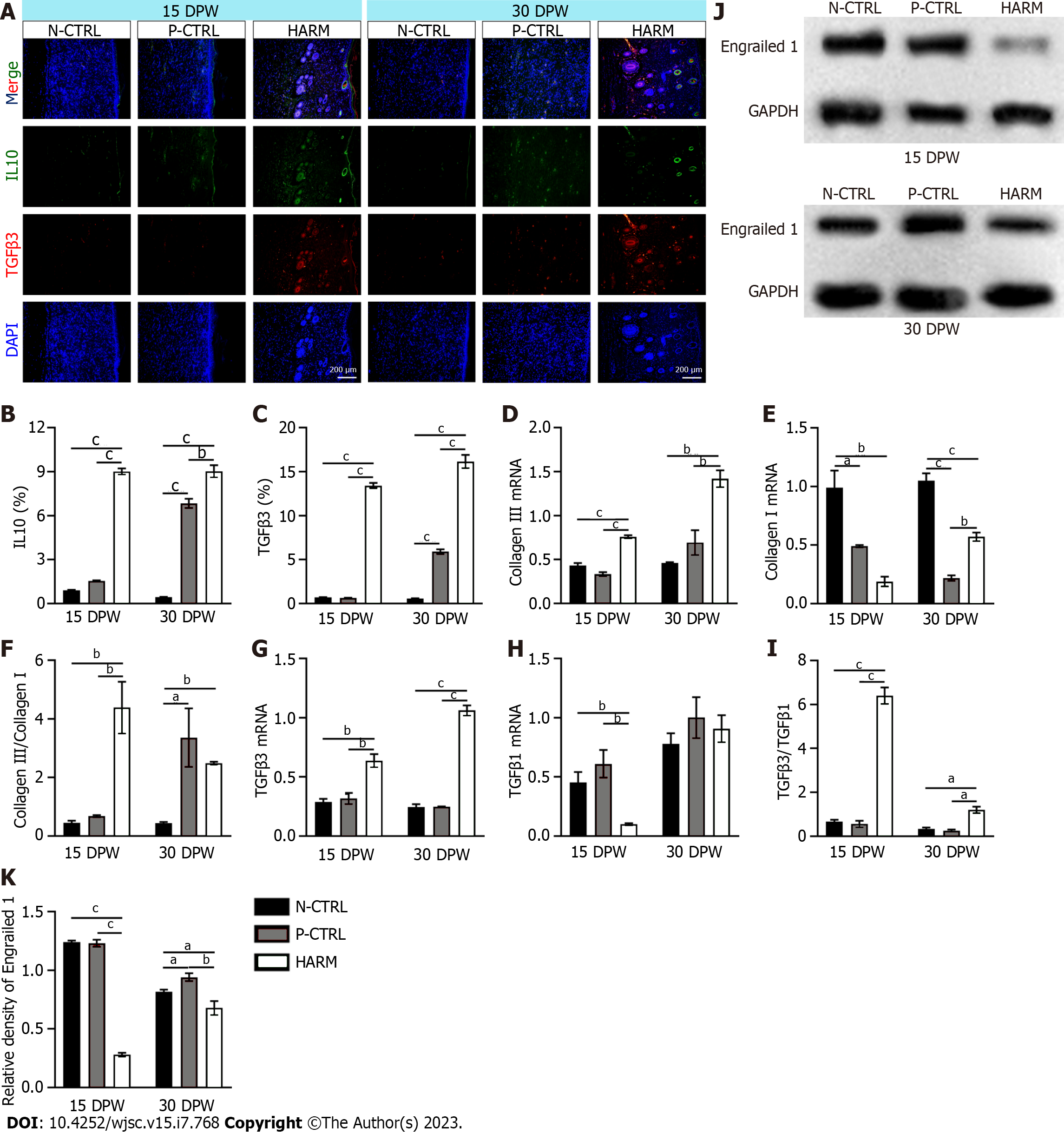
Figure 4 Effects of hydrogels from the antler reserve mesenchyme on the expression of typical genes related to fetal wound healing in healed skin of rat wounds.
A: IL 10 and TGFβ3 immunofluorescence staining; B and C: Expression percentages of IL-10 and TGFβ3; D-I: mRNA expression levels of collagen III (D), collagen I (E), TGFβ3 (G), and TGFβ1 (H) via quantitative real time PCR assay; the ratios of collagen III/collagen I and TGFβ3/TGFβ1 (F and I); J and K: Protein expression of Engrailed 1 in the healed skin via Western blot assay. Note that hydrogels from the antler reserve mesenchyme up-regulated the expression levels of IL10, TGFβ3, and collagen III; but down-regulated the expression levels of collagen I, TGFβ1, and Engrailed 1. IL 10: Interleukin 10; TGFβ1: Transforming growth factor β1; TGFβ3: Transforming growth factor β3; DPW: Days post-wounding; HARM: Hydrogels derived from antler reserve mesenchyme; N-CTRL: Negative control; P-CTRL: Positive control. n = 3; mean ± SEM; statistical significance set at aP < 0.05, bP < 0.01, cP < 0.001.
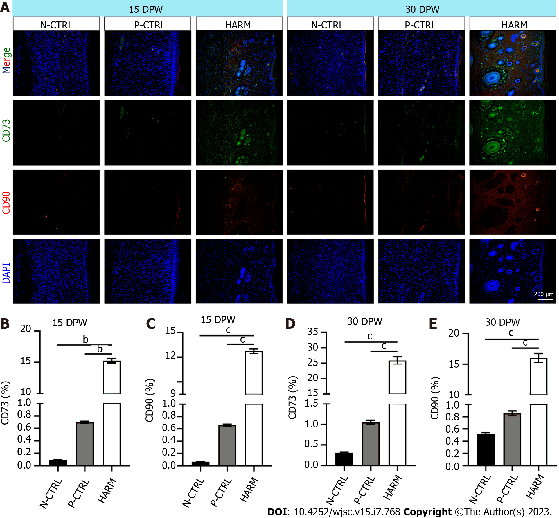
Figure 5 Effects of hydrogels from the antler reserve mesenchyme on the expression of stem cell markers in healed skin of rat wounds.
A: CD73 and CD90 immunofluorescence staining; B and C: Expression percentages of CD73 and CD90 on 15 DPW; D and E: Expression percentages of CD73 and CD90 on 30 DPW. Note that the HARM group had the highest percent of CD73 and CD90 compared to the other two groups. DPW: Days post-wounding; HARM: Hydrogels derived from antler reserve mesenchyme; N-CTRL: Negative control; P-CTRL: Positive control. n = 3; mean ± SEM; statistically significant at bP < 0.01, cP < 0.001.
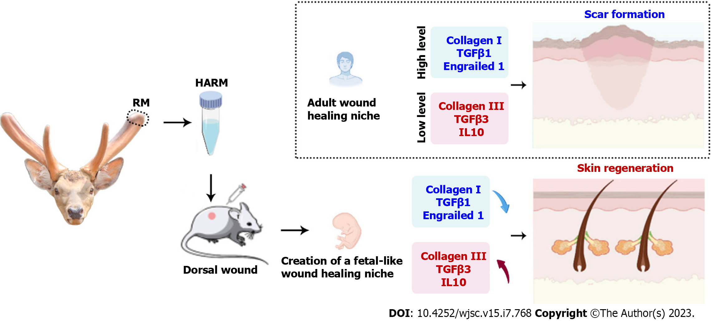
Figure 6 Hydrogels from the antler reserve mesenchyme promoted regenerative wound healing via creating a fetal-like niche.
FMT inhibition. hydrogels from the antler reserve mesenchyme up-regulated the expression levels of fetal wound healing-related genes [interleukin 10, transforming growth factor β3 (TGFβ3), and collagen III]; but down-regulated the expression levels of adult wound healing-related genes (collagen I, TGFβ1, and Engrailed 1). HARM: Hydrogels derived from antler reserve mesenchyme; IL 10: Interleukin 10; TGF: Transforming growth factor.
- Citation: Zhang GK, Ren J, Li JP, Wang DX, Wang SN, Shi LY, Li CY. Injectable hydrogel made from antler mesenchyme matrix for regenerative wound healing via creating a fetal-like niche. World J Stem Cells 2023; 15(7): 768-780
- URL: https://www.wjgnet.com/1948-0210/full/v15/i7/768.htm
- DOI: https://dx.doi.org/10.4252/wjsc.v15.i7.768


