©The Author(s) 2023.
World J Stem Cells. Jul 26, 2023; 15(7): 713-733
Published online Jul 26, 2023. doi: 10.4252/wjsc.v15.i7.713
Published online Jul 26, 2023. doi: 10.4252/wjsc.v15.i7.713
Figure 1 Screening based on GEO datasets identified high expression of COL7A1 in human induced pluripotent stem cells derived keratinocytes and low expression in burn wounds.
A: Volcano plot of differentially expressed genes (DEGs) in GSE140926 dataset, which included 3 normal skin tissue samples and 3 burn eschar tissue samples. Abscissa axis indicates log2 (Fold change); B: Volcano plot of DEGs in GSE27186 dataset, which included 2 human induced pluripotent stem cells (hiPSCs) samples and 2 hiPSCs- keratinocytes samples. Abscissa axis indicates log2 (Fold change); C: The venn diagram exhibited the intersection of the downregulated DEGs in the GSE140926 dataset and the upregulated DEGs in the GSE27186 dataset; D: Gene ontology (GO) entry enrichment bar plot. Abscissa is the proportion of genes identified and ordinate is the GO entry name; E: Visualization of significant DEGs enriched in GO-biological pathway (BP) entries; F: Visualization of significant DEGs enriched in GO-cellular component entries. G: Visualization of significant DEGs enriched in GO-MF entries; H: The Venn diagram exhibited the intersection of GO-BP, GO-CC, and GO-MF; I: Heatmap of the 4 DEGs in GSE140926.
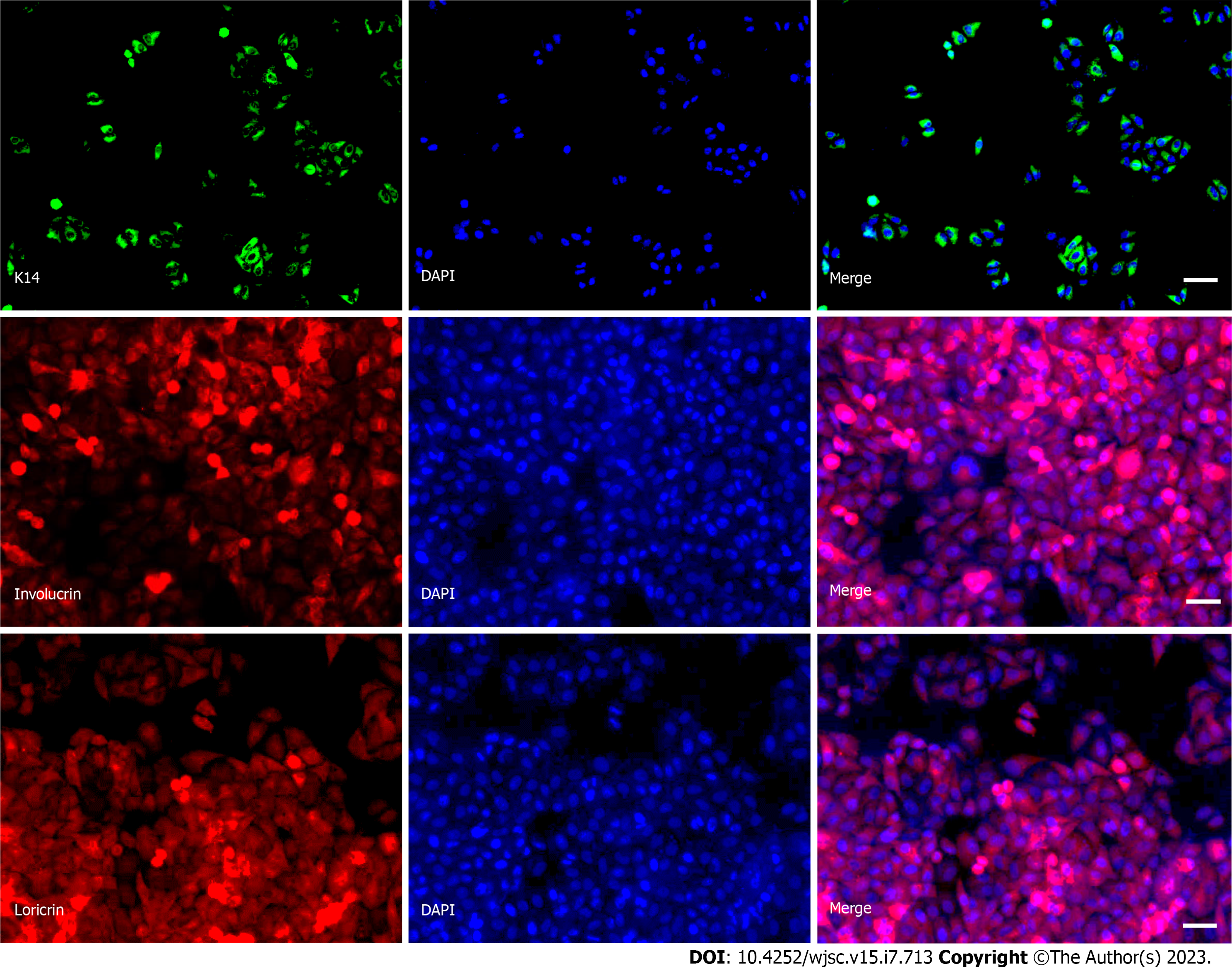
Figure 2 Human induced pluripotent stem cells successfully differentiated into keratinocyte.
Retinoic acid (RA) and BMP-4 were used to induce differentiation of hiPSCs into keratinocyte. The images as immunofluorescence staining of keratinocyte marker proteins K14, involucrin and loricrin at 14 d of induced differentiation, scale bar: 25 µm.
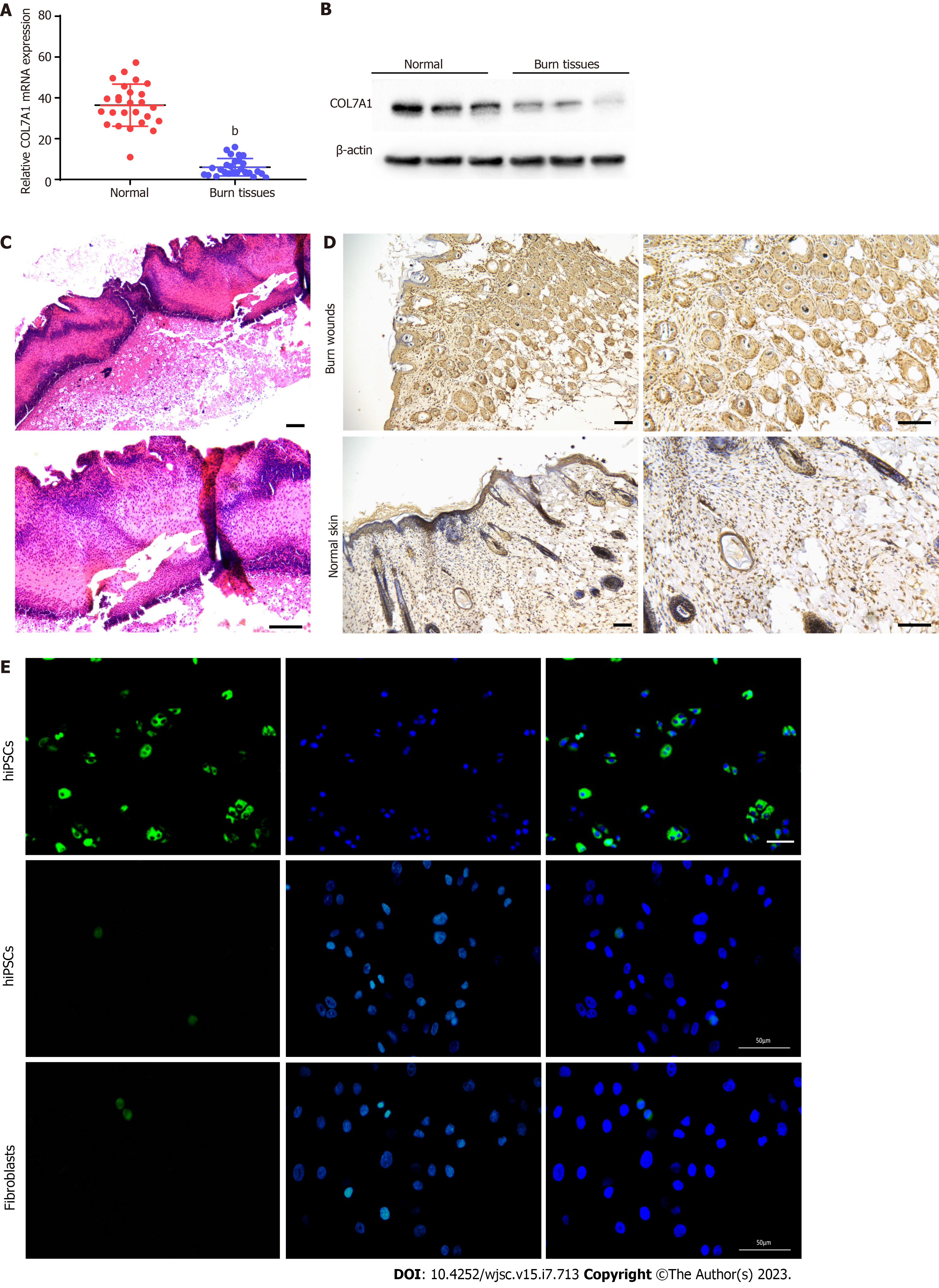
Figure 3 COL7A1 was significantly under-expressed in deep II burn tissues.
A: RT-qPCR analysis of COL7A1 mRNA expression in 26 humans deep second degree burn wound tissues and 26 normal skin tissues; B: Western blotting analysis of COL7A1 protein expression in 3 human deep second degree burn wound tissues and 3 normal skin tissues; C: Representative photomicrographs of hematoxylin-eosin stained burned skin of mice at 24 h post burn, scale bar: 100 µm; D: Immunohistochemical detection of COL7A1 expression levels in burn wounds and surrounding normal skin of mice, scale bar: 100 µm; E: Immunofluorescence detection of COL7A1 expression levels in keratinocytes, hiPSCs and fibroblasts, scale bar: 50 µm. Data: Mean ± SEM. bP < 0.01 vs Normal group.
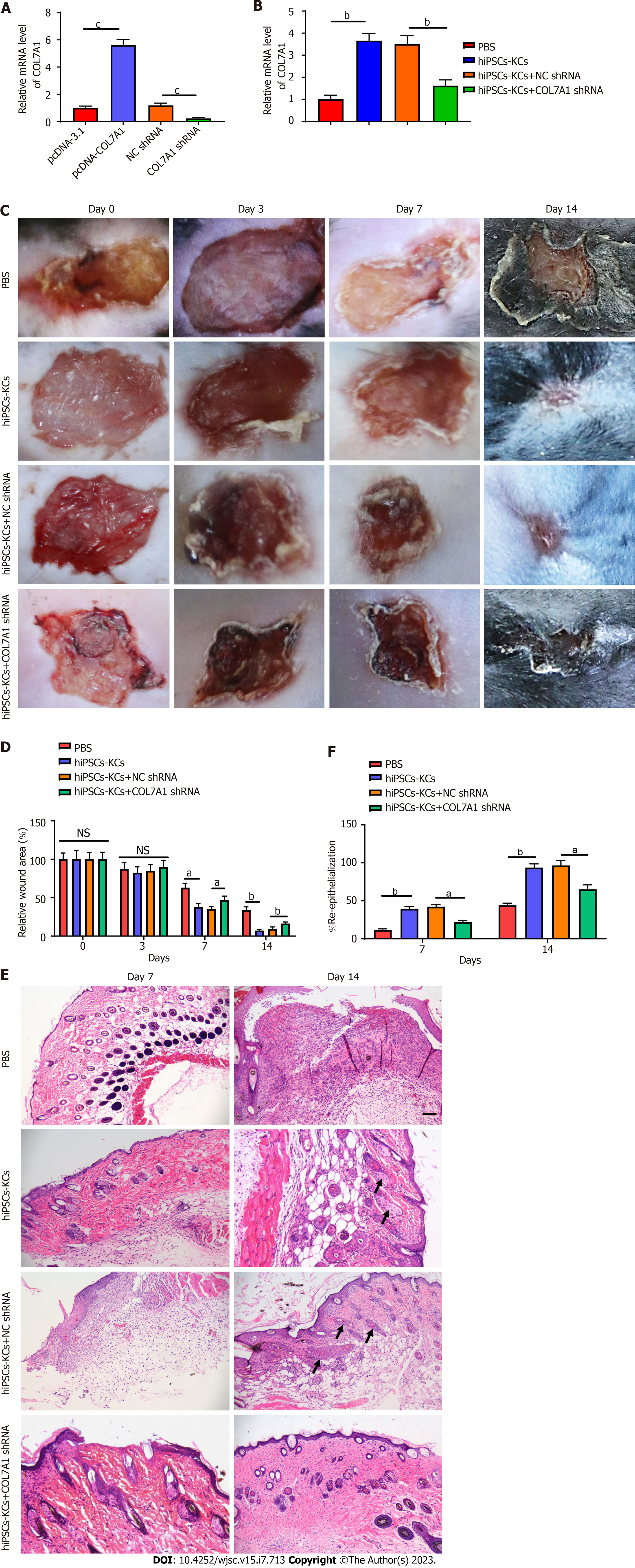
Figure 4 COL7A1 inhibition abolished the promoting effects of human induced pluripotent stem cells derived keratinocytes on burn healing and re epithelialization in mice.
A: RT-qPCR detection of the COL7A1 mRNA levels in human induced pluripotent stem cells derived keratinocytes (hiPSCs-KCs) after transfection of pcDNA- COL7A1 or COL7A1 shRNA; B: RT-qPCR detection of the COL7A1 mRNA levels in burned skin of mice after transplantation of hiPSCs-KCs or COL7A1 knock-downed hiPSCs-KCs; C: At 3, 7, 14 d after hiPSCs-KCs transplantation, the healing of burn wounds in mice was recorded by taking photographs with a digital camera; D: Statistical analysis of relative size of burn wounds in each group of mice; E: Hematoxylin-eosin staining was performed after burn tissues were paraffin sectioned at 7 and 14 d after hiPSCs-KCs transplantation, and then wound healing as well as re-epithelialization were observed by histological sections in each group, scale bar: 200 µm; F: Re-epithelialization ratio of the burned window in mice of each group. Data: Mean ± SEM. aP < 0.05, bP < 0.01, cP < 0.001. n = 10. NS: Not significant; PBS: Phosphate buffered saline; hiPSCs: Human induced pluripotent stem cells; KCs: Keratinocytes.
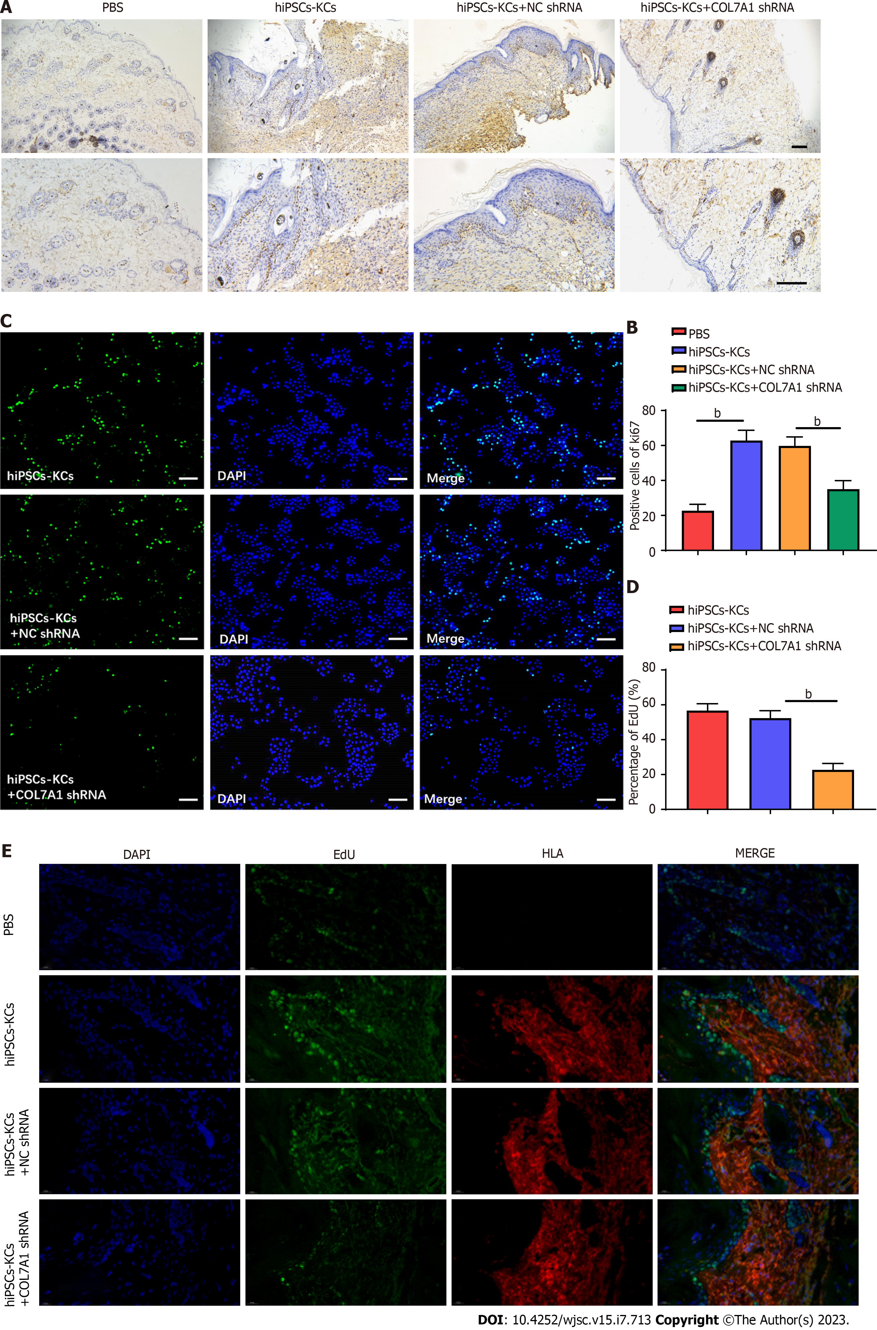
Figure 5 COL7A1 knockdown abolishes the facilitative effect of human induced pluripotent stem cells derived keratinocytes on keratinocyte proliferation in mouse burn wounds.
A: The Ki67 positive cells in burn wound tissues of each group were detected by immunostaining on the 14th postoperative day. scale bar: 200 µm; B: The statistical graph of the number of Ki67 positive cells; C: The proliferation ability of keratinocytes in each group was detected by EdU staining, scale bar: 100 µm; D: The proliferation ability of keratinocytes in each group was detected by EdU staining; E: Immunofluorescence estimate of the EdU and HLA levels in burn tissue of mice in each group. aP < 0.05, bP < 0.01. n = 10. PBS: Phosphate buffered saline; hiPSCs: Human induced pluripotent stem cells; KCs: Keratinocytes; HLA: Human leukocyte antigen.
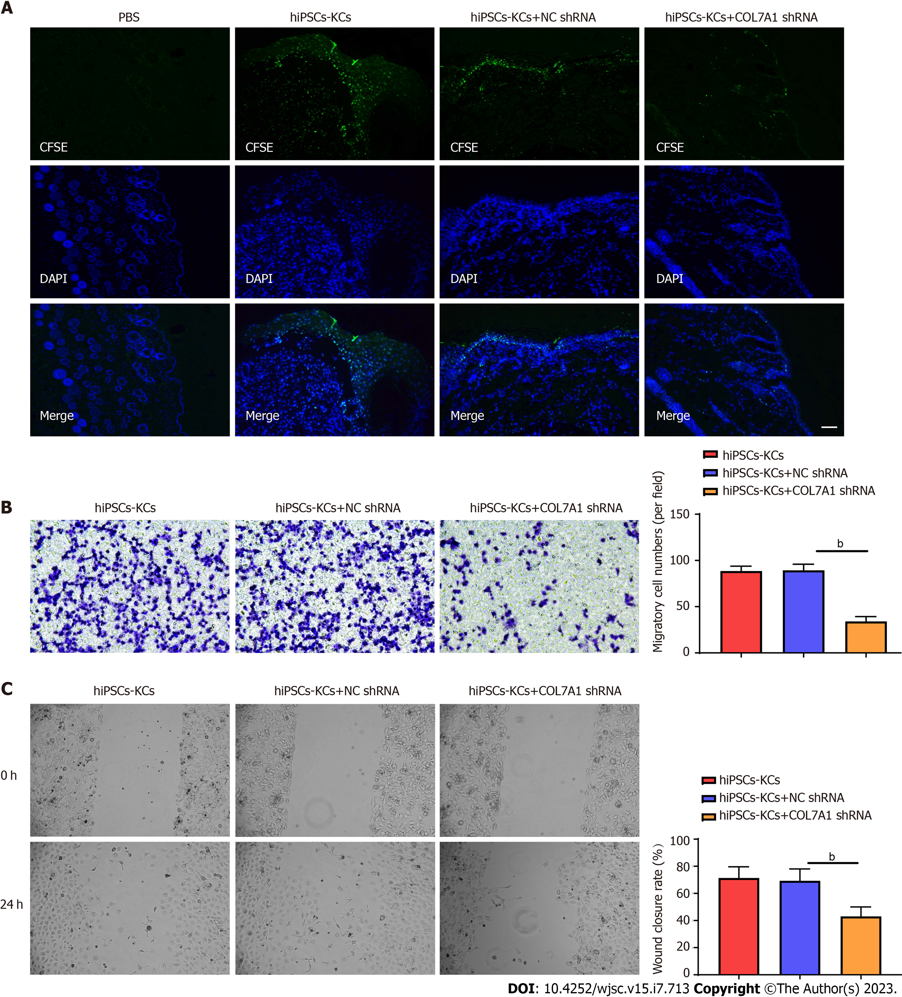
Figure 6 COL7A1 knockdown abolishes the facilitative effect of human induced pluripotent stem cells derived keratinocytes on keratinocyte migration in mouse burn wounds.
A: On the 14th day after operation, frozen sections showed that carboxifluorescein diacetate succinimidyl ester labeled human induced pluripotent stem cells derived keratinocytes (hiPSCs-KCs) migrated to the epithelium of the wound healing site scale bar: 100 µm; B: Transwell assay was employed to determine the migration capability of hiPSCs-KCs transfected with COL7A1 shRNA, scale bar: 100 µm; C: Damage repair assay was applied to test the migration capability of hiPSCs-KCs transfected with COL7A1 shRNA. Data: Mean ± SEM. aP < 0.05, bP < 0.01, cP < 0.001. n = 10. PBS: Phosphate buffered saline; hiPSCs: Human induced pluripotent stem cells; KCs: Keratinocytes; CFSE: Carboxifluorescein diacetate succinimidyl ester.
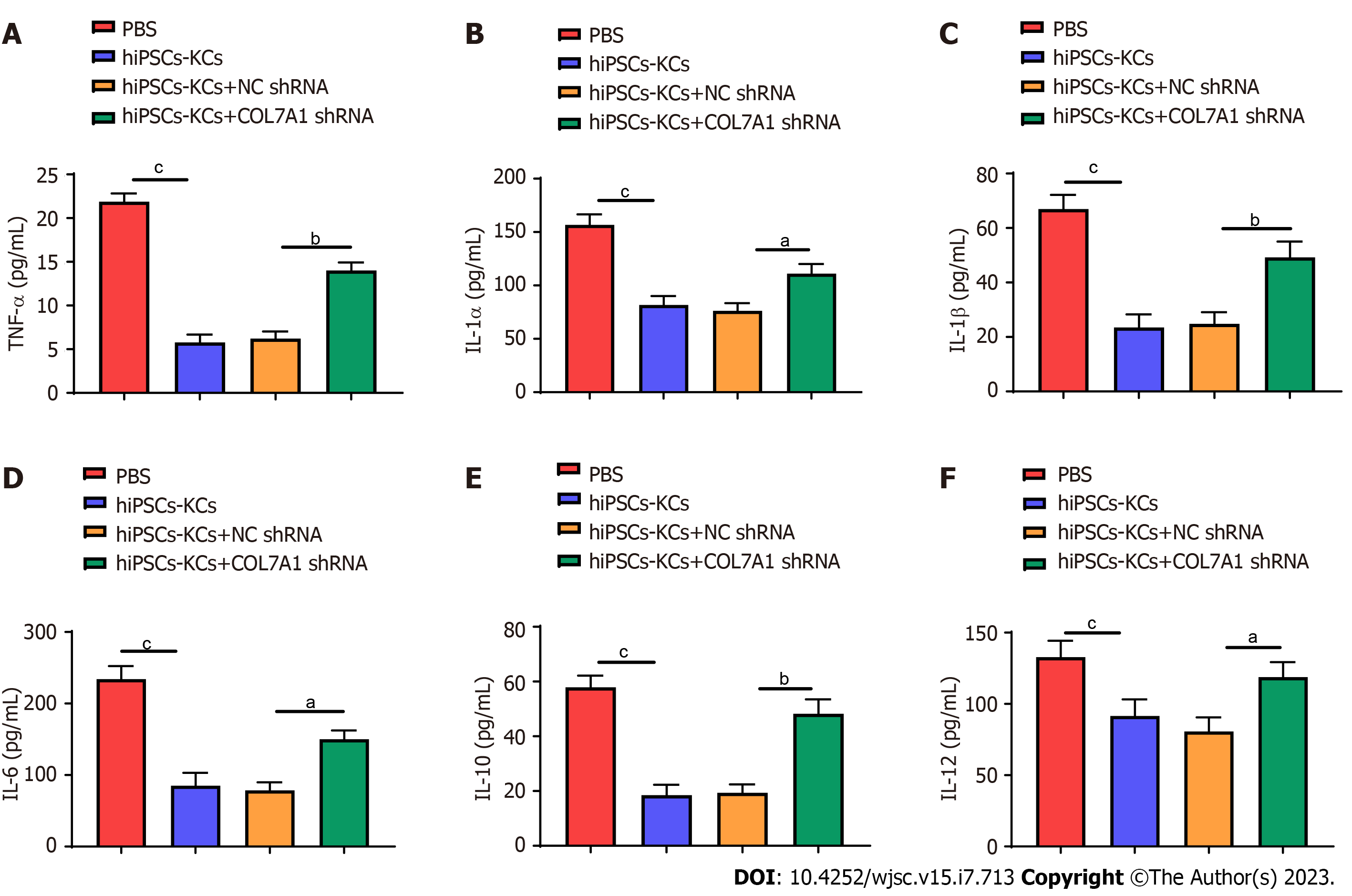
Figure 7 COL7A1 inhibition counteracted the inhibitory effects of human induced pluripotent stem cells derived keratinocytes on the inflammatory response to burn injury in mice.
A: Enzyme linked immunosorbent assay (ELISA) detection of the tumor necrosis factor α contents in the plasma of mice in each group; B: ELISA detection of the interleukin (IL)-1α contents in the plasma of mice in each group; C: ELISA detection of the IL-1β contents in the plasma of mice in each group; D: ELISA detection of the IL-6 contents in the plasma of mice in each group; E: ELISA detection of the IL-10 contents in the plasma of mice in each group; F: ELISA detection of the IL-12 contents in the plasma of mice in each group. aP < 0.05, bP < 0.01, cP < 0.001. n = 10. PBS: Phosphate buffered saline; hiPSCs: Human induced pluripotent stem cells; KCs: Keratinocytes.
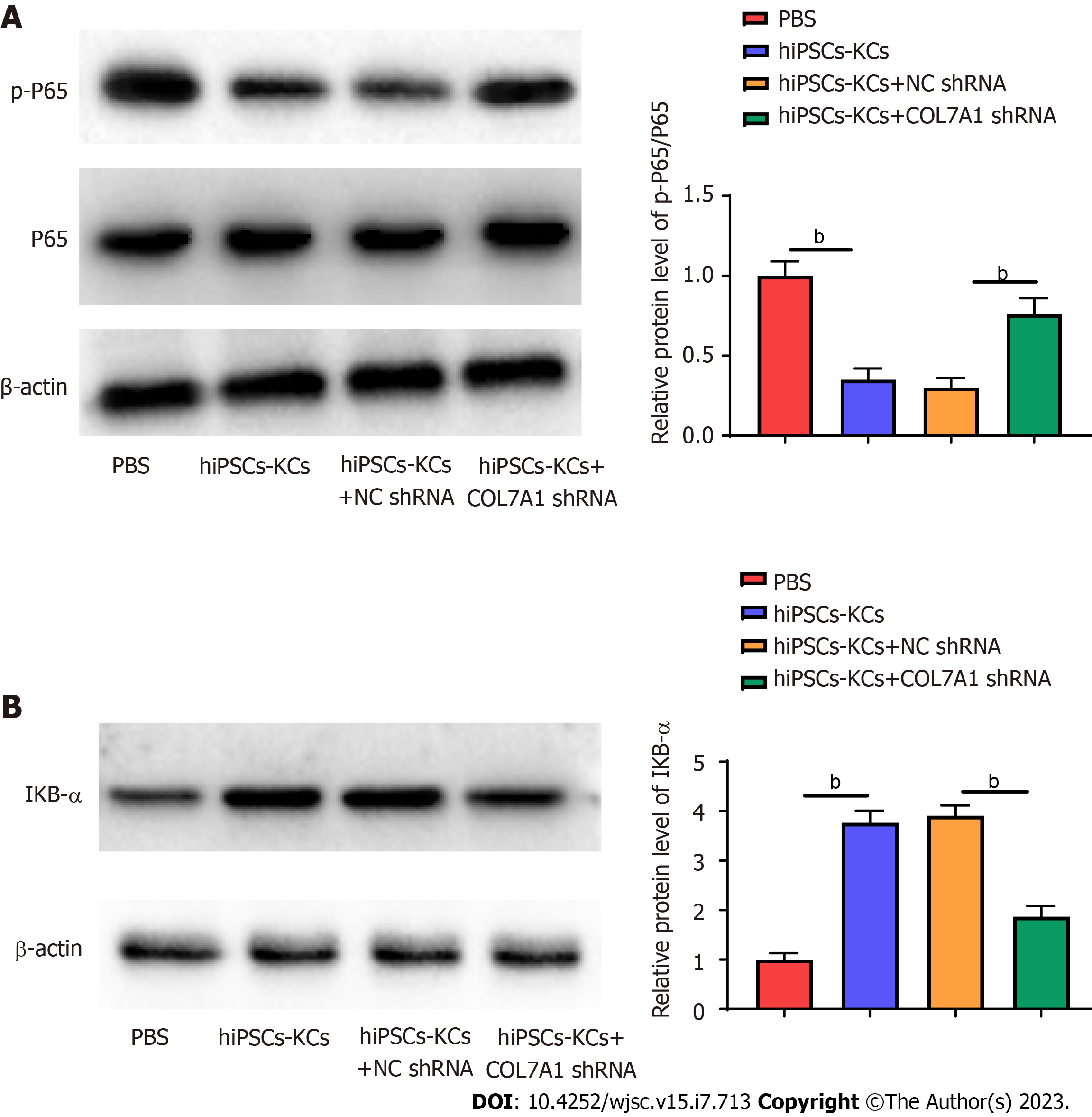
Figure 8 COL7A1 inhibition reversed the inhibitory effect of human induced pluripotent stem cells derived keratinocytes on the NF-κB pathway in mouse burn wounds.
A: The protein level of p-p65 in mouse wound tissues treated with hiPSCs-KCs, with or without COL7A1 shRNA transfection, was employed by Western blotting; B: The protein level of IκBα in mouse wound tissues treated with hiPSCs-KCs, with or without COL7A1 shRNA transfection, was employed by Western blotting. bP < 0.01. n = 10. PBS: Phosphate buffered saline; hiPSCs: Human induced pluripotent stem cells; KCs: Keratinocytes.
- Citation: Wu LJ, Lin W, Liu JJ, Chen WX, He WJ, Shi Y, Liu X, Li K. Transplantation of human induced pluripotent stem cell derived keratinocytes accelerates deep second-degree burn wound healing. World J Stem Cells 2023; 15(7): 713-733
- URL: https://www.wjgnet.com/1948-0210/full/v15/i7/713.htm
- DOI: https://dx.doi.org/10.4252/wjsc.v15.i7.713


